

Detalles de Imagen
![]() Home Banco de Imágenes
Home Banco de Imágenes
![]() Búsqueda Imágenes
Búsqueda Imágenes
![]() Imágenes Recientes
Imágenes Recientes
![]() Home BJORL
Home BJORL
![]()
Código de la Imagen : 3669
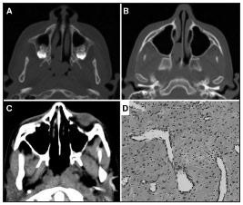
Figure 1. A: Preoperative axial CT scan showing the tumor inserted into the nasal septum and extending to the choanae
Imagen publicada en: 2013 Vol.: 79 Ed.: 5 - 23º
1
Descripción: Figure 1. A: Preoperative axial CT scan showing the tumor inserted into the nasal septum and extending to the choanae; B: Axial CT scan showing preoperative pterygopalatine fossa without disease involvement; C: Axial CT scan postoperatively; D: HE histological section showing spindle cell proliferation with hyalinization areas intermingled with vessels - sometimes arched.
Autor (es) del artículo de origen: Felipe Gustavo Correia1; Juliana Caminha Simões2; José Arruda Mendes-Neto3; Maria Teresa de Seixas-Alves4; Luis Carlos Gregório5; Eduardo Macoto Kosugi6
Título y link del artículo: Extranasopharyngeal angiofibroma of the nasal septum - uncommon presentation of a rare disease
oldfiles.bjorl.org/conteudo/acervo/acervo_english.asp?id=4509