 |
|
| Código de la Imagen : 3669 | |
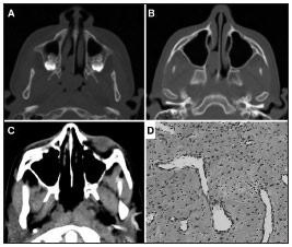
| Figure 1. A: Preoperative axial CT scan showing the tumor inserted into the nasal septum and extending to the choanae | |
Imagen publicada en: |
|
|
|
| 1 | |
| Descripción: | |
|
|
| Autor (es) del artículo de origen: | |
| Felipe Gustavo Correia1; Juliana Caminha Sim§es2; JosÚ Arruda Mendes-Neto3; Maria Teresa de Seixas-Alves4; Luis Carlos Gregˇrio5; Eduardo Macoto Kosugi6 | |
| Título y link del artículo: | |
| Extranasopharyngeal angiofibroma of the nasal septum - uncommon presentation of a rare disease | |
| oldfiles.bjorl.org/conteudo/acervo/acervo_english.asp?id=4509 |
All rights reserved - 1933 /
2025
© - Associação Brasileira de Otorrinolaringologia e Cirurgia Cérvico Facial

