

Detalles de Imagen
![]() Home Banco de Imágenes
Home Banco de Imágenes
![]() Búsqueda Imágenes
Búsqueda Imágenes
![]() Imágenes Recientes
Imágenes Recientes
![]() Home BJORL
Home BJORL
![]()
Código de la Imagen : 3607
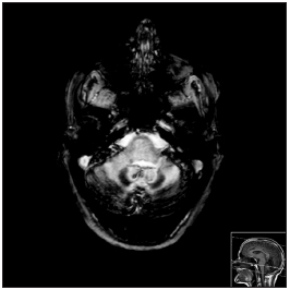
Figure 1. MRI - CE, axial slices in T2*
Imagen publicada en: 2013 Vol.: 79 Ed.: 2 - 21º
1
Descripción: Figure 1. MRI - CE, axial slices in T2*, where we see intense hypo signal in T2* of the pial coating of all the structures in the posterior fossa, in the internal vertent of the occipital lobe and the Sylvian fissures, marked cerebellar atrophy, translating into superficial siderosis of the CNS.
Autor (es) del artículo de origen: Diana Cunha Ribeiro1; Joana Nunes2; Ana Cláudia Ribeiro3; Felisberto Maricato4; Carlos Ribeiro5
Título y link del artículo: Superficial siderosis of the central nervous system: an usual cause of sensorineural hearing loss
oldfiles.bjorl.org/conteudo/acervo/acervo_english.asp?id=4435