|

| Código de la Imagen : 3607 | |
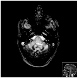
| Figure 1. MRI - CE, axial slices in T2* | |
Imagen publicada en: |
|
|
|
| 1 | |
| Descripción: | |
|
|
| Autor (es) del artículo de origen: | |
| Diana Cunha Ribeiro1; Joana Nunes2; Ana Cláudia Ribeiro3; Felisberto Maricato4; Carlos Ribeiro5 | |
| Título y link del artículo: | |
| Superficial siderosis of the central nervous system: an usual cause of sensorineural hearing loss | |
| oldfiles.bjorl.org/conteudo/acervo/acervo_english.asp?id=4435 |
All rights reserved - 1933 /
2026
© - Associação Brasileira de Otorrinolaringologia e Cirurgia Cérvico Facial