

Detalles de Imagen
![]() Home Banco de Imágenes
Home Banco de Imágenes
![]() Búsqueda Imágenes
Búsqueda Imágenes
![]() Imágenes Recientes
Imágenes Recientes
![]() Home BJORL
Home BJORL
![]()
Código de la Imagen : 3585
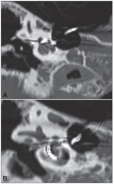
Figure 3. A-B: Right temporal bone high-resolution CT scan.
Imagen publicada en: 2013 Vol.: 79 Ed.: 2 - 5º
3
Descripción: Figure 3. A-B: Right temporal bone high-resolution CT scan. Coronal view, bone window, showing the placement of the array from the basal (A) to the apical (B) turn of the cochlea.
Autor (es) del artículo de origen: Aline Gomes Bittencourt1; Robinson Koji Tsuji2; João Paulo Ratto Tempestini3; Alfredo Luiz Jacomo4; Ricardo Ferreira Bento5; Rubens de Brito6
Título y link del artículo: Cochlear implantation through the middle cranial fossa: a novel approach to access the basal turn of the cochlea
oldfiles.bjorl.org/conteudo/acervo/acervo_english.asp?id=4419