|

| Código de la Imagen : 3585 | |
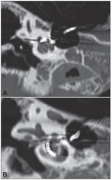
| Figure 3. A-B: Right temporal bone high-resolution CT scan. | |
Imagen publicada en: |
|
|
|
| 3 | |
| Descripción: | |
|
|
| Autor (es) del artículo de origen: | |
| Aline Gomes Bittencourt1; Robinson Koji Tsuji2; João Paulo Ratto Tempestini3; Alfredo Luiz Jacomo4; Ricardo Ferreira Bento5; Rubens de Brito6 | |
| Título y link del artículo: | |
| Cochlear implantation through the middle cranial fossa: a novel approach to access the basal turn of the cochlea | |
| oldfiles.bjorl.org/conteudo/acervo/acervo_english.asp?id=4419 |
All rights reserved - 1933 /
2025
© - Associação Brasileira de Otorrinolaringologia e Cirurgia Cérvico Facial