

Detalles de Imagen
![]() Home Banco de Imágenes
Home Banco de Imágenes
![]() Búsqueda Imágenes
Búsqueda Imágenes
![]() Imágenes Recientes
Imágenes Recientes
![]() Home BJORL
Home BJORL
![]()
Código de la Imagen : 3554
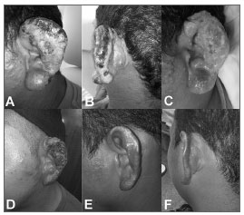
Suppurative lesion
Imagen publicada en: 2012 Vol.: 78 Ed.: 5 - 23º
1
Descripción: Figure 1. A: Suppurative lesion involving the ear pinna, with necrotic areas in the helix and, in a lesser degree in the anti-helix, infiltrative aspect in the lobe and papules in the pre-auricular region. Notice the lack of lesion in the external auditory canal inlet. B: Necrosis extending to the helix, infiltrative aspect in the adjacent skin and papule with central necrosis behind the ear, similar to the inoculation lesion. C: Granulomatous aspect seen in the bottom of the lesion after improvement in the suppurative process. Notice the persistence of papulae behind the year and the infiltrative aspect in the lobe and adjacent skin. D: Initial state of the ulcer healing process, about 20 days after treatment onset. E-F: Ear pinna reepithelialisation, 60 days after the end of treatment. Notice the improvement in the ear lobe and in the pre and retro auricular regions.
Autor (es) del artículo de origen: Márcia dos Santos da Silva1; Renato Telles de Sousa2; Eucides Batista da Silva3; Jorge Augusto de Oliveira Guerra4; Nathália Matos Gomes5; Renata Farias de Santana6; Rebecca Souza Mubarac7
Título y link del artículo: Primary lesion of Mucocutaneous Leishmaniasis simulating external otitis
oldfiles.bjorl.org/conteudo/acervo/acervo_english.asp?id=4367