

Detalles de Imagen
![]() Home Banco de Imágenes
Home Banco de Imágenes
![]() Búsqueda Imágenes
Búsqueda Imágenes
![]() Imágenes Recientes
Imágenes Recientes
![]() Home BJORL
Home BJORL
![]()
Código de la Imagen : 3672
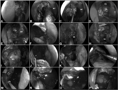
Figure 2. A-B: Intraoperative photographs demonstrating the resection of a right ethmoidal adenocarcinoma.
Imagen publicada en: 2013 Vol.: 79 Ed.: 6 - 18º
2
Descripción: Figure 2. A-B: Intraoperative photographs demonstrating the resection of a right ethmoidal adenocarcinoma. Firstly, the tumor is debulked to identify it origin; C-D: Subperiosteal dissection of the tumor separates it from the medial orbital wall, nasoethmoidal complex and nasofrontal recess; E: The dissection continues along the maxillary line and posteriorly, along the lamina papyracea, to reach sphenoid sinus; F: The left middle turbinate is removed to establish an adequate margin and to expand the space for instrumentation; G-H: Wide sphenoidotomies establish the posterior margin; I: Residual tumor at the anterior aspect of the medial wall of the maxillary sinus cannot be adequately removed via a midmeatal antrostomy; J: An endoscopic medial maxillectomy is performed with the resection of the inferior turbinate; K: The resection extends from the orbit down to the floor of nose; however, its' exposure is insufficient. Therefore, an endoscopic Denker's approach is deemed necessary for a full exposure; L: The piriform aperture and ascending process of the maxilla are removed, dissecting the nasolacrimal duct and transecting it sharply. Exposure of the piriform aperture requires a vertical incision on the edge of the aperture; M-N: This edge can be palpated with a blunt dissector to optimize the placement of the incision, which is then carried through the periosteum down to bone. A subperiosteal lateral dissection exposes the anterior maxilla. The medial maxillectomy is then extended to remove the piriform aperture and sufficient anterior maxillary wall to expose the entire confines of the antrum; O-P: This corridor facilitates the adequate resection of tumor with negative margins; T: Tumor; MT: Middle turbinate; FS: Frontal sinus; SS: Sphenoid sinus; MS: Maxillary sinus; IT: Inferior turbinate.
Autor (es) del artículo de origen: Pornthep Kasemsiri; Daniel Monte Serrat Prevedello; Bradley Alan Otto; Matthew Old; Leo Ditzel Filho; Amin Bardai Kassam; Ricardo Luis Carrau
Título y link del artículo: Endoscopic Endonasal endonasal technique: treatment of paranasal and anterior skull base malignancies
oldfiles.bjorl.org/conteudo/acervo/acervo_english.asp?id=4531