

Detalles de Imagen
![]() Home Banco de Imágenes
Home Banco de Imágenes
![]() Búsqueda Imágenes
Búsqueda Imágenes
![]() Imágenes Recientes
Imágenes Recientes
![]() Home BJORL
Home BJORL
![]()
Código de la Imagen : 3666
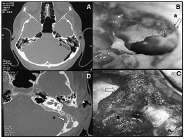
Figure 1. A: CT scan showing the communication between the mastoid cavity
Imagen publicada en: 2013 Vol.: 79 Ed.: 5 - 20º
1
Descripción: Figure 1. A: CT scan showing the communication between the mastoid cavity and the posterior fossa; B: Intraoperative image of a mastoidectomy showing the communication between mastoid air cells and the posterior fossa (A); C: occlusion with a temporal muscle pedicled flap; D: Six months after surgery: closure of the communication, occlusion of the mastoid and middle ear (A) and absorption of the pneumocephalus (B).
Autor (es) del artículo de origen: Fabio Augusto Rabello1; Eduardo Tanaka Massuda2; Jose Antonio Apparecido de Oliveira3; Miguel Angelo Hyppolito4
Título y link del artículo: Otogenic Spontaneous pneumocephalus: case report
oldfiles.bjorl.org/conteudo/acervo/acervo_english.asp?id=4506