

Year: 2003 Vol. 69 Ed. 6 - (16º)
Artigo de Revisão
Pages: 829 to 837
 PDF PT
 Tuberculous otitis media
Author(s):
Márcia M. Pinho1,
Arthur Octavio de A. Kós2
Keywords: ear, otitis, tuberculosis, extrapulmonary, otorhinolaryngologic
Abstract:
This paper is the result of a literature review since the beginning of the 20th century about Tuberculous Otitis Media (TOM). It includes historic, epidemiologic, physiopatologic, clinical, diagnostic and treatment aspects, emphasizing their evolution throughout time considering the several modifications of the population and medicine. It also reinforces the predominance of extrapulmonary forms of tuberculosis, as TOM, in HIV infected patients, which, in present times, is of great interest.
![]()
INTRODUCTION
Tuberculosis is a necrotizing bacterial disease caused by Mycobacterium tuberculosis. It primarily affects the lungs but it can be secondarily disseminated to other organs. Primary infections of other organs, such as the ear for example, without detection of the primary pulmonary focus, are rare presentations.
Despite the advances in clinical treatment of tuberculosis, from pasteurization of milk to vaccination with BCG, tuberculosis remains as of the most common infectious diseases of our times. Its incidence gained new ascension in 1989. This tendency seems to be due to different factors, especially the increase of risk populations to the disease, including HIV patients, homeless people, victims of hunger and malnutrition, and intravenous drug users. The difficulty to maintain effective treatment of tuberculosis, owing to short governmental funds, and difficulty to manage and monitor treatment, especially in developing countries, have also contributed to the current increase in tuberculosis. On top of that, there is the increase in drug-resistant mycobacteria.
Tuberculous otitis media (TOM) is not a frequent disease but when present, it causes significant morbidity. It is the second most frequently disease of tuberculous etiology seen in ENT, second only to laryngeal tuberculosis. There are indications that its frequency is actually higher than estimated, owing to a large number of undiagnosed cases because of lack of suspicion or difficulty to confirm the etiology.
The existence of effective therapy makes early diagnosis essential to have a satisfactory final outcome, starting clinical treatment as soon as possible and preventing the occurrence of severe complications such as hearing loss, facial paralysis, intracranial involvement and unnecessary surgical interventions.
LITERATURE REVIEW
History of tuberculous otitis media
The involvement of the temporal bone in tuberculosis was first described by Jean Louis Petit in the 18th century, much before the discovery of tuberculosis bacilli 1. In 1835, Romberg and Geissler associated tuberculous mastoiditis with pulmonary tuberculosis 2. The clinical signs of the disease were described for the first time by Wilde, who in 1853, described the occurrence of non-painful otorrhea and tympanic membrane characteristics 3. In 1882, Koch demonstrated the bacillus of tuberculosis and in 1883 Esche isolated the bacillus in middle ear secretion4,5.
Incidence comparing to otitis media
TOM was a common disease in the first decades of the 20th century, a time when the condition was known as a clinical entity but to which there were no antimicrobial agents.
According to Turner and Fraser, in a study conducted between 1907 and 1914, among the 1,797 cases of otitis media in subjects younger than 15 years, 51 (2.8%) were tuberculosis6. Out of these 51, 50% were aged below 1 year and 27.9% of the patients were in the second year of life, being that in adolescents, the number went down to 2%. The authors assumed that this high incidence in the first years of life was caused by intake of contaminated milk.
Proctor & Lindsay7 conducted in 1942 a survey whose source were 8,555 cases of otitis media reported by many authors and they found an incidence of tuberculous otitis media that ranged from 1.3% to 15.4% (men of 2.7%). This wide variation in percentage was probably owed to the different age ranges studied, the difference in local incidence of tuberculosis in different parts of the world and the many employed techniques used to define the diagnosis.
After the advent of antimicrobial agents, pasteurization of milk and better understanding of the epidemiology of tuberculous disease, the incidence of TOM reflected in the decrease of incidence of pulmonary tuberculosis, that is, the occurrence of TOM declined. Between 1950 and 1959, Jeanes and Friedmann found only 12 cases (0.05%) among the 23,000 cases of otitis media studied in one single hospital in London8. These figures were of great contrast with those found in the pre-antibiotic era, but the highest incidence in children was still present, being that 6 out of 12 cases were detected in children below 12 years.
Plester et al.4, in 1980, had histology confirmation of tuberculosis in 14 of the 4,000 biopsies of middle ear that they performed (0.35%).
Maitre9, for example, says that at present, the incidence of TOM does not go beyond 0.1%, meaning that this entity is practically unknown for young physicians.
According to Bento et al.5, this is a fact in all regions of the world, and they stated that: "Even though in Brazil there are no statistics about it, we have reasons to believe that the incidence of TOM in Brazil is even higher than that reported by Maitre".
The statements of these authors make us conclude that lack of cases, at least in our country, can result from lack of knowledge of the disease as well as difficulties to scientifically report the diagnosed cases.
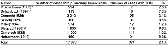
Incidence comparing to pulmonary tuberculosis
Some authors reported the frequency of TOM relative to the presence of lung disease (Table 1).
Etiology
The infectious agent of TOM is Mycobacterium tuberculosis, whose varieties bovis and hominis are the ones that normally affect the ears. Other rare species of Mycobacteria can cause atypical infections in special situations, mainly in cases of immunodeficiency.
M bovis is the one that is currently least found, but it used to be very common in the past. It causes infection by contaminated milk in regions in which milk is not pasteurized. The contaminations take place through gastrointestinal route.
M. tuberculosis hominis (Koch's bacillus) is currently more common and the lungs are the main input route to the human body. Other areas of the airways can be receptors of the bacillus.
Pathophysiology
Tuberculosis in the tympanic cavity follows the same course of the disease in other locations, but the results are modified by the peculiarities of the ear structure, such as the air spaces that are recovered by mucosa in the bone; the ossicle; the close relation of the connection of the mucosa membrane and the bone, and the unique structure of the tympanic membrane.
Primary x secondary onset
Primary TOM is the one that is detected in the ear without detection of another distant focus that could have reached the ear. Many cases, however, can be truly secondary and brought to the blood from an undetected origin, such as infected tonsil or lymph node.
Proctor & Lindsay7, in 1942, did not confirm the existence of any TOM of primary origin. They stated that the lung lesion might not be fully clinically or radiologically demonstrated when the ear disease was manifested.
To present, primary TOM is rarely reported. It is necessary to consider that in ancient times, when the primary form was more frequently diagnosed, there was probably less capability of detecting lung foci and mainly extrapulmonary foci 2.
When the primary cases are detected, normally they are found in children and neonates.
According to Birrel2, secondary TOM is normally found in adults and, in most cases, it is secondary to pulmonary tuberculosis, with the presence of Koch's bacillus in sputum. There may be tuberculous associated lesions in the larynx, pharynx and rhinopharynx.
De Paep et al.14 disagreed and said that 30 years before, the identification of TOM was based on a completely different picture. The patient that had pulmonary tuberculosis developed tuberculous otitis media as a complication of the lung pathology. The mucosa of the middle ear showed an inflammatory pattern of hyperemia, with granulation and otorrhea that was sanguinolent in many occasions According to the author, TOM has changed its manifestations and it is now more chronic than acute. It is normally very difficult, if not impossible, to find evidence of the correlation with pulmonary tuberculosis. The history of the patient can reveal lung infection in childhood, contact with tuberculosis people, or even contact with bovine bacillus.
Infection routes
Another extremely controversial topic is the possible infection routes to the middle ear. They are: auditory tube, hematogenic route, external auditory canal (EAC) route, and intracranial focus close to the temporal bone.
The most frequent routes are auditory tube and hematogenic route, being that the occurrence of EAC infection through a previously existing tympanic perforation is rare and difficult to confirm.
Modern authors believe that hematogenic route is the main one.
Skolnik et al.3, in 1986, detected the modification of the disease pathophysiology throughout the time and added: "the hematogenic route has gained space as being the most common one for middle ear infection by tuberculosis, when the bacillus of hominis type started to be the predominant pathogen and many studies of autopsy support this statement."
Despite being rare, auditory tube contaminations may happen. In 1982, two cases had histology confirmation of ear tuberculosis associated with adenoid tissue tuberculosis, a fact that suggested auditory tube contamination5.
Congenital form
Tuberculosis can also be congenital and even though it is rare, it occurs sporadically. The fetus or the newborn is susceptible to many forms of contamination: directly through the placental circulation; by aspiration of infected amniotic liquid, or at birth, by contact of the infected genital mucosa.
TOM can also be detected in the congenital form by mother-fetus transmission of infection. Two cases of congenital TOM without involvement of other organs were reported for the first time in history in 1989 10, despite the fact that congenital tuberculosis is normally a multi-systemic disorder.
Ear Pathogenesis
The initial lesion of ear tuberculosis can occur from a single focus, or more frequently, from multiple foci in bone barrow spaces, in mastoid cells and tympanic cavity mucosa or in both.
Subepithelial tuberculosis is developed. This tuberculum in the mucosa can lead to dissemination of bacillus through lymphatic network under the mucosa; or the initial mucosal tuberculum, which is developed by producing caseation and necrosis can transform into an ulcer and excrete contaminated material in the air space of the mastoid and middle ear. Once the contaminated exsudate invades the air spaces, it spreads and can be reimplanted in the mucosa in many sites.
If the tuberculum are implanted in the internal aspect of the tympanic membrane, its growth and caseation can produce multiple perforations, which end up leading to a single perforation.
Granulation tissue surges can gradually cause ossicle chain osteitis, many times with extrusion of ossicles. When the disease progresses, the mastoid cell system is replaced by granulation tissue.
Clinical aspects
There is no typical clinical picture for TOM, but authors normally give much attention to the combination of signs and symptoms so that a diagnostic suspicion can be created. The wide variation of clinical manifestations of TOM hinders the diagnosis in clinical basis, and diagnostic confirmation depends on laboratory tests.
The characteristics between adults and children are divergent. The first symptoms are not immediately detected in children and infections are easily spread2. One of the characteristics that differentiate children from adults is the tendency of the former to have mastoid involvement.
The clinical manifestation includes initial otorrhea, which is the most frequent finding, being observed in almost all patients even in those whose tympanic membrane (TM) is intact, but granulous and thickened. It is initially scarce and aqueous, but it later becomes abundant, thick and purulent, as a consequence of secondary bacterial infection, which can result in an undistinguishable clinical picture from nonspecific chronic otitis media. A common trait found in TOM otorrhea is its resistance to conventional therapy. Characteristically, otorrhea is present without pain. The occurrence of pain can be associated with periosteum involvement. When the granulation tissue involves the mastoid cavity and is under pressure, there may be pain 4. The observation of the granulation tissue under tension in the mastoid during the surgical act can be considered a characteristics aspect of otological tuberculosis.
An increase in pre-auricular lymph nodes can be detected. In the past, this characteristic plus suppurative chronic otitis media used to be considered pathognomonic of auricular tuberculosis 11.
In children, it is common to find skin fistulae. The most common site is the post-auricular region, together with the pre-auricular region 12.
Other very common occurrences are early profound hearing loss, characteristically not proportional to otoscopic findings, and peripheral facial palsy 13.
Otoscopy can reveal thickness and sometimes bulging of TM very early in the pathology, erasing landmarks. After that, it normally progresses to tympanic perforation that can occasionally be multiple, but quickly coalesce to a single wide perforation, with purulent otorrhea. In rare cases, there is no perforation of the TM and the exam shows thickened and abnormal membrane with exuberant granulation tissue.
The tympanic cavity is frequently taken by polyps and abundant pale or pink granulation tissue, which can be observed through the intact tympanic membrane or, more frequently, through the perforated membrane.
Granulation tissue can extend to the mastoid cavity and it looks spongy during the surgical act. Romages and Gertler 15 referred to granulous tissue as the most frequent finding.
It is quite common to observe osteitis with necrosis of the ossicles of the temporal bone area 16, especially during surgical exploration 15.
Audiometric aspects
The hearing loss presented in tuberculous otitis media is a common and early symptom. In many situations reduced hearing precedes TM inflammation and the onset of any other symptom. MacAdam and Rubio 17, however, reported a case of slow development of hearing loss, indicating, thus, that hearing loss can be variable.
The main audiological characteristic of TOM is deafness not proportional to the apparent degree of disease development seen at otoscopy. Hearing loss can occur at different levels, but normally it ranges from moderate to severe since its onset. The audiometric type can be conductive, sensorineural or mixed 18.
According to Harbert and Riordan 19 deafness is normally conductive owing to the accumulation of fluid in the middle ear, necrosis of the ossicles and formation of fibrous and dense connective tissue in the tympanic cavity. There may be sensorineural impairment if the labyrinth is affected.
Radiological aspects
Radiological aspects of the middle ear and mastoid in TOM from simple x-rays or CT scan do not reveal specific characteristics, but together with clinical examination and other complementary tests, they can reinforce diagnostic suspicion. In addition, it helps to define the level of involvement of the structures, allowing better surgical planning when the surgery is necessary.
Many authors stated that the detection by x-ray of well pneumatized mastoid, sometimes filled with soft tissue (opacified) in patients with chronic otitis media suggests the possibility of tuberculous otitis media 1, 5, 7, 16, 19.
Sometimes, we can detect cases of bone destruction and mastoid erosion 20.
It is also essential to bear in mind that normal chest x-rays do not rule out the possibility of ear tuberculous infection.
Skin tests
The purified protein derivate of tuberculin (PPD) is used as a routine in screening tests for tuberculosis.
Tuberculin test indicates previous contact of the subject with the bacillus, and not necessarily the presence of active disease. It is, therefore, a complementary test that can help but does not allow the differentiation between infected and sick people. In areas in which vaccination with BCG is performed as a routine its interpretation is hindered, since BCG induces the reaction to tuberculin test as much as the natural infection. It is important to bear in mind that even though skin tests help defining the diagnosis, some young subjects with confirmed active lung tuberculosis can fail to react to up to 250 units of tuberculin. Therefore, a PPD negative result never excludes the presence of tuberculosis 20.
Odetoyinbo 21 presented 23 cases of TOM and they were all PPD negative, except those that had associated pulmonary disease.
In patients with AIDS, the skin test can be performed but the absence of reaction is not significant, since immunosuppression associated with HIV infection can also cause false negative results. If, however, the reaction is positive, directed diagnostic tests should be conducted 22.
Bacteriological and histology studies
The diagnosis of TOM can be obtained through lab tests of secretion or tissues obtained for biopsy. The latter provides a higher rate of positive results.
The experience confirms low sensitivity of bacterioscopy to BAAR (bacillus alcohol acid resistance) in clinical specimen other than sputum. It occurs owing to the scarce amount of mycobacteria present in such specimens and the interference of secondary germs that end up developing in the secretion.
The presumed diagnosis of TOM can be made by tissue studies collected from biopsy that shows granulation tissue. However, the isolation of M. tuberculosis in tissue or secretion is necessary for the definite diagnosis 23. Therefore, the tissues obtained in suspicion cases should be observed directly as well as stained with BAAR and cultivated in special mycobacteria means 23.
It is difficult to demonstrate the presence of tuberculosis bacillus in ear secretion. Positivity of the cultures range from 5 to 35%, being that repeated exams improve the rate up to 50% of positive responses, whereas direct exam (bacterioscopy) has a mean positive range of 20% 12.
The study of tissues can show granulations with epithelioid cells and multinucleated giant cells (Langhans giant cells), caseous necrosis zone, lymphocytarian infiltrate, fibrous tissue, presence of BAAR and, more rarely, ulceration and signs of bone absorption.
Additionally to the culture, we can conduct an antibiogram to determine sensitivity of bacillus to different anti-tuberculosis drugs. It has gained importance in recent years, in which bacterial resistance has increased.
Polymerase chain reaction (PCR) is a technology that can detect even few organisms in a clinical specimen. In a study of tuberculous meningitis, the PCR test had 75% greater sensitivity than conventional methods. This technique is an important alternative to culture, to make diagnosis of extrapulmonary tuberculosis definitive and quick 23.
Diagnosis
Tuberculous otitis media is not a very frequent disease, but when it occurs, it causes significant morbidity. It should be suspected even in patients without evidence of the pathology in another body segment 24.
There is evidence that its occurrence is in fact higher than estimated, because of a large number of undiagnosed cases, be it because of lack of suspicion or difficulty to have etiology confirmation.
Currently, owing to the large number of HIV patients, we should always be attentive to TOM, since many studies indicate that extrapulmonary forms of tuberculosis are much more frequent among these patients.
The diagnosis of TOM is a challenge in many situations, since there are no specific clinical characteristics that indicate the tuberculous nature of the disease.
There are some factors of relevance that should make us think about the tuberculus etiology in patients with suppurative chronic otitis media (COM). A past morbid history of contact or tuberculosis per se, and resistance to habitual treatment of non-cholesteatoma COM should be valued: development of peripheral facial palsy, early disproportional severe hearing loss, retroauricular fistula, Mantoux strong reaction; normal pneumatization of mastoid in the x-ray; caseous granuloma of the ear; aural polyp in infants, regional lymphadenitis, especially in children.
In view of suspicion of middle ear tuberculosis, the following tests are important: bacterioscopy and culture of middle ear secretion, specifically for BAAR; polyp biopsy or middle ear mucosa and histopathology studies with tissue culture; tuberculin test (Mantoux); chest x-ray. Other tests can clarify some details. Radiological studies and CT scan of the mastoid and audiogram.
The main problem of TOM is early diagnosis. This difficulty is caused by the nonspecific character of signs and symptoms. The characteristics described in ancient times are rarely observed today owing to the changes in peculiarities of the disease.
Diagnosis delay can lead to high morbidity, including hearing loss, facial paralysis, etc.
Unfortunately, in most cases, the correct diagnosis ends up being made only after the therapy fails and there is chronic recurrence of symptoms. In summary, the development of complications and absence of response to conventional treatment are part of tuberculous etiology.
If the tests are all negative, but there is strong clinical suspicion, we should repeat the exam many times, trying to have its confirmation. The lack of aggressiveness in diagnostic investigation ends up leading to delay in appropriate management.
In cases in which it is difficult to demonstrate the presence of the bacillus, despite all efforts, and the suspicion persists when other disease have been ruled out, we should conduct a therapeutic trial 5.
Complications
Many complications can result from tuberculous otitis media: mastoiditis, abscesses, skin fistulae, tuberculus lymphadenitis, osteitis, bone sequestration, sigmoid sinus phlebitis, facial paralysis, labyrinthine fistula, labyrinth pathology, hemorrhage, meningitis, extradural abscesses, tuberculomas, involvement of cranial nerves 9th, 10th and 11th, and petritis.
Tuberculosis and AIDS
The association of tuberculosis (including TOM) with AIDS has been observed at varied frequencies, taking great importance in regions of high prevalence of tuberculosis.
In Brazil, tuberculosis occupies the second rank between the diseases associated with AIDS, second only to oral candidiasis. According to the data analyzed in 1996 by the Ministry of Health and the State Department of Health in the states of Sao Paulo and Rio de Janeiro, there were 23% and 29.5%, respectively, of cases of tuberculosis associated with AIDS.
AIDS speeds up the dynamic of transmission and progression of tuberculous disease 25.
The Department of Health of the city of New York estimated in 1993 that at least 40% of the subjects with tuberculosis in the city were also infected with HIV, but they remembered that it was a conservative estimate, since people with tuberculosis were not tested for the presence of HIV.
The subjects with AIDS are hundreds of time more prone to active tuberculosis than the general population. The most prone population to be doubly infected is users of intravenous drug. Currently, owing to the problem of multidrug-resistance bacilli strains, there is no other potentially lethal and contagious disease as tuberculosis, especially for HIV-infected patients 26.
According to Pitchenik et al. 27, tuberculosis is normally preceded by the diagnosis of AIDS within 1 to 17 months, and it is frequently extrapulmonary or disseminated.
Treatment
Treatment with drugs to patients in general
TOM treatment suffered great transformation, since before the advent of drugs, the only alternative for cure was wide and destructive surgery, which rarely led to success.
Currently, anti-tuberculosis chemotherapy is the procedure of choice in view of the diagnosis of TOM. The first cures for TOM using antibiotic therapy were reported by Grief and Gould, in 1948; Adams in 1948; Siirala and Lhi-Kainem, in 1949, and Titche in 1950 2. The first successful therapy with TOM used only streptomycin, but current standard chemotherapy uses a combination of drugs.
To present, the surgery is indicated when tuberculous mastoiditis is complicated by subperiosteal abscess, facial palsy, labyrinthitis, persistent post-auricular fistula, involvement of the central nervous system, or when there is associated cholesteatoma, and it normally consists of incision and drainage with removal of the sequestration present. Mastoidectomy whose purpose is to eradicate the disease is not indicated as a means to complete resolution of the granulation tissue and otorrhea. Anti-tuberculosis chemotherapy can fully solve the problem of granulation and otorrhea 8.
As to local treatment, many different therapies have been tried. Local cleaning is important and should be frequently performed.
Since the advent of anti-tuberculosis chemotherapy, two basic principles are recommended for treatment:
a) Treatment schemes should be of multiple drugs, to which organisms are more susceptible;
b) Intake of drugs should be maintained for a sufficient period of time (to promote and monitor compliance is essential for treatment success).
There is a great number of possible drug combinations and administration schemes but the objective of treatment should be to provide the most effective management, within the shortest period of time. The resources available should also be considered upon selecting the treatment regimen.
Currently, out of the 16 drugs known for its effective action against tuberculosis bacilli, six are preferably used: streptomycin (SM); rifampicyn (RMP); etambutole (EMB); pyrazinamide (PZA), etionamide (ETH) and isoniazide (INH).
In our country, the manual of norms to control tuberculosis - DNPS/MS-1984 recommends two types of basic regimens for tuberculosis 28. Regimen 1, for 6-month duration, is indicated for the treatment of all forms of pulmonary or extrapulmonary tuberculosis, except for tuberculous meningitis and AIDS. At physician's discretion, isoniazide can be maintained for six months more (up to 12 months).
Regimen 2 is indicated in the cases of treatment failure, characterized by persistence of direct bacilloscopic positive response up to 4th months of treatment or by onset of strongly positive results of bacilloscopy during treatment, after the negative period.
Some authors advocate treatment duration of 18 months 18 and others, of 24 months 29.
The used drugs can vary according to the authors, and many combinations can be employed. Regardless, it is necessary to bear in mind that early interruption of treatment allows multiplication of germs that are in state of semi-latency, causing tuberculosis recurrence.
Currently, bacterial resistance to anti-tuberculosis drugs is an important problem and one of the main factors that hinder the fight against the disease.
A study conducted in New York in the 1970s found evidence of multi-drug resistant organisms and resistance rate to isoniazide of 8%. In 1991, 19% of the cases were resistant to isoniazide and rifampicyn. In January 1992, four hospitals of the city reported occurrence of multi-drug resistant strains, causing 80 to 90% mortality 26.
The increase in drug resistance occurred especially in HIV positive patients, as well as in those with incomplete and inappropriate treatment that favored the survival of resistant strains.
Owing to the onset of these multi-drug resistant strains, there is today no disease as contagious and potentially lethal as tuberculosis, especially for HIV infected subjects. As the development of therapies for resistant strains should probably take many years, inappropriate prevention measures can lead to a large-scale outbreak of tuberculosis 26.
Treatment with drugs to HIV positive patients with TOM
In Brazil, according to the commission of specialists of the Ministry of Health and the Brazilin Society of Pneumology and Tisiology who got together in July 1989, the recommendations were 28:
a) patients with tuberculosis (all forms) with positive HIV serology should be treated with regimen 1 for 6 months;
b) patients with tuberculosis (all forms) with AIDS should be treated with the same drugs and doses of regimen 1, being that the second phase should last for 7 months, rather than 4 months (total of 9 months).
Surgical treatment
The role of surgery changed throughout time. In oldest reports the surgeries were radical, especially in young people, in which the destruction of the disease was more extensive, owing to its little resistance. In that time, radical mastoidectomy was a practice used for treatment of TOM. The criteria for surgical indication were most varied.
Today, surgical treatment ranges in size. It ranges from simple exeresis of pale granulation tissue of the tympanic membrane through tympanic perforation or polyp that is project from the external auditory canal, to radical mastoidectomy, including simple mastoidectomy.
Since histology is essential for the diagnosis, some interventions intend to collect samples for the exam 30.
In the middle ear affected by tuberculosis, even in the very advanced lesions of the temporal bone, it can be reverted and scarred through specific chemotherapy 15. Not knowing this fact can tempt people to conduct radical surgery, in view of the aspect of extensive and destructive lesion found during the exploration.
In some cases, reconstructive tympanoplasty can be conducted later.
The typical TOM surgical findings are pale granulation tissue in the mastoid cavity, middle ear and EAC, extensive bone destruction with evidence of bone necrosis and formation of sequestrations; and caseous material that can be present or not.
Prognosis
The prognosis of TOM was seen as very poor in the past, especially in very young people, and many died of the disease and its complications before the advent of streptomycin. In the best case scenario, the patients had deafness, which was always severe and frequently led to facial palsy.
With antibiotic therapy, the results were better. With the use of specific drugs for tuberculosis, drainage of the ear ceased immediately and the otological lesions scarred without complications, but generally there was no hearing improvement 16.
The repair of conductive hearing loss can be reached after cessation of otorrhea, by reconstructive tympanoplasty. The recovery of sensorineural hearing loss does not happen by curing the process 30.
Facial palsy can regress partially or completely depending on the duration of evolution and degree of nerve impairment. The speed and degree of recovery were directly related to the time interval between installation of facial palsy and the beginning of treatment. According to Singh 8, the prognosis of facial palsy does not depend on decompression, as stated by Legent and Baron.Table 1. Relative frequency of TOM in patients with pulmonary tuberculosis (Birrell2).
* Reported by Ormerod (1931); **Reported by Proctor & Lindsay (1942).
Discussion
Middle ear tuberculosis is rare, but it is present and has great morbidity. Currently, it grows in incidence because of the increase in tuberculosis in general, especially in HIV positive patients.
The etiology is Mycobacterium tuberculosis, which invades the middle ear.
The clinical aspect is nonspecific and has been modified throughout times, but in general it is present with chronic and non-painful otorrhea, tympanic membrane perforation, hearing loss, presence of granulation tissue and polyps in the middle ear and occasionally, peri-auricular lymph nodes increase.
The audiometry detects early and disproportional hearing loss, considering the apparent degree of disease. There may be conductive, sensorineural and mixed hearing loss.
Radiological studies of the middle ear and mastoid do not reveal specific characteristics, but the detection of well-pneumatized mastoid in patients with chronic otorrhea suggests the possibility of TOM.
The diagnosis of TOM is difficult, requiring high level of suspicion and knowledge of the disease by the physician. For diagnostic confirmation, we can use direct bacterioscopy, secretion culture, histopathology studies and tissue culture using specific methods for BAAR. Currently, new exams such as PCR can also facilitate the diagnosis.
The existence of effective therapy makes early diagnosis extremely important, preventing complications and unnecessary surgical interventions.
Treatment can be conducted with anti-tuberculosis chemotherapy, surgery or their association. Currently, the treatment with drugs leads to cure, and surgery is reserved for the cases of complication or restoration of the middle ear.
The earlier the treatment, the better the prognosis. Hearing loss is difficult to reverse, especially sensorineural loss. Conductive hearing loss can improve partially after reconstructive surgery of the tympanic cavity. Facial paralysis can disappear partially or completely.
Closing Remarks
By better understanding this topic, we can have early diagnosis of TOM and appropriate management in order to reduce the morbidity of the disease.
REFERENCES
1. Granato L & Lima e Silva LA. Otite média tuberculosa. Rev Bras Otorrinolaringologia 1973; 39(3):125-32.
2. Birrell JF. Aural tuberculosis in children. Proc Roy Soc Med 1973; 66:331-8.
3. Skolnik PR, Nadol Jr JB & Baker AN. Tuberculosis of the middle ear: Review of the literature with an instructive case report. Rev infect dis 1986;8(3):403-10.
4. Plester D, Pusalkar A & Steinbach E. Clinical records - Middle ear tuberculosis. J Laryng Otol 1980; 94:1415-21.
5. Bento RF, Cruz OLM, Morimoto E, Ramos CC, Siebert D & Miniti A. Tuberculose do osso temporal. Estado atual e apresentação de 2 casos. Rev Bras Otorrinolaringologia 1987; 53(3):90-5.
6. Emmett JR, Fischer ND & Biggers WP. Tuberculousmastoiditis. Laryngoscope 1977; 87:1157-63.
7. Proctor B & Lindsay JR. Tuberculosis of the ear. Arch Otolaryngol 1942; 35:221-49.
8. Singh SP & Nath H. Early radiology of pulmonary tuberculosis. AJR 1994; 162(4):846.
9. Maitre B, Conraux C, Reichling P, Bailly P & Boidevezi D. La tuberculose de l'oreille moyenne-Etat actuel. J Fr Oto Rhino Laryng1984; 33(2):75-81.
10. Naranbhai RC, Mathiassen W & Malan AF. Congenital tuberculosis localized to the ear. Arch Dis Child 1989; 64:738-40.
11. Craig DH. Tuberculous mastoiditis - A review of eight cases. J Laryngol Otol 1962; 76:623-38.
12. Chaturvedi VN & Chaturvedi P. Tuberculosis of the middle ear. Indian Pediatr 1986; 23(3):199-204.
13. Sellars SL & Seid AB. Aural tuberculosis in childhood. S Afr Med J 1973; 4(6):216-8.
14. De Paep K, Offeciers FE, Van De Heyning P, Claes J & Marquet J. Tuberculosis in the middle ear: 5 case reports. Acta Oto-Rhin-Laryng.Belg 1989; 43(4):321-6.
15. Ramages LJ & Gertler R. Aural tuberculosis: A series of 25 pacients. Jour Laryngol Otol 1985; 99:1073-80.
16. Wallner LJ. Tuberculous otitis media. Laryngoscope 1953; 63:1058-77.
17. MacAdam AM & Rubio T. Tuberculous otomastoiditis in children. Am J Dis Child 1977; 131(2):152-6.
18. Lucente FE, Tobias GW, Parisier SC & Som PM. Tuberculous otitis media. Laryngoscope 1978; 88(7):1107-16.
19. Harbert F & Riordan D. Tuberculosis of the middle ear. Laryngoscope 1964; 74:198-204.
20. Saltzman SJ & Feigin RD. Tuberculous otitis media and mastoiditis. J Pediatr 1971; 79(6):1004-6.
21. Odetoyinbo O. Early diagnosis of tuberculous otitis media. J Laryngol Otol 1988; 102:133-5.
22. EUA, Centers For Disease Control, U.S. Department Of Health And Human Services. Diagnosis and management of mycobacterial infection and disease in persons with Human Immunodeficiency Virus infection. Ann Intern Med 1987; 106:254-6.
23. Lee PYC & Drysdale AJ. Tuberculous otitis media: A difficult diagnosis. J Laryngol Otol 1993; 107(4):339-41.
24. Barrionuevo CE, Bayer E & De Masi E. Otite média tuberculosa. Estado atual. Rev Bras Otorrinolaringologia 1991; 57(2):61-4.
25. Landesman SH. Comentary: Tuberculosis in New York City - The consequences and lessons of failure. Am J Public Health 1993; 83(5):766-8.
26. Lerner BH. N.Y. City's tuberculosis control efforts: The historical limitations of the "War on Consumption". Am J Public Health 1993; 83(5):758-66.
27. Pitchenik AE, Cole C, Russell BW, Fischl MA, Spira TJ & Snider D.E. Tuberculosis atypical mycobacteriosis and the acquired immunodeficiency syndrome among Haitian and non-Haitian patients in South Florida. Ann Intern Med 1984; 101:641-5.
28. BRASIL. Ministério da Saúde. Secretaria Nacional de Programas Especiais de Saúde. Divisão de Pneumologia Sanitária. Campanha Nacional Contra a Tuberculose. Controle da tuberculose: Uma proposta de integração ensino-serviço/ CNCT/ NUTES. 4a ed. Brasília 1994;1-155.
29. Sahn SA & Davidson PT. Mycobacterium tuberculosis infection of the middle ear. Chest 1974; 66(1):104-6.
30. Cruz NA, Cruz Filho NA & Campilongo M. Tuberculose do ouvido médio. F méd (BR) 1995; 110:17-24.
1 Master in Otorhinolaryngology, Federal University of Rio de Janeiro.
2 Former Faculty professor, Discipline of Otorhinolaryngology, Federal University of Rio de Janeiro; Vice-President of SBORL
for the states of Rio de Janeiro and Espírito Santo.
Affiliation: Federal University of Rio de Janeiro, University Hospital Clementino Fraga Filho, Medical School.
Address correspondence to: Márcia Pinho - Rua Lagoa das Garças 200, ap. 706 Barra da Tijuca Rio de Janeiro 22793-400
Tel (55 21) 2499-5516 - E-mail: marciapinho@openlink.com.br
This paper is part of the master dissertation submitted to the Medical School, Federal University of Rio de Janeiro,
Master degree in Medicine, major in Otorhinolaryngology.
Article submitted on April 23, 2003. Article accepted on July 07, 2003.
Print: ![]()