
Year: 2002 Vol. 68 Ed. 5 - (15º)
Artigo Original
Pages: 692 to 697
Distortion product otoacustic emissions in Bell´s palsy
Author(s):
Cristiane A. Kasse 1,
José R. G. Testa 2,
Yotaka Fukuda 3,
Oswaldo L. M. Cruz 4
Keywords: Bell's palsy, otoacoustic emissions, idiopathic facial paralysis
Abstract:
Introduction: The facial nucleus and olivar nucleus are connected with fibers, then a lesion in this conection could interfer in the outer cell function changing the result of otoacoustic emission product distortion test (DPOE). Objective: To observe the possibility of Bell's palsy affect the function of outer hair cells, using the DPOE test. Study Design: Prospective clinical. Material and Method: Forty patients with Bell's palsy were compared with 69 patients without symptons (control group) using DPOE. Results: The patients with Bell's palsy without DPOE response were 17.5% and in the control group, 7.2%, without statistical difference between them. We did not observe a correlation with stapedium reflex and degree of palsy and DPOE. Conclusion: There was no correlation with Bell's palsy and DPOE.
![]()
Introduction
Bell's palsy, also called idiopathic peripheral facial paralysis, is a prevalent disease nowadays, ranging from 13 to 34 cases in every 100,000 people1. It is characterized by affecting the face unilaterally, causing partial or total paralysis owing to an acute dysfunction of the facial nerve and it is considered a diagnosis of exclusion, because its etiology is not totally understood yet.
The most likely hypothesis resultant from recent studies is that of viral etiology, caused by herpes simplex virus2, 3, 4, 5, leading to direct aggression to the nerve, edema and reduction of conduction.
The fact that some patients also report complaints of hearing loss, hyperacusis or tinnitus following the clinical picture, in addition to its topographic proximity with the cochlear nerve and its nucleus, has generated many investigations about the correlation between facial palsy and cochlear nerve.
The fact that the facial nerve nucleus is intimately related to the superior olivary nucleus has raised our interest in studying the modifications caused to this system by Bell's palsy. The stapedial reflex, whose afferent impulse is given by the cochlear nerve, is located at the superior olivary nerve, which sends efferent fibers to the facial motor nucleus, responsible for the contraction of the stapedial muscle6.
In Bell's palsy, some studies reported that the disease is not restricted to the nerve and the facial nucleus, and there is the possibility of it affecting the olivary nucleus or the cochlear nerve, without damaging it, but generating abnormalities detected at the auditory brainstem response audiometry7, 8, 9, 10 and high frequency audiometry11.
Despite that, Hendrix12 and Uri13 did not confirm the above referred facts, and did not observe abnormalities in brainstem evoked responses in their patients. Experimental studies5, histopathology studies and virus detection using PCR (polymerase chain reaction)2, have also classified the disease as being restricted to the nerve and the facial nucleus. Therefore, there is still controversy in the literature about the extension of the disease.
Since the olivocochlear tract (efferent pathway), specifically the medial olivocochlear bundle, is originated from the olivary nucleus, making synapses with the outer hair cells to modulate its contractility14, a possible abnormality caused by Bell's palsy at the level of the nucleus would be expected to cause an abnormality of the cochlear efferent path, confirming the idea that the disease would not be restricted to the facial nerve and nucleus. This abnormality would possibly be detected by otoacoustic emissions investigation, by checking the function of outer hair cells.
The objective of the present study was to assess whether Bell's palsy impaired the functioning of outer hair cells by studying distortion product otoacoustic emissions (DPOAE).
Material and Method
We analyzed 40 patients with Bell's palsy at acute phase, both genders, seen at the Ambulatory of Otorhinolaryngology, UNIFESP-EPM, between April 1999 and July 2000, comparing the group of patients without symptoms (control normal group). This study was approved by the Medical Research Ethics Committee at Escola Paulista de Medicina - UNIFESP.
We conducted a complete ENT examination, observing the grade of facial palsy from its onset to complete recovery.
Auditory tests were pure tone audiometry, vocal discrimination, immitanciometry and DPOAE, which were performed at the beginning and after complete recovery. In those cases that did not progress satisfactorily, we considered the final grade as the grade after 2 months from the beginning of the clinical picture (end).
Patients with facial palsy over 15 days of evolution and/or definite cause and/or abnormal pure tone and vocal audiometry were excluded from the study.
The control group consisted of 69 patients of both genders, no hearing complaints, with normal audiometric thresholds (thresholds below or equal to 25 dB HL), normal immitanciometry and no facial palsy.
The classification of grade of facial palsy followed the criteria by House & Brackmann (1985)15.
Gráfico 1. Distribuição dos pacientes (n = 40) segundo o grau inicial de paralisia de Bell.
Graph 2: Distribution of patients (n = 40), according to grade of final facial paralysis.
Results
1. Control Group
We analyzed 138 ears of normal subjects, no otological history or symptoms.
The minimum value of distortion product amplitude for each frequency corresponded to the percentile 5. As to noise floor, we adopted the percentile 95 as maximum value, being the value -6 common for most frequencies of the control group, leading us to the adoption of this value as cut-off value.
In order to consider the otoacoustic emissions as present at a specific frequency, the values should be within these limits.
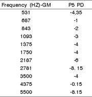
In the analysis of the distortion product, we selected as the cut-off values the percentile 5 (P5) for each tested frequency, according to Table 1.
We classified the distortion product as present or absent according to the criteria in Table 2, which we considered as the result of each frequency, noise floor and distortion product.
According to the results, the tests in each ear were classified as normal or abnormal, depending on number of frequencies with absent distortion product. The ears with up to 2 absent frequencies were considered normal, and over that, they were considered abnormal.
Therefore, in the control group, 7.2% of the subjects had an abnormal test.
2. Sample
We analyzed 40 patients with diagnosis of Bell's palsy, with normal pure tone and vocal audiometry. The mean age was 27.9 years, ranging from 10 to 52 years; 22 subjects were female and 18 were male, with a female-male ratio of 1.22.
The main race found was Caucasian in 65% of the cases, followed by Africans in 35%, and there were no Asian patients.
As to affected side, 24 were on the right and 16 on the left, mean duration of presentation was 6.25 days from onset of symptoms to medical search, and 4 had progressive onset whereas 36 had sudden onset.
2.1 Correlation between grade of facial palsy and DPOAE at onset of symptoms
The initial grade of facial palsy, according to House & Brackmann classification, followed the distribution of Graph 1, with 10 patients as grade VI, 8 as grade V, 17 as grade IV, 4 as grade III and 1 as grade II.
Upon the analysis of the correlation between initial grade of Bell's palsy and the result of DPOAE, we did not observe evidence of correlation between them, and owing to low frequency of some classifications, there was no applicable test, as observed in Table 3.
2.2 Correlation between initial stapedial reflex and response to DPOAE
Patients with reflex present at the onset of the clinical presentation were 5, one had grade II palsy, 3 had grade III facial palsy and I had grade IV. DPOAE did not show abnormal results in any patient.
Table 4 illustrates the correlation between stapedial reflex and DPOAE; 17.5% of the patients with absent stapedial reflex presented abnormal response to DPOAE. Upon the submission of the sample to Fischer's test, there was no statistically significant difference (p=0.565).
2.3 Correlation between grade of facial palsy and DPOAE after 2 months
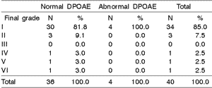
The final grade (after 2 months) of facial palsy was: one case with grade VI, one case with grade V, one case with grade IV, 3 with grade II and 34 with grade I (Graph 2).
Most of the cases progressed to normal status (85%). Some cases improved concerning the initial grade, one maintained the same grade and one case worsened.
Table 5 related final facial palsy grade (after 2 months from onset of the condition) and response to DPOAE.
2.4 Correlation between final stapedial reflex and response to DPOAE
After the recovery of the palsy and reflex, we observed that all patients that had initial abnormal response to DPOAE recovered the reflex and improved the grade of facial palsy.
The final stapedial reflex was 97% present and we did not observe correlation between this fact and DPOAE (p=1.000) at Fisher's test, as shown in Table 6.
2.5 Response to DPOAE concerning the control group
By comparing the percentage of patients with facial palsy with abnormal DPOAE (n=7) and normal control group, we noticed 17.5% of the patients with Bell's palsy with absent OAE, which greater than in the control group (7.2%) (Table 7). Upon the submission of these samples to the statistical test (chi-square test), we did not observe statistically significant difference (Observed value = 1.77 and Critical value = 3.84).
Considering only the patients with facial palsy that did not have stapedial reflex in the acute phase, but had abnormal DPOAE, this abnormality was present in 20% of the cases. Even though we observed a greater amount of abnormal cases in the group with Bell's palsy, the incidence was not enough to determine statistical significance. The test employed was chi-square test, with observed value of 1.77 and critical value of 3.84 (Table 8).Table 1: Values of distortion product (DP) in each frequency in the control group, for percentile values of 5 (P5).
Table 2: Assessment of DPOAE results according to noise floor (NF) and distortion product (DP).
Table 3: Correlation between initial grade of facial palsy and DPOAE in 40 patients with Bell's palsy.
Table 4: Correlation between DPOAE and initial stapedial reflex in 40 patients with Bell's palsy (p =0.565).
Table 5: Correlation between final grade of facial palsy and response to DPOAE in 40 patients.
Table 6: Relation between presence or absence of stapedial reflex and DPOAE in 40 patients with Bell's palsy (p= 1.000).
Table 7: Comparison of patients with facial palsy (n = 40) and the control group (N = 69). Chi-square test with Observed Value: 1.77 and Critical Value: 3.84.
Table 8: Correlation between DPOAE in the facial palsy group (n = 35) with absent initial reflex and the control group (n = 69). Chi-square test with Observed Value of 1.77 and Critical Value of 3.84.
Discussion
The facial nucleus is located on the posterior portion of the pons, in a 4mm long column, laterally to the reticular formation, dorsally to the superior olivary complex and medially to the spinal nucleus of the trigeminal nerve, with the connection between them through nervous fibers, enabling that high intensity sounds, harmful to the cochlea, reach the auditory nerve through the olivary nucleus, send stimuli to the facial nucleus, making the efferent fibers to contract the stapedial muscle, keep the ossicle chain more rigid and hinder the passage of loud sounds. This phenomenon is named stapedial reflex and it is measurable through an impedanciometer, clinically useful to diagnose different diseases.
This reflex affects simultaneously the contralateral ear, because the fibers of the olivary nucleus connect with the contralateral facial nucleus6.
The medial olivary complex originates from the subnucleus of the superior medial olivary complex, sending efferent myelin fibers to the cochlea, predominantly and contralaterally to the outer hair cells16. The fibers of the medial efferent system are responsible for the modulation of contractility of the outer hair cells, since their inhibition affects the function17.
Can the closeness and the interaction of the facial nucleus and the olivary nucleus influence the contractility of outer hair cells in Bell's palsy?
The results of DPOAE assessments conducted in patients with Bell's palsy showed 17.5% (7/40) of the patients with abnormalities in the exam, whereas 82.5% were normal (Table 7).
Qiu, Stucker & Welsh (1998)18 observed that most of the analyzed patients with Bell's palsy (19.4%) had normal OAE assessment. The authors analyzed the OAE through transient evoked otoacoustic emissions (TEOAE).
The possibility of having superior olivary nucleus affected owing to its anatomical proximity with the facial nucleus and its tracts, as suggested by Kamani & Jafary (1995)7 in Bell's palsy, through the observation of abnormal condition in ABR of patients with the disease, and the possibility of affecting the brainstem by herpes simplex virus and its nucleus, suggested by Rosenhall et al. (1983)10, Shanon, Himelfarb & Zikk (1985)19, were not confirmed in this study, since the abnormality of the nucleus would take to abnormal contractility of outer hair cells and consequent abnormality of DPOAE.
Of the 7 patients who had initially presented abnormal DPOAE, 4 of them had grade IV facial palsy and only 2 had grades greater than V. By comparing to the group that had no abnormality of DPOAE,. 28 presented facial palsy greater than IV (Table 3). After recovery, patients that remained with sequelae, that is, with paralysis grades over II, and 27 patients that had complete recovery did not present abnormal DPOAE at any time (Table 5). These facts probably indicate that fiber and nucleus affection is not necessarily associated with the olivary nucleus damage by contiguity.
The patients with stapedial reflex initially present did not show abnormal results of DPOAE during recovery, but the sample was too small (5 patients), so it is not possible to correlate presence of reflex and normal test (Table 4). The patients that initially presented absent stapedial reflex with abnormal DPOAE recovered the reflex and the only patient that did not recovery the reflex did not present any abnormality in the DPOAE exam (Table 4). These results suggest that the reflex does not show complete recovery of facial paralysis.
The absence of correlation between stapedial reflex and OAE was also observed by Qiu, Stucker & Welsh (1998)18, even though they studied TEOAE. The study by Wormald, Rogers & Gatehouse (1995)20 related stapedial muscle paralysis with abnormal discrimination of these patients. The outer hair cells participate in selectivity of frequency (discrimination) through contraction, increasing the movement of the basilar membrane, thus, the absence of stapedial reflex could directly influence over the function of the outer hair cells, according to the above referred study. Wormald, Rogers & Gatehouse (1995)20 explained the influence of the reflex in discrimination by the fact that this contraction attenuates more low frequency sounds than high frequency sounds, thus high frequency sounds would increase discrimination. In Bell's palsy, absence of reflex hinders the discrimination and the authors correlated this abnormality with the affection of the cochlear nerve.
The percentage of patients with Bell's palsy with abnormal DPOAE was greater (17.5%) than the control group (7.2%), as shown in Table 7. Upon the analysis of patients with Bell's palsy with absence of reflex, which suggested greater affection of the nucleus and the nerve, and by comparing to the control group, the percentage increased to 20% (Table 6). Despite the fact that the data were not statistically significant, there is evidence that these patients can have abnormal results of DPOAE.
After recovery from palsy (Table 3) and restoration of the reflex, we observed that the percentage of patients with abnormal DPOAE was 10% (4/40), close to the percentage of the control group (7.24%), suggesting the association of recovery and normal result in the test.
Conclusion
The results of the study led us to the conclusion that the analyzed sample did not show correlation between Bell's palsy and abnormal result of DPOAE comparing with the control group, concerning initial or final grade of facial palsy and stapedial reflex.
References
1. Bleicher JN, Hamiel S, Gengler JS & Antimarino J. A survey of facial paralysis: etiology and incidence. Ear Nose Throat J 75(6):355-358.
2. Murakami S, Mizobuchi M, Nakashiro Y, Doi T, Hato N & Yanagihara N. Bell palsy and herpes simplex virus: identification of viral DNA in endoneural fluid and muscle. Ann Int Med 1996;124(1):27-30.
3. McCormick DP. Herpes simplex virus as a cause of Bell's palsy. Lancet 1972;1(7757):937-939.
4. FurutaY, Fukuda S, Chida E, Takasu T, Ohtani F, Inuyama Y & Nagashima K. Reactivation of herpes simplex virus type 1 in patients with Bell's palsy. J Med Virol 1998;54:162-166.
5. Ishii K, Kurata T, Sata T, Hao MV & Nomura Y. An animal model of type-1 herpes simplex virus infection of facial nerve. Acta Otolaryngol 1988 (Stockh), 446:157-164.
6. Borg E. On the neuronal organization of acoustic middle ear reflex. A physiological and anatomical study. Brain Res 1973;49(1):101-123.
7. Kamani M & Jafary AH. Auditory brain-stem response audiometry in patients with Bell's palsy. Clin Otolaryngol 1995;20:135-138.
8. Uri N, Schuchman G & Pratt H. Auditory brain-stem evoked potentials in Bell's palsy. Arch. Otolaryngol 1984;110:301-304.
9. Welkoborsky HJ, Amedee RG, Elkhatieb A & Mann WJ. Auditory-evoked brain-stem responses auditory disorders in patients with Bell's palsy. Eur Arch Otorhinolaryngol 1991;248(7):417-419.
10. Rosenhall U, Edström S, Hanner P, Badr G & Vahlne A. Auditory brainstem response abnormalities in patients with Bell's palsy. Otolaryngol Head Neck Surg 1983;91(4):412-416.
11. Rahko T & Kama P. High frequency audiometry in facial paralysis. Acta Otolaryngol Suppl 1988;449:161-163.
12. Hendrix RA & Melnick W. Auditory brain stem response and audiology tests in idiopathic facial nerve paralysis. Otolaryngol Head Neck Surg 1983;91(6):686-690.
13. Uri N, Schuchman G & Pratt H. Auditory brain-stem evoked potentials in Bell's palsy. Arch Otolaryngol 1984;110:301-304.
14. Guinan JJ Jr, Warr WB, Norris BE. Topographic organization of the olivocochlear projections from the lateral and medial zones of superior olivary complex. J Comp Neurol 1984;226:21-27.
15. House JW & Brackmannn DE. Facial nerve grading systems. Otolaryngol Head Neck Surg 1985;93(2):146-147. 5
16. Guinan JJ Jr, Warr WB & Norris BE. Differential olivocochlear projections from lateral versus medial zones of superior olivary complex. J Comp Neurol 1983;221: 358-370. 6
17. Zheng XY, McFadden SL, Henderson D, Ding DL & Burkard R. Cochlear microphonics and otoacoustic emissions in chronically de-efferented chinchilla. Hear Res 2000;143(1-2):14-22. 7
18. Qiu WW, Stucker FJ & Welsh LW. Clinical interpretations of transient otoacoustic emissions. Am J Otolaryngol 1998;19(6): 370-378. 8
19. Shanon E, Himelfarb MZ & Zikk D. Measurement of auditory brain stem potentials in Bell's palsy. Laryngoscope 1985;95(2):206-209. 10
20. Wormald PJ, Rogers C & Gatehouse S. Speech discrimination in patients with Bell's Palsy and a paralysed stapedius muscle. Clin Otolaryngol 1995;20(1):59-62.
1 Master in Otorhinolaryngology, UNIFESP-EPM.
2 Ph.D., Guest Professor, Discipline of Pediatric Otorhinolaryngology, UNIFESP-EPM.
3 Joint Professor, Discipline of Otorhinolaryngology and Head of the Sector of Otology, UNIFESP-EPM.
4 Full Professor, Guest Professor, Discipline of Pediatric Otorhinolaryngology, UNIFESP-EPM.
Address correspondence to: Rua David Eid 1907 bloco 2 apto 103 - São Paulo - 04438-000
E-mail: cakasse@bol.com.br
Article submitted on November 29, 2001. Article accepted on August 29, 2002


