

Year: 2003 Vol. 69 Ed. 1 - (21º)
Relato de Caso
Pages: 128 to 130
 PDF PT
 Relapsing polichondritis: a case report
Author(s):
Emerson M. Rodrigues,
Roberta C. N. Silveira,
Nocy Leite,
Marcelo Mendes Tepedino
Keywords: synovial sarcoma, neoplasia, head and neck.
Abstract:
Relapsing polychondritis is an inflammatory systemic disease leading to cartilage destruction. It is thought to be an auto-imune pathology. The auricular, nasal and articular condrous tissue are most affected. Condrous biopsy can be diagnostic. The authors review the literature and describe one case that was well controlled with corticoid therapy.
![]()
Introduction
Relapsing polychondritis (RP) is an uncommon systemic disease described for the first time in 1923 by Jaksch Wartenhorst. The etiology is unknown, but it is believed to be an autoimmune process1. It is characterized by recurrent inflammation and destruction of cartilaginous tissues resulting from the inflammation cartilage that generates the formation of granulation tissue and fibrosis.
The purpose of the present study was to describe a case diagnosed as RP.
Case Report
Female 40 -year-old patient, cleaning woman, living in Rio de Janeiro, had complaints of right hip joint pain for 2 weeks, constant but normally more severe in the morning, causing difficulty to walk. She reported bilateral pinna pain, with local phlogistic signs for 8 months, initially sporadic and then worsened in the past 2 months. She did not present auditory, vestibular, ocular, cardiovascular or neurological abnormalities. She had diabetes type 2, without appropriate clinical control. She did not report alcohol abuse or smoking. At the physical examination, she was thin, had fever, was hypoblushed (++/+4), dehydrated, restrained to bed, keeping the right hip joint in flexion, which was painful upon passive movement; no other joints had signs of arthritis. Auricular pinna presented heat, edema and red color, but the symptoms spared the lobes (Figures 1 and 2), articular cartilage of softened consistency, edema on the lateral 1/3 of the external auditory canal, with no other ENT affection. She was submitted to x-rays of the femur joint, soft tissue ultrasound and bone scintigraphy, and all the results were within the normal range. Auricular cartilage and nasal septum biopsy revealed histology pattern of chronic inflammatory process, with degenerative abnormalities. We started treatment with prednisone 7.5 mg/day, and there was improvement of all clinical symptoms. Six months after, she presented reactivation of auricular symptoms; we associated azathioprin 100mg/day and she managed to reach symptom control.
Discussion
It is a rare disease that affects the population between 20 and 60 years, with an incidence peak on the 4th decade of life2; there is no predominance of gender and ethnic group and no clear genetic predisposition and strong association with HLA-DR43.
Approximately 30% of the cases are associated with hematological or autoimmune diseases. It is estimated that the 5-year survival rate is 74%4, being that infectious and respiratory impairment contributes to reducing survival.
Auricular chondritis is the most frequent and characteristic clinical aspect; it can be unilateral or bilateral, which is more characteristic, showing phlogistic signs in the auricular pinna with no affection of the earlobes. There are repetitive episodes that progressively reduce the structural integrity of the external ear causing laxity. The external auditory canal edema can lead to conductive hearing loss. Vasculitis of the inner auditory artery may cause cochlear and/or vestibular affection.
The nasal impairment can result in deformity in saddle nose. The respiratory symptoms are not common and can be potentially lethal, owing to the collapse of the airways by dissolution of the cartilaginous rings of the trachea and bronchi.
RP arthritis generally leads to asymmetrical non-erosive polyarthritis, with predominance of large extremity joints, sternoclavicular joint, costochondral and manubriosternal joints. It is seronegative and episodic and not related to the disease activity.
The cardiovascular system is involved in 10% of the cases5; thoracic aorta and abdominal aneurysm can occur. The aortic and mitral valves can be sites of implantation, leading to valve incompetence.
The whole ocular structure and the adjacent tissues can be affected. Episcleritis and uveitis are the most common ones5. Complications can include cataract, optical neuritis, keratitis, proptosis, corneal ulceration and extra-ocular muscle paralysis.
Dermatological findings are variable. Neurological manifestations can include cranial neuropathies, headache, seizures, encephalopathy, hemiplegia and ataxia.
In 1976, McAdam et al.6 defined the following criteria, which require three or more clinical findings to confirm the diagnosis of RP: recurrent bilateral auricular chondritis; nasal chondritis; non-erosive and seronegative inflammatory polyarteritis; inflammation of ocular structures; respiratory tract chondritis involving the laryngeal and tracheal cartilages; cochlear and/or vestibular dysfunction.
Biopsy of auricular chondritis is made to confirm the diagnosis of RP; it shows the presence of mononuclear cells and occasional polymorphonuclear cells, with fibrosis on the fibrochondral junction. Lab abnormalities are nonspecific and normally reflect inflammatory status.
The auricular lesion of RP should be differentiated from the traumatic or chemical lesions, acute infection by streptococcus, fungi, syphilis and Hansen's disease. The nasal effects can result from fungi, tuberculosis, syphilis and Hansen's disease or granulomatous lesions such as Wegener's granulomatosis, polyarthritis nodosa, Behçet's syndrome and Cogan's syndrome. RP aortitis should be differentiated from Ehlers-Danlos' syndrome, Marfam's syndrome and medial cystic necrosis that is idiopathic or associated with ankylosing spondilitis.
The initial treatment can be made using non-steroidal anti-inflammatory drugs (NSAID) or glycocorticoids at low doses for cases of auricular and/or nasal chondritis or arthritis. Glycocorticoid in immunosuppressant doses is indicated in more severe clinical manifestations, such as: laryngotracheal, ocular, inner ear, intense nasal and auricular, systemic vasculitis, aortitis or glomerulonephritis. The use of immunosuppressants with methotrexate, azathioprin, hydroxychloroquine, chlorambucil and cyclophosphamide are recommended to spare corticoids or for non-responsive cases. Dapsone, a drug that blocks the lisosomal activity of polymorphonuclear cells, has also been successful used at RP.
Laryngotracheal involvement should be followed by laryngoscopy and serial computed tomography. Tracheostomy is necessary to treat the symptoms of subglottic stenosis. In cases of diffuse airway involvement, causing collapse due to laxity, there is no effective therapy and the use of tracheal stent provides limited results. Surgical reconstruction of collapsed nasal septum is not recommended, since there is frequent relapse after the surgery. Aortic insufficiency may result in need for valve replacement.
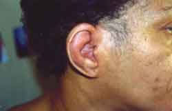
Figure 1. Right ear with hyperemia and edema on the skin of the cartilaginous portion, with no earlobe impairment. Before treatment.
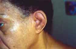
Figure 2. Left ear with hyperemia and skin edema on the cartilaginous region, without earlobe impairment. Before treatment.
CLOSING REMARKS
RP is a rare disease in which clinical manifestations should be differentiated from other diseases in order to define the appropriate therapeutic approach.
REFERENCES
1. Rampelberg O, Gerard JM, Namias B, Gerard M. Ent manifestations of relapsing polychondritis. Act otorhinolaryngol Belg 1997;51(2):737.
2. Molina JF, Espinoza LR. Relapsing polychondritis. Baillieres best pract res clin rheumatol 2000 Mar;14(1):97-109.
3. Gunaydin I, Daikeler T, Jacki S, Mohren M, Kanz L, Kotter I. Articular involvement in patients with relapsing polychondritis. Rheumatol Int 1998;18(3):93-96.
4. Michet CJJr, Mckenna CH, Luthra HS, O'Fallon WW. Relapsing polychondritis; survival and predictive role of early disease manifestations. Ann Intern Med 1986;104:74-78.
5. Isaak BL, Liesegang TJ, Michet CJJr. Ocular and systemic findings in relapsing polychondritis. Ophthalmology 1986;93:681-689.
6. Mcadam LP, O'Hanlon MA, Bluestone R, Pearson CM. Relapsing polychondritis: prospective study of 23 patients and a reviw of the literature. Medicine 1976;55;193-215.
1 Physician Resident in Otorhinolaryngology, Hospital dos Servidores do Estado-RJ.
2 Physician, Resident in Rheumathology, Hospital dos Servidores do Estado-RJ.
3 Physician, Head of the Service of Rheumathology, Hospital dos Servidores do Estado-RJ.
4 Assistant Professor in Otorhinolaryngology, Federal University of Rio de Janeiro.
Study conducted at Hospital dos Servidores do Estado do Rio de Janeiro.
Address correspondence to: R: Vereador Alacy Costa nº 204 Centro - Barra de São Francisco ES 29800-000 - Tel. (55 27) 3756-1105 - E-mail: emersonmr@bol.com.br
Article submitted on February 27, 2002. Article accepted on April 18, 2002.
Print: ![]()
All rights reserved - 1933 /
2026
© - Associação Brasileira de Otorrinolaringologia e Cirurgia Cérvico Facial