

Year: 2003 Vol. 69 Ed. 1 - (6º)
Artigo Original
Pages: 29 to 33
 PDF PT
 Assessment of pyriform sinus carcinoma treatment
Author(s):
Claudiney C. Costa,
José Francisco S. Chagas,
Maria Beatriz N. Pascoal,
José G. T. Camargo3,
José Luiz B. Aquino3
Keywords: treatment, pyriform sinus, survival.
Abstract:
Introduction: Laryngeal and pharyngeal tumors exhibit a high incidence in Brazil, occupying the sixth position in the most frequent cancer sites in males. The initial diagnosis is usually made with the lesion at an advanced clinical stage, hindering success of any treatment employed. Aim: To evaluate the evolution and prognosis of patients with epidermoid carcinoma of the hypopharynx, considering treatment of choice, complications and estimated 5-year survival. Methods: This is a retrospective study using Kaplan-Meier and the exact Fisher test. 60 patients with advanced epidermoid carcinoma of the hypopharynx, with the epicenter located at thepyriform sinus, were enlisted. Results: 43 of the 60 patients were submitted to surgical treatment followed by radiotherapy. Currently 27.9% of the patients are alive without disease, 11.6% are alive with disease, 9.4% died from other causes, 34.8% died due to the disease and 16.3% are lost to follow-up. The most frequent post-operative complication was salivary fistula. Five patients evolved with local recurrence, 6 with regional recurrence, three with loco-regional recurrence and six with distant metastasis. There was no correlation between small surgical safety margins and survival in the 20 patients exhibiting recurrences (Fisher test). Applying the actuarial survival curve with the Kaplan-Meier method we obtained a survival average of 23.3 months in five years. Conclusion: The major post-surgical complication in hypopharyngeal carcinoma treatment was salivary fistula, being successfully treated without surgery. A small surgical safety margin did not influence the prognosis, even though this happens to be at fault with one of the fundamental cancer surgery predicaments. The major feature of treatment failure was loco-regional recurrence. The actuarial five-year survival curve (Kaplan-Meier) averaged 23.3 months.
![]()
INTRODUCTION
Malignant hypopharynx tumors are not common and account for 5% to 10% of the upper aerodigestive tract and for 0.5% of all cancer types. Malignant neoplasias that affect the hypopharynx (pyriform recesses, posterior wall and posterior cricoarytenoid area)1 present the worst outcome among those in the head and neck region.
Brazil has a high incidence of malignant tumors in the larynx and hypopharynx, being the sixth most common site among malign tumors in male, with 2,300 deaths in 1996. According to the American Cancer Society, 10,600 new cases of larynx and hypopharynx cancer were reported in the United States in 1999, with 4,200 deaths2.
The pharynx is the upper segment in the aerodigestive tract through which air, food and drink passes. The hypopharynx is divided in three anatomic sites: pyriform sinus, posterior wall and post-cricoid area. In the United States, the incidence of tumors in this region is distributed as follows: 66-75% in the pyriform sinus and 20-25% in the posterior wall and post-cricoid area1,3.
Tumors in the pyriform sinus often have submucosal spread, invading contiguous structures with early involvement of cervical lymphatic drainage chains located ipsilaterally and contra laterally to the primary tumor, as well as an insidious onset, which leads to the initial diagnosis and advanced stage in most cases1,3.
Today, with the combination of radical surgery and post-operative radiotherapy, the local-regional control of these tumors has increased. However, overall survival was not changed due to distant metastasis. This does not represent a failure in the treatment, but the natural evolution of the disease4.
Regularly, a multidisciplinary team is required to treat tumors in the pyriform sinus, composed of head and neck surgeon, plastic surgeon, chest surgeon, oncologist, radiotherapist, speech therapist, nutritionist and psychologist in an attempt to improve the survival and quality of life of these patients1,3,5,6.
This study aims to evaluate the outcome of 60 patients with epidermoid carcinoma of the hypopharynx, epidemiological characteristics, treatment of choice, surgical procedures associated with laryngopharyngectomy, post-operative complications and survival of these patients.
MATERIAL AND METHOD
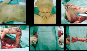
Sixty patients with squamous cell carcinoma of pyriform sinus were enrolled in the study. Seventeen of these patients had unresectable lesions due to adherence to the carotid artery or to the spine and were referred to treatment with radiotherapy and chemotherapy. Forty-three patients underwent total laryngopharyngectomy with uni-or bilateral neck dissection, total glossectomy when necessary, and reconstruction of the upper digestive tract with muscle flap or isoperistaltic gastric tube of the greater gastric curvature (Figure 1).
Figure 1. Preparation of isoperistaltic tube of greater gastric curvature to reconstruct the upper digestive tract in patients submitted to total laryngopharyngectomy with bilateral radical neck dissection.
All patients were referred from the Head and Neck Department, Hospital Celso Pierro, Pontifical Catholic University of Campinas (HMCP/ PUCCAMP) in Campinas, São Paulo.
Graph 1. Distribution of cases according to TNM staging (E).
Graph 2. Surgical procedures associated with laryngopharyngectomy.
Graph 3. Current status of 43 patients submitted to laryngopharyngectomy.
These patients were treated by the medical team of the Head and Neck Service of HMCP/ PUCCAMP from January, 1993 through July, 2001, with mean and median of 24 months, ranging from 12 to 60 months.
Of the 43 patients who underwent surgery, two (4.6%) were female and 41 (95.4%) were male.
Patients were distributed in five age groups with a ten-year variation within each group. The youngest patient was 38 years old and the oldest one was 76, with a mean patient age of 55.7 years and median age of 55 years.
Of the 43 patients enrolled in the study, 41 (95.3%) were at an advanced stage (III and IV) and two (4.7%) at stage II, according to the International Union Against Cancer classification from 1997 (Graph 1).
All 43 patients underwent laryngopharyngectomy combined with other surgical procedures, as follows: unilateral neck dissection in 14 patients; bilateral neck dissection in 13 patients; bilateral neck dissection and reconstruction of the upper digestive tract with the creation of an isoperistaltic gastric tube of greater gastric curvature in 11 patients; total glossectomy and bilateral neck dissection and rotation of the myofascial flap of pectoris major in 4 patients; bilateral neck dissection and rotation of the trapezius flap in one patient (Graph 2).
METHODS
Data were obtained from hospital records and files from the Head and Neck Department, HMCP/ PUCCAMP after being evaluated by the Research Ethics Committee from January, 1993 through July, 2001, with follow-up ranging from 12 to 67 months and average of 24 months. The statistical tests employed were: a) Kaplan-Meier method to obtain the actuarial survival curve, and b) exact Fisher test, to study the relationship between the size of surgical safety margins and the survival rates of patients.
RESULTS
Out of the 43 patients who underwent surgery, today there are 12 (27.9%) alive and free from disease, 5 (11.6%) are alive with disease, 4 (9.4%) died from other causes, 15 (34.8%) died of the disease and 7 were lost to follow-up (16.3%) (Graph 3).
Of the 12 patients who progressed without recurrence, 10 patients underwent the following procedures in addition to laryngopharyngectomy: reconstruction of the upper digestive tract with isoperistaltic gastric tube of greater gastric curvature (3 patients); total glossectomy (3 patients); rotation of the myofascial flap of pectoris major (3 patients) and rotation of the trapezius flap (1 patient).
Thirty-three patients (76.7%) had post-operative complications, the most common being the presence of pharyngocutaneous fistula (Table 1).COMPLICATION / NUMBER
Pharyngocutaneous fistula - 11
CCervical cellulitis - 4
Gastric tube fistula - 4
Hypothyroidism - 2
Pharyngeal stricture - 2
Bronchopneumonia (BCP) - 2
Gastric tube stenosis - 2
Cerebral vascular accident - 2
Sepsis, jugular vein rupture, pneumothorax, Claude Bernard Horner syndrome, bronchopneumonia
with pneumothorax, bronchopneumonia with pneumothorax and gastric tube dehiscence, lymphatic fistula - 1 in each patient (7)
Table 1. Post-operative complications in 33 (76.7%) of the 43 patients submitted laryngopharyngectomy.
Table 2. Time and type of treatment failure in 20 patients submitted to laryngopharyngectomy.
Table 3. Correlation between surgical margins and current status of 20 patients with recurrence and metastases.
Of all complications, 3 led to post-operative death: cerebral vascular accident (2 patients), sepsis and rupture of the internal jugular vein.
Follow-up of alive patients ranged from 12 to 60 months with mean and median of 24 months.
Of those 20 patients who had tumor recurrence, 15 died and five remained alive with the disease. All these patients were at an advanced clinical stage (11 at IV and 9 at III). Local recurrence occurred in 5 patients, regional recurrence in 6, local-regional in 3 and distant metastasis in 6 patients (Table 2). Distant metastasis sites were: bone in 3 patients, lungs in 2, liver and bone in 1 patient.
As to the surgical margins in patients with tumor recurrence, 8 of those who died had compromised surgical margins, while 1 of the 5 patients alive with the disease had compromised surgical margin. The Fisher test indicated no correlation between compromised surgical margins and survival in 20 patients with tumor recurrence (Table 3).
Applying the actuarial 5-year survival curve using the Kaplan-Meier method, we obtained the following values:
- Mean: 23.2 m.
- Standard deviation error: 3.2 m.
- Confidence interval: 16.9 to 29.5 m.
DISCUSSION
Prognosis is poor in Stage III and IV tumors of the larynx and pharynx, but since survival is usually extended, an aggressive therapy is justified in an attempt to resect the tumor and mitigate problems arising from such treatment. Reconstruction should go beyond simply placing a tube to remove saliva, in order to allow vocal rehabilitation and swallowing as close as possible to that attained after a traditional total laryngectomy primary closure5,6.
Cancer of the hypopharynx and cervical esophagus have the worst outcome among primary sites in the upper aerodigestive tract. This is largely due to the high incidence of metastasis for regional lymph nodes and advanced stage of the disease at diagnosis and treatment4,7. The presence of cervical adenopathy at the first consultation occurred in about 66% of the cases, while the presence of micrometastasis was observed in 41% of the clinically negative neck who underwent elective neck dissection7.
Hypopharynx carcinoma is a devastating disease that still has poor outcomes. Two- and five-year survival rates for patients with hypopharynx carcinoma at stages III and IV may range from 0% to 50%8,9. Of the patients eligible for radical treatment, only 1 in 3 survives after two years and less than 1 in 5 survives for over 5 years. Some of the survivors may have to live with a gastrostomy, salivary fistula or need to have periodic sessions of stricture dilation10. Survival rates following radiotherapy alone are discouraging, since they range from 4% to 10%, and also because this kind of therapy is not sufficient to manage patients with cervical metastasis4,11.
In our study, by applying the actuarial survival curve according to the Kaplan-Meier method, we reached an average of 23.2 months in a 5-year projection, which is compatible to the literature 1,3,4,7,11,12.
Gastric interposition is a good method for hypopharynx reconstruction. This procedure presents a low incidence of fistula and stenosis and allows large resection margins. Today, we have to perform major abdominal surgery and dissection of the posterior mediastinum to deviate the stomach to the cervical region, leading to a large volume in this area and possible compression of the structures in that region and in the lungs, as well as a section of the diaphragm and vagus. Among the several methods that use the stomach to reconstruct the digestive tract, a tube with the greater gastric curvature seems to be easier to be made and to be transposed to the cervical region through the retrosternal route, thus avoiding mediastinum handling as well as maneuvers for full mobilization of the stomach and other problems that arise from gastric transposition, as previously mentioned4.
Fifty-eight percent of the patients managed at the Memorial Sloan-Kettering Cancer Center between 1975 and 1985 failed treatment, with similar rates for local failure, regional failure and distant metastasis 3.
In a study conducted by the Head and Neck Department of Heliópolis Hospital, the authors reported the following distribution of recurrences: local (8.3%), regional (22.2%), distant metastasis (6.2%), local-regional (3.7%) and local-regional recurrence and distant metastasis (0.6%), with 5-year overall survival of 70%, 60%, 32% and 14% for stages I through IV, respectively1,12.
In our study, recurrences were distributed as follows: local (25%), regional (30%), local-regional (15%) and distant metastasis (30%). According to the Fisher test, there was no correlation between compromised surgical margins and the survival of patients with tumor recurrence. The surgical margin was deemed compromised when the margin of tissue without tumor in the surgical specimen was smaller than 5 mm in the pathology analysis.
The most common post-operative complication in the surgical treatment of advanced hypopharynx tumors is the presence of pharyngocutaneous fistula. It usually shows between the first and the sixth week following the surgery and it is more frequent in malnourished patients and those who present compromised surgical margins. According to Shemen, Spiro13, it affects 30% to 50% of surgically treated patients. In our study, 76.7% of the patients who underwent surgery presented post-operative complications, the most common being pharyngocutaneous fistula (34.3%).
Most fistulas end up closing spontaneously in two or three weeks, applying sterile wound care and local compressive dressing. When necessary, fistulas can be closed surgically, with primary closure and/or flap rotation1,4.
CONCLUSIONS
The most common post-operative complication was pharyngocutaneous fistula and it was clinically treated.
Compromised surgical margins did not change the outcome, despite being one of the oncologic surgery principles.
The major treatment failure was local-regional recurrence.
The five-year actuarial survival curve (Kaplan-Meier) averaged 23.2 months.
REFERENCES
1. Fava AS. In: Carvalho MB. Tratado de Cirurgia de Cabeça e Pescoço e Otorrinolaringologia. 1ª edição. São Paulo, Rio de Janeiro, Belo Horizonte: Editora Atheneu; 2001. p. 491-512.
2. INCA; MINISTÉRIO DA SAÚDE; GOVERNO FEDERAL. Estimativa da incidência e mortalidade por câncer no Brasil 2000. Rio de Janeiro, 2000.
3. Clayman GL, Weber RS. In: Myers EN, Suen JY. Cancer of the Head and Neck. 3rd edition. Philadelphia: Saunders Company; 1996. p. 425-438.
4. Chagas JFS. Reconstrução da faringe e do esôfago com tubo isoperistáltico da curvatura gástrica maior após faringolaringoesofagectomia cervical. São Paulo, 2001, p. 3-6 (Tese de Doutorado apresentada à Universidade Federal de São Paulo - Escola Paulista de Medicina).
5. Blom ED. Evolution of Tracheoesophageal Voice Prostheses In: Tracheoesophageal Voice Restoration Following Total Laryngectomy. 1st ed. San Diego: Singular Publishing Group; 1998. p.1-8.
6. Costa CC. Reabilitacão vocal de laringectomizados com prótese traqueoesofagica. São Paulo, 2000, p. 57 (Tese de Mestrado apresentada ao Complexo Hospitalar Heliópolis, publicada na Rev Bras Otorrinolaringol 2001;67(5):707-714.
7. Shah JP, Kowalski LP. In: Cirurgia de Cabeça e Pescoço. 2ª edição. Rio de Janeiro: Editora Reter Ltda; 2000. p. 235-265.
8. Fabian RL. Pectoralys major myocutaneous flap reconstruction of the laryngopharynx and cervical esophagus. Laryngoscope 1988;98:1227-1231.
9. Keane TJ. Carcinoma of the hipopharynx. J Otolaryngol 1982;11:227-231.
10. Fairmann HD, John HT. Treatment of cancer of the pharynx and cervical oesophagus. J Laryngol Otol 1966;80:1091-1101.
11. Pingree TF, Davis RK, Reichman O, Derrick L. Treatment of hypopharyngeal carcinoma: a 10-year review of 1,362 cases. Laryngoscope 1987;97:901-904.
12. Rapoport A. Fatores determinantes da sobrevida no cancer da hipofaringe: Variáveis relacionadas ao estadiamento, terapêutica e evolução. São Paulo, 1987 (Tese de Livre Docência apresentada a Faculdade de Medicina da Universidade de São Paulo).
13. Shemen LJ, Spiro RH. Complications following laryngectomy. Head Neck Surg 1986;8:185.
1 Head and Neck surgeon (volunteer physician), Service of Otorhinolaryngology, Hospital das Clinicas, Federal University of Goiás.
Doctorate studies under course, Federal University of São Paulo - Escola Paulista de Medicina.
2 Head of the Service of Otorhinolaryngology and Head and Neck Surgery, Hospital e Maternidade Celso Pierro, Pontifical Catholic University of Campinas (HMCP-PUC).
3 Assistant Surgeon, Service of Head and Neck Surgery, HMCP-PUC. Campinas-SP.
Affiliation: Service of Head and Neck, Hospital e Maternidade Celso Pierro, PUC, Campinas-SP.
Address correspondence to: Claudiney Candido Costa - Rua 16 A., 262, Ed. Carajás, apto 201 Setor Aeroporto Goiânia GO 74075-150 - Tel (55 62) 229-1924 - E-mail: orlccp@uol.com.br
Study presented as free communication at XVIII Congresso Brasileiro de Cirurgia de Cabeça e Pescoço, on September 03 - 06, 2001, in Recife-PE.
Article submitted on September 09, 2002. Article accepted on November 07, 2002.


Print: ![]()