Year: 2004 Vol. 70 Ed. 5 - (17º)
Relato de Caso
Pages: 692 to 694
 PDF PT
 Ear-piercing perichondritis: case reports and literature review
Author(s):
Ernesto N. Takahashi1,
Adriana G. Chaves2,
Fernanda Akaki2,
Anne K.S.L.Nunes1,
Maria Carmela C.Boccalini3,
Cícero Matsuyama4
Keywords: perichondritis, ear piercing, nickel allergy.
Abstract:
Auricular perichondritis is a complication related to ear piercing, caused by the hypersensitivity to nickel. The diagnosis is essentially based on clinical findings. The most pathogen frequently involved is Pseudomonas aeruginosa. Culture is important to determine the antibiotic susceptibility. We report 3 cases of ear piercing associated perichondritis and present a literature review.
![]()
Introduction
Perichondrites are infections of slow evolution located in the auricular cartilage that lead to laceration, contusions, surgeries or other affections.
Auricular perichondritis by piercing has been increasingly observed, particularly among youngsters. 1-4
The first signs and symptoms appear within a few days or weeks after ear piercing. Patients present intense pain, erythema, edema and hardening of the affected region.1-3,5-8
The etiological agent most frequently found in perichondritis by ear piercing is Pseudomonas aeruginosa, whose inoculation results from exposing the perichondrium and pinna cartilages to the bacteria, during or after the piercing procedure. 1,2,4-6 Other bacteria may also be associated with ear piercing complications, particularly Staphylococcus aureus.1,2,8,9
The main complications include contact dermatitis and high sensitivity to nickel.2,6,7,9-12
Investigation on the etiology of this condition is mandatory, which can be done through culture and antibiogram of the secretion1.
Treatment includes surgical drainage, antibiotic therapy and systemic antiinflammatory treatment.
The present study describes 3 cases of perichondritis by ear piercing admitted at the Otorhinolaryngology ambulatory of Cema Hospital.
Case Report
Case 1
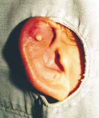
D. M. C., 15-year-old Caucasian female with piercing placed on the right ear 14 days before being admitted to the hospital.
Local pain, erythema and progressive edema were presented by the patient 4 days after piercing the ear. Four days before arriving at our Service, she had initiated oral antibiotic therapy with Cephalexin. Two days later, her clinical status got worse, with spontaneous drainage of purulent secretion (Figure 1).
The patient was seen at our Service, where puncture and collection of the purulent secretion was performed and sent for culture and antibiogram. Following, surgical drainage with debridement of necrotic tissue and placement of Penrose drainage were performed.
Intravenous antibiotic therapy with Ciprofloxacin and steroidal antiinflammatory treatment were initiated, to which the patient responded well, leaving hospital within 48 hours. She maintained oral treatment with 500 mg Ciprofloxacin, BID, for 10 days.
In the follow-up visit, minimal scar deformity was observed. Culture result confirmed the presence of Pseudomonas aeruginosa.
Case 2
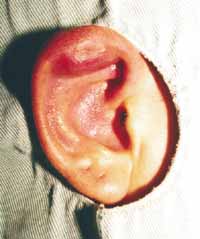
D. H., 15-year-old Caucasian female with piercing placed on the right ear 9 days before being admitted to the hospital.
The patient had presented local pain, edema and swelling of helix cartilage for 5 days, despite use of Cephalexin for the last 4 days (Figure 2).
The patient came to our Service, where puncture and collection of the purulent secretion was performed and sent for culture and antibiogram.
Afterwards, surgical drainage followed by debridement of necrotic tissue and placement of a Penrose drainage were performed. The patient stayed at the hospital and started antibiotic therapy with 500 mg Ciprofloxacin BID plus steroidal antiinflammatory treatment. She responded well to treatment, leaving the hospital within 48 hours.
Oral Ciprofloxacin treatment was maintained for 10 days.
In the follow-up visit, minimal scar deformity was observed. Culture result confirmed the presence of Pseudomonas aeruginosa.
Case 3
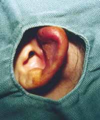
A. M. L., 14-year-old Caucasian female with intense edema, erythema, local pain and purulent secretion on the left ear, as a result of ear piercing 7 days before arriving at our Service (Figure 3).
Surgical drainage was performed and a sample of secretion was sent for culture and antibiogram. Oral antibiotic therapy with 500 mg Ciprofloxacin BID was initiated and maintained for 10 days.
Patient recovered well and did not show any deformities by scar.
Culture results confirmed the presence of Pseudomonas aeruginosa.
Discussion
The first case of perichondritis by Pseudomonas aeruginosa immediately after ear piercing is quite recent. It generally occurs during hot seasons (mainly Summer), when excessive body perspiration hinders healing and predisposes to infection. 2,4,10 Lack of arterial irrigation in the cartilaginous tissue favors infection perpetuation. 2-4,8 Potential complications occur within days or weeks (average time: 1 week) 2-4,7, which are as follows: local (cysts, edema, granuloma, hematoma, deformities, lipoma and abscess), or systemic (toxic shock, osteomyelitis, chondritis, diffuse acute glomerulonephritis and superficial cervical lymphadenopathy).1-3,6,9,13
Nickel seems to be the main responsible for hypersensitivity to the piercing. 7,11-13 It induces allergic reactions and leads to development of a nickel-protein complex, yielding an immunological response. 12 Several authors describe gold as the second material involved in this type of reaction. 2,6,7,12
The diagnosis is essentially clinical, while culture plus antibiogram should be performed. Generally, cultures reveal Gram-negative bacteria, among which Pseudomonas aeruginosa is the most frequently found.
Figura 1
Figura 2
Figura 3
Clinical treatment consists of antimicrobial agents (Cephalosporins, Ceftazidime, quinolones) seeking Pseudomonas aeruginosa erradication.1,2,4,8,10
Hospital admission and intravenous therapy should be considered in refractory cases. 2,10
Use of 70% isopropyl alcohol and iodine solution proved to be effective for local asepsis.2,6,8,10
Extensive surgical incision with necrotic tissue removal is determinant for good healing and to avoid deformities. 2,4,8,10 The most frequent sequel is auricular pinna deformity, which usually comes as a cauliflower-like aspect.
Perichondritis by ear piercing presents high morbidity, leading to complications in up to 35% of the cases, according to Simplot and Hoffman.3
Due to risk of esthetical deformities, immediate antimicrobial treatment, surgical drainage and local care were instituted.
Closing Remarks
The use of piercing is related to severe complications, mainly at lower vascular supplied portions of the ear (auricular cartilage). According to our experience and relevant literature, nickel is mainly responsible for such high sensitivity reaction. Treatment is based on oral antibiotic therapy, and, in non-responsive cases, intravenous therapy should be adopted.
Surgical drainage is imperative in the presence of perichondrial abscess. The otorhinolaryngologist should make early diagnosis and start effective treatment in order to minimize esthetical deformities.
References
1. Cossette JE. High Ear-Piercing. Otolaryngology- Head and Neck Surgery 1993; 107: 967-8.
2. Cumberworth VL, Hogarth TB. Hazards of ear-piercing procedures which traverse cartilage: a report of Pseudomonas perichondritis and review of other complications. BJCP 1990; 44 (11): 512-3.
3. Hanif J et al. High ear piercing and the rising incidence of perichondritis of the pinna. BMJ 2001; 322: 906-7.
4. Hendricks W M. Complications of Ear Piercing: Treatment and Prevention. CUTIS 1991; 48: 386-94.
5. Landeck A, Newman N, Breadon J, Zahner S. A simple technique for ear piercing. Journal of the American Academy of Dermatology 1998; 39 (5): 795-6.
6. Matarasso SL, Glogau RG. Surgical Pearl: Ear piercing facilitated by magnetic earrings. Journal of the American Academy of dermatology 1994; 31 (3): 485-6.
7. More DR, Seidel JS, Bryan PA. Ear-piercing techniques as a cause of auricular chondritis. Pediatric Emergency Care 1999; 15 (3): 189-92.
8. Nakada T, Iijima M, Nakayama H, Maibach HI. Rôle of ear piercing in metal allergic contact dermatitis. Contact Dermatitis 1997; 36: 233-6.
9. Nielsen NH, Menne T. Nickel sensitization and ear piercing in an unselected Danish population. Contact Dermatitis 1993; 29: 16-21.
10. Rogero SO, Higa OZ, Saiki M, Correa V, Costa I. Cytotoxicity due to corrosion of ear piercing studs. Toxicology in vitro 2000; 14: 497-504.
11. Simplot TC, Hoffman HT. Comparison between cartilage and soft tissue ear piercing complications. American Journal of Otolaryngology 1998; 19 (5): 305-10.
12. Staley R, Fitzgibbon JJ, Anderson C. Auricular Infections Caused by High Ear Piercing in Adolescents. Pediatrics 1997; 99 (4): 610-1.
13. Widick MH, Coleman J. Perichondrial abscess resulting from a high ear-piercing-case report. Otolaryngology- Head and neck surgery 1992; 107: 803- 5.
Print: