

Year: 2004 Vol. 70 Ed. 5 - (16º)
Relato de Caso
Pages: 688 to 690
 PDF PT
 Rhinolith: case report and literature review
Author(s):
Heráclio Villar R. Cavalcanti1,
Giordania Gomes Campos2,
Tatiana Silveira Velasco2,
Polliana Ferraz Rego2,
Alencar Polimeni Benetti3
Keywords: rhinolith, chronic sinusitis, nasal obstruction, unilateral rhinorrhea.
Abstract:
The authors report one case of a 57-year-old woman who for many years had a unilateral nasal obstruction; purulent rhinorrhea and chronic infection both caused by rhinolithiasis. The disease most frequently seen in association with rhinolithiasis is chronic sinusitis. The rhinolithiasis is a calcareous concretion formed in layers visible by X-rays. The rhinolith was discovered incidentally in some patients. Rhinoliths are rare and can have various clinical presentations. The treatment of choice is surgical removal. A high index of suspicion is required for the diagnosis of such a forgotten entity.
![]()
INTRODUCTION
Rhinolithiasis is a rare infection characterized by the presence of calcareous deposits in the nasal fossa that are progressively built up around an undiagnosed foreign body 1-3. There are physical-chemical and mechanical factors (pH affections, supersaturation and secretion stasis, airflow abnormalities, infection and inflammation) involved in the calcification process.4
Many children have the habit of placing small objects into the nasal cavity, leading to obstruction and rhinorrhea 5. These foreign bodies can normally be exogenous, such as vegetables, metals, plastic materials, etc, or more rarely from endogenous origin, such as mucous, teeth, clots, etc.
As the rhinolith increases in volume, there are nasal obstruction and purulent rhinorrhea, sometimes followed by epistaxis, foul odor, headache, etc. 6, 7. Unilateral purulent fetid rhinorrhea is pathognomonic of nasal foreign body until proven otherwise.
The purpose of the present study was to present a case of rhinolith in an adult woman seen in our center and to make a brief review of the literature.
CASE REPORT
MJS, female 57-year-old patient, born in Niterói, RJ, was seen in our center with complaints of nasal obstruction on the left, dyspnea, cacosmia and purulent rhinorrhea on the left for many years. She reported different treatment approaches in the past directed to sinusitis (sic). During childhood, at the age of about 7, the patient informed she had been taken to the emergency room because of bilateral rhinorrhea and they tried to remove a foreign body, but with no success. At that time, the physician referred her to surgical removal under general anesthesia, but the family refused to perform the procedure.
Anterior rhinoscopy revealed purulent rhinorrhea on the left and visualization of grey mass. Nasal endoscopy, in addition to the referred rhinorrhea, evidenced amorphous mass, of hardened consistency in the topography of the inferior meatus, close to areas 4 and 5 of Cottle; the other visualized structures had not affections.
We ordered computed tomography scan of paranasal sinuses as routine exam.
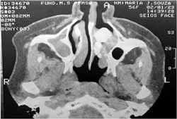
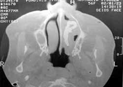
CT scan revealed presence of calcareous density mass on the posterior third of the left nasal fossa and normal transparency paranasal sinuses (Figures 1 and 2).
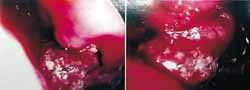
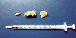
In view of the results, we confirmed the diagnostic hypothesis of rhinolith, referring the patient to surgical removal. The procedure was performed via endoscopic access (Figures 3 and 4).
After 2 months of ambulatory clinical follow-up, we conducted nasal endoscopy and the result was normal appearing cavity. The patient has been asymptomatic to present (Figure 5).
DISCUSSION
Rhinolith develops in the lumen of the nasal fossa from a nucleus whose origin is exogenous or endogenous. Around this nucleus, there is production of an inflammatory process and suppuration, causing trapping of the foreign body.
Rhinoliths are normally unilateral but not necessarily single, of grey or dark color, rounded, with surface irregularities that convey a coral-like shape. Size and weight are variable. The composition is equally variable: calcium phosphate, organic compounds, calcium carbonate, magnesium phosphate and water. It is subject to the influence of patients' lifestyle, such for example the composition of air inhaled and environmental pollution, and the nature of the foreign body.
The diagnosis is conducted with anterior rhinoscopy, which may sometimes induce to mistakes, since the rhinolith is surrounded by purulent exudates and recovered by mucous vegetating and congestive buds, simulating the sequestration of syphilis nature, malignant tumor or fungal sinusitis. Sometimes the rhinolith may be asymptomatic, found in complementary routine tests8,9. It may be diagnosed through complications such as for example septal perforation, nasal mucosa ulceration, osteomyelitis of nasal bones and/or mucosa synechiae.
The main differential diagnoses are osteogenic or odontogenic lesions. It is necessary to exclude the following affections as well: osteoma, granuloma, osteomyelitis sequestration, carcinoma, chondrosarcoma, and osteosarcoma.
Rhinoliths are uncommon in pediatric patients 10, whereas nasal foreign bodies are frequently found in children. In most cases, children do not say they have introduced something into their noses, fearing their parents' reaction. Thus, there is high likelihood that such foreign bodies become rhinoliths in the adults.
Treatment is based on removal of rhinolith. Surgical removal indicated under general anesthesia is reserved for large volume rhinoliths or when there is the need to repair nasal fistula.
The possible surgical complications are nasal epithelium ulcerations that progress to complete healing and formation of synechia between the turbinate and nasal septum 11.
The diagnosis in our case was facilitated by the clinical history, careful and detailed ENT examination and complementary exams.
CONCLUSION
Rhinolithiasis is a rare pathology that can have different forms of presentation. It requires high level of clinical suspicion for the correct diagnosis, as well as use of radiological exams. Treatment of choice is surgical removal.
REFERENCES
1. Davy-Chédauté F, Jézéquel JA. In: Encyclopedie Medico Chirurgicale. Edição 2000; Paris: Ed. Scientifiques et Médicales Elsevier SAS. p. 20-390-A-101 a 5.
2. Moulonguet L, Brette M D, Monteil JP. Ann Otolaryngol Chir Cervicofac 1995; 112(8):406-9.
3. Celikkanat S, Turgut S, Ozcan I, Balyan FR, Ozden C. Rhinolithiasis. Rhinology 1997 Mar; 35(1):39-40.
4. Aguayo V, Martín A, Soto C, Araceli I. Rinolitiasis: presentación de un caso. Rev Méd IMSS mayo-jun 1996; 34(3):207-9.
5. Endo LH, Montenegro MCS. Tratado de Otorrinolaringologia. V. 3. 1ª ed. Editora Rocca; 2003:175-80.
6. Hungria H. Otorrinolaringologia. 8ª ed. Editora Guanabara Koogan; 2000:79-85.
7. Balatsouras D, Eliopoulos P, Kaberos A, Economou C. Rhinolithiasis: an unusual cause of nasal obstruction. Rhinology 2002 Sep; 40(3):162-4.
8. Royal AS, Gardner RE. Rhinolithiasis: an unusual pediatric nasal mass. Pediatr Radiol 1998 Jan; 28(1):54-5.
9. Kharoubi S. Rhinolithiasis associated with septal perforation. A case report. Acta Otorhinolaryngol Belg 1998; 52(3):241-5.
10. Hadi U, Ghossaini S, Zaytoun G. Rhinolithiasis: a forgotten entity. Otolaryngol Head Neck Surg 2002 Jan; 126(1):48-51.
11. Brown WBS, Ballantyne J, Groves J. Diseases of Ear, Nose and Throat, Volume 1, 2ª Ed. London: Editora Butterwortlis; 1965.
Figure 1. Preoperative CT scan, coronal section: calcareous density material in the inferior meatus of the left nasal fossa.
Figure 2. Preoperative CT scan, axial sections: calcareous density material occupying the posterior third of the left nasal fossa.
Figure 3. Intraoperative follow-up: grayish rhinolith surrounded by polypoid mucosa, bleeding upon touch.
Figure 4. Rhinolith after removal.
Figure 5. Two months after surgery -nasal fossa with normal aspect.

Print: ![]()