
Year: 2002 Vol. 68 Ed. 6 - (16º)
Artigos Originais
Pages: 883 to 890
Distortion product otoacustic emissions in individual with tinnitus complaint
Author(s):
Fernanda A. R. Burguetti 1,
Angela G. Peloggia 2,
Renata M. M. Carvallo 3
Keywords: hearing deficits, tinnitus, otoacoustic emissions
Abstract:
Tinnitus is a sensation of sound without external stimulation. Tinnitus doesn't have a cause described precisely, however some authors relate that tinnitus is caused by damaged outer hair cells and normal inner hair cells. Aim: Distortion Product Otoacustic Emissions (DPOAE) evaluate outer hair cells of Organ of Corti, so this study objectives to investigate DPOAE amplitude in patients with tinnitus, in comparison with the results obtained with a control group. Study design: Clinical prospective. Material ant method: 66 female patients (132 ears) constituted the sample. Two subgroups were determined: Study Group with 27 patients (54ears) with tinnitus and without pharmacological treatment, exhibiting audiometric thresholds better than 25 dB HL across octave frequencies 500 to 4000 Hz; Control Group with 39 patients (78 ears) without tinnitus and exhibiting audiometric thresholds better than 25 dB HL across octave frequencies 500 to 8000 Hz. DPOAE was collected by Otodynamics ILO 92 V4.9 and the amplitude of responses were investigated at 1257, 2002, 3174, 4004 and 5042 Hz of f2 frequencies. Results: Study Group presented lower DPOAE amplitude than Control Group (p <0.01) in all frequencies except 1257 Hz at the left ear and 2002 Hz at the right ear did. Conclusion: Results suggest reduction of outer hair cells activity in patients with tinnitus.
![]()
Introduction
Tinnitus is a sound sensation not related to an external source of stimulation. Some authors refer to tinnitus as an auditory illusion (Bento et al., 1995, Sanchez, 1998, Fukuda, 1998, Rosanowski et al. 2001, Hall III & Haynes 2001).
It can be defined as a sound impression in one or both ears that seems to be present without any acoustic impulse from the environment or as a morbid symptom caused by different parts of the auditory system (from the external acoustic canal to the cerebral cortex) or other organs (Chelminsk & Chelminska, 2001).
Tinnitus can transform the life of a person, characterized as a disabling problem. It is a ghost auditory perception, noticed in most cases exclusively by the patient. Since it is subjective, investigations are difficult and limited concerning physiology.
Many methods have been used in order to define the etiology of tinnitus. Little is known about the physiological mechanisms that cause it, but there are various theories that try to explain the genesis of tinnitus. Most of those theories related tinnitus and hyperactivity of hair cells or nervous fibers, activated by chemical imbalance in the cell membranes or by disarrangement of hair cells steriocilia.
According to Sanchez et al. (1997), in most cases, tinnitus is associated with a problem of cochlea or auditory nerve nature. The lesion of outer hair cells can cause an increase in firing rate, stimulating the nervous fibers similarly to real sound. In addition, cochlear lesion of any nature can reduce the inhibition created by the central nervous system, enabling greater spontaneous neuronal activity in the auditory system (Schleuning, 1993).
Hall III & Haynes (2001) considered that tinnitus can result from the presence of damaged outer hair cells and normal inner hair cells.
The foundation for distortion product otoacoustic emissions (DPOAE), considering the non-linearity of the inner ear, is to evaluate the hair cells with absolute selectively of frequencies, conveying the idea of sectors for cell vitality throughout the basilar membrane (Munhoz et al. 2000).
Therefore, DPOAE assess the integrity of outer hair cells in Corti’s organ, and they can detect a cochlear dysfunction before it is evidenced by pure tone audiometry. Thus, they can confirm the presence of cochlear dysfunction in patients with tinnitus and normal pure tone audiometry (Hall III & Mueller, 1997).
The objective of the present study was to check the amplitude of distortion product otoacoustic emissions in female subjects complaining of tinnitus in order to assess the cochlear function of the outer hair cells of the patients.
METHOD
Material
A group of 54 ears of 27 female subjects aged 13 to 60 years with complaints of tinnitus but without using medication, with auditory thresholds of up to 25dB HL in frequencies of 500Hz to 4,000Hz, were selected among the population treated at the Service of Clinical Audiology, in the Center for Research and Education in Speech Therapy and Audiology, FMUSP, characterized as the studied group.
For the control group, we selected in the Service of Clinical Audiology a group of 78 ears of 39 female subjects aged 18 to 30 years without tinnitus complaints and with auditory thresholds of up to 25 dB HL in frequencies of 250 Hz to 8000 Hz.
All subjects were volunteers and agreed to the terms of the study by signing the informed consent term approved by the Ethics Committee for Research Project Analysis, Hospital das Clínicas, Medical School, University of São Paulo - USP, under protocol CAPPESQ Nº 368/98.
Equipment
• Audiometer GSI 61 – Grason Stadler – The equipment allows the investigation of pure tone thresholds in frequencies of 250 to 20000 Hz and is in accordance with ANSI S 3,6-1989; ANSI S3,43-1992; IEC 645-1(1992); IEC 645-2(1993); ISO 389; UL 544 standards. We used regular Telephonics TDH 50P phones with impedance of 80 ohms to perform conventional pure tone audiometry (250 Hz to 8000 Hz).
• Middle Ear Analyzer GSI 33 – Grason Stadler Version 2 – microprocessed and counting on three frequencies of tones in the immittance probe: 226 Hz, 678 Hz and 1000 Hz. The equipment makes automatic tympanometric measures, in the speed of 50decapascals per second (dPa/s), and the results are recorded in graphs and printed by the printer coupled to the system. We used thermal-sensitive paper for printing. The Middle Ear Analyzer was calibrated for the altitude conditions of the city of São Paulo, and we followed all the manufacturers’ requirements concerning electrical installation.
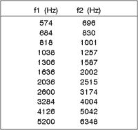
• Cochlear Emission Analyzer ILO 92 – OTODYNAMICS ILO 92 DPOAE version 1.35 – The equipment is programmed to produce two tones, one is a low frequency (f1) and the other is a higher frequency (f2), paired in a f2/f1 = 1.2 ratio. We maintained the intensities of 65dB SPL for f1 and 55 dB SPL for f2 (N1-N2=10, where N = intensity in dB SPL). We selected the responses recorded in 2f1-f2. The frequencies assessed with the equipment are listed in Chart 1.
Procedure
The subjects were submitted to:
• Meatoscopy to identify the presence of cerumen or other abnormalities that would prevent the conduction of the tests.
• Tympanometry with Compensated Admittance modality at the level of the tympanic membrane (Ymt) with probe frequency of 226Hz; ipsilateral and contralateral acoustic reflexes with stimuli of 500Hz, 1000Hz, 2000H, 4000Hz and Broad Band Noise. The procedure was conducted in order to detect the presence of middle ear affection that would prevent the conduction of the other tests.
• Pure tone audiometry (250 to 8000Hz) that helped the selection of patients whose auditory thresholds were up to 25dB HL for the conduction of distortion product otoacoustic emissions (DPOAE).
• Speech Reception Thresholds (SRT) to match the thresholds obtained with pure tone audiometry.
• Speech Recognition Index (SRI) at 40dB SL over the auditory thresholds of 500 Hz, 1000 Hz and 2000 Hz, adopted as an additional normal parameter. We accepted results equal or greater than 88%.
• Distortion product otoacoustic emissions in all frequencies. The test was conducted with the patient in a sound-proof booth. The first ear was randomly assessed and the responses were collected after verification of the appropriateness of the probe. We accepted responses obtained with signal/noise ratio greater than 3dB. To analyze statistically, we used the distortion products (2f1-f2) of frequencies (f2) of 1257 Hz, 2002 Hz, 3174 Hz, 4004 Hz and 5042 Hz.
We compared the data from subjects in the control group and the studied group in order to define whether the data were statistically different or not.
We used two techniques in the study: the first one was a comparison of the groups altogether, that is, all variables considered as a whole, which is called Multivariate Mean Comparison (MANOVA); the second one allowed a more specific analysis and it is known as Univariate Mean Comparison (ANOVA).
These techniques were used to compare the results of amplitude of DPOAE between the right and left ears of the control group and the studied group, and the left ear of the studied group and the control group and the right ear of both groups.
The statistical analysis conducted for each comparison was p value. This kind of statistics helps us conclude how the test was performed. If the value is greater than the significance level adopted, it is concluded that there is no statistically significant difference between the means of the groups. We adopted a significance level of 5%. Therefore, if p value was greater than 5%, there was statistically significant difference between the means of the groups. The statistically significant differences were marked with asterisks on the tables of results.Chart 1 – Frequencies (f1 and f2) assessed by the analyzer otoacoustic emissions ILO 92 – OTODYNAMICS ILO 92 DPOAE version 1.35.
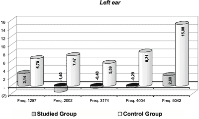
Graph 1. Amplitude of DPOAE on the left ear for the studied and the control groups.
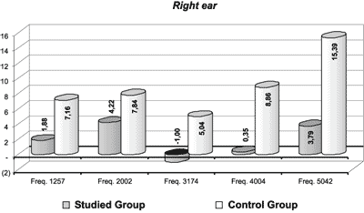
Graph 2. Amplitude of DPOAE on the right ear for the studied and the control groups.
Results
I. Analysis of distortion product otoacoustic emissions (DPOAE):
Table 1 shows the distribution of amplitude values of DPOAE on the right and left ears of both groups. According to these results, we noticed decreased amplitude of DPOAE in the studied group compared to the control group. Graphs 1 and 2 illustrate such findings.
II. Comparison between the groups:
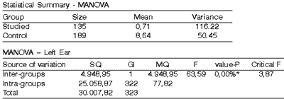
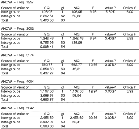
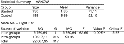
The analysis of results according to p-value referring to the left ear (Table 2) and right ear (Table 4) isolated demonstrated that there is statistically significant difference between the studied and the control groups. Thus, the analysis was detailed in order to define the frequencies (Hz) that had generated such difference.
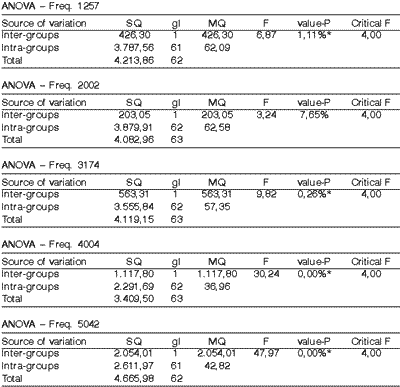
According to Table 3, on the left, all frequencies except for frequency of 1257Hz showed statistically significant differences between the studied and the control group. As to the right ear, as demonstrated in Table 5, all frequencies, except for 2002Hz, had statistically significant differences in mean.
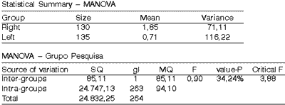
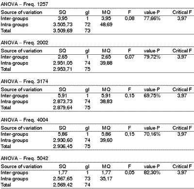
By comparing the left ear and the right ear of the studied group, we observed that there was no statistically significant difference, because p value was equal to 34.24% (Table 6). Upon the performance of univariate analysis to observe if the situation of similarity was remained, we noticed that the results for the left ear were statistically identical to those obtained for the right ear and that it occurred for each frequency isolated, as shown by Table 7.
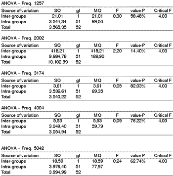
As to the comparison between the left and right ear in the control group, we observed that there was no statistically significant difference between the ears, that is, the left ear produced the same effect as the right ear and that was noticed in all frequencies (Table 8).
Based on these results, we concluded that there was no statistically significant difference between the distortion product otoacoustic emissions responses in left and right ears.Table 1. Amplitude in dB of DPOAE on the right ear (RE) and left ear (LE) in the studied and control groups.
Table 2. Comparison between Amplitude of DPOAE on the left ear in the Studied group and the Control group using the multivariate analysis.
Table 3. Univariate analysis for amplitude of each frequency on the Left Ear in the Studied group and Control group.
Table 4. Comparison between amplitude of DPOAE on the Right Ear in the Studied group and the Control group using multivariate analysis.
Table 5. Univariate analysis of amplitude of each frequency on the Right Ear of the Studied group and the Control group.
Table 6. Comparative analysis between amplitude of DPOAE on the Left Ear and on the Right Ear (Studied group).
Table 7. Univariate comparison for each frequency of DPOAE on the Right Ear and Left Ear (Studied group).
Table 8. Univariate comparison for each frequency of DPOAE on the Right Ear and Left Ear (Control group).
Discussion
The focus of the present study was directed to the analysis of differences in amplitude of DPOAE in groups of female subjects, being that 27 of them had tinnitus complaint and 39 did not have hearing complaints. Both groups presented auditory thresholds below 25dB HL between 500Hz and 4000Hz. The selection criteria of the group focused on normal hearing and not age as a factor of interference in amplitude of DPOAE, similarly to the studies by Barreñas & Wikström (2000). Quaranta et al. (2001) conducted a study in which they did not find statistically significant difference among amplitude of DPOAE in five different age ranges between 20 and 58 years, although they had observed reduced amplitude of otoacoustic emissions as a result of increased thresholds.
Similarly, the similarity in auditory profile in the two groups enabled the comparison of amplitudes of DPOAE regardless of age, as described below.
The smaller amplitude of DPOAE in the studied group compared to the control group and the statistically significant differences between DPOAE in the two groups can reflect subtle cochlear differences, caused probably by the tinnitus present in the studied group subjects.
Such findings confirmed the data in the study performed in patients with tinnitus submitted to acupuncture by Chami et al. (2001), in which the amplitude of distortion product otoacoustic emissions was abnormal before treatment.
According to Fukuda (1998), cochlear dysfunction commonly generates tinnitus and hearing loss. However, this dysfunction is sometimes not enough to cause a hearing loss detected at pure tone audiometry, but in some clinical tests, such as otoacoustic emissions, the dysfunction can be characterized.
In addition, whereas the precise cause is not established for the tinnitus and the audiometry shows normal clinical limits, the presence of cochlear dysfunction is almost invariably documented with more sensitive measures, such as otoacoustic emissions. Hall III & Haynes (2001) referred that in cases of patients with tinnitus, in addition to conventional pure tone audiometry, it is important to perform distortion product otoacoustic emissions to assess the function of outer hair cells. DPOAE abnormalities and cochlear deficiencies can be detected in patients with normal hearing, because not all patients with cochlear dysfunction will show deficits in the frequencies between 250Hz and 8000Hz in the audiogram.
According to Sanchez et al. (1997), the occurrence of tinnitus in patients without hearing loss should be explained by diffuse damage to up to 30% of the outer hair cells in the whole cochlear spiral, with no hearing threshold impairment. The presence of damaged outer hair cells and intact inner hair cells can characterize the tinnitus with frequencies close to the site of cochlear lesion. According to this fact, it is suggested that the approximate frequency of tinnitus would vary widely, since all analyzed frequencies presented statistically significant differences, except for 1257Hz on the left and 2002Hz on the right. However, these data are suggestive since no further tests were conducted in the present study.
Conclusion
The study of amplitude of distortion product otoacoustic emissions (DPOAE) in subjects who have tinnitus evidenced statistically significant differences in amplitude of DPOAE, both in the left ear and the right ear, when compared to the amplitude of DPOAE in patients without tinnitus complaints. The responses in the studied group presented amplitudes of DPOAE at intensity levels below those in the control group, suggesting a reduction of the level of activity of outer hair cells in the group with tinnitus complaint, compared to those without complaint.
References
1. Barrenäs ML, Wikström I. The Influence of Hearing and Age on Speech Recognition Scores in Noise in Audiological Patients and in the General Population. Ear and Hearing. 2000;21(6):569-577.
2. Bento RF, Caetano MHU, Rezende VA, Sanchez TG. Mascaramento do zumbido rebelde ao tratamento clínico. Rev Bras de Otorrinolaringologia 1995;61(4): 290-297.
3. Chami FAI, Onishi ET, Fukuda Y, Yamamura Y et al. Alterações das Emissões Otoacústicas por Produto de Distorção em Pacientes Portadores de Zumbido Submetidos a Acupuntura. Estudo Preliminar. Arquivos da Fundação Otorrinolaringologia 2001;5(2): 78-85.
4. Chelminski K, Chelminska M. Tinnitus. Pol Merkuriusz Lek mar 2001;10(57):195-197.
5. Fukuda Y. Zumbido e suas correlações otoneurológicas. In: Ganança MM. Vertigem tem cura? São Paulo: Lemos Editorial; 1998. p.171-176.
6. Hall III JW, Haynes DS. Audiologic Assessment and Consultation of the Tinnitus Patient. Seminars in Hearing 2001;22(1):37-49.
7. Hall III JW, Mueller HG. Otoacoustic Emissions. In: ________. Audiologists’ Desk Reference. Vol. 1: Diagnostic Audiology Principles Procedures and Practices. California: Singular Publishing Group Inc.; 1997. p.235-288.
8. Munhoz MSL, Silva MLG, Frazza MM, Caovilla HH, Ganança MM, Carvalho P. Otoemissões Acústicas. In: Munhoz et al. Audiologia Clínica. São Paulo: Editora Atheneu; 2000. p.121-148.
9. Quaranta N, Debole S, Di Girolamo S. Effect of ageing on otoacoustic emissions and efferent suppression in humans. Audiology 2001;40(6):308-312.
10. Rosanowski F, Hoppe U, Kullner V, Weber A, Eysholdt U. Interdisciplinary management of chronic tinnitus. Versicherungsmedizin jun 2001;53(2):60-66.
11. Sanchez TG. Zumbido. In: Bento RF, Miniti A, Marone, SAM. Tratado de Otologia. São Paulo: Editora USP; 1998. p.322-330.
12. Sanchez TG et al. Controvérsias sobre a Fisiologia do Zumbido. Arquivos da Fundação Otorrinolaringologia 1997;1(1): 2-8.
13. Schleuning AJ. Tinnitus. In: Bailey BJ. Head and Neck Surgery. Otolaryngology. Philadelphia: J.B. Lippincott Company; 1993. p. 1826-1832.
1 Audiologist, Specialist in Clinical Audiology, FMUSP.
2 Audiologist, Specialist in Clinical Audiology, FMUSP.
3 Audiologist, Ph.D., Professor, School of Speech and Language Pathology and Audiology FMUSP.
Affiliation: Medical School, University of São Paulo (FMUSP)
Address correspondence to: Fernanda A. R. Burguetti – Rua Barão de Tatuí 673, apto 72 18030-150
Tel (55 15) 211-3524 (55 15) 9789-1010 Fax (55 15) 231-7951
E-mail: farb@splicenet.com.br
Article submitted on June 19, 2002. Article accepted on September 19, 2002
