

Year: 2002 Vol. 68 Ed. 6 - (8º)
Artigos Originais
Pages: 821 to 824
Diagnosis of anterior comissure microwebs by laringovideostroboscopy
Author(s):
Adriana da Silva Lima 1,
Domingos H. Tsuji 2,
Natasha M. A. Braga 1,
Rui Imamura 3,
Luiz Ubirajara Sennes 4
Keywords: anterior comissure microwebs, laringovideostroboscopy
Abstract:
The microweb is a very thin mucusal membrane with 23mm located on the infraglotic surface of the anterior comissure of glottis. Because of its small size and location it is a lesion usually difficult to detect with routine clinical examination of the larynx. Aim: To estimate of microwebs in patients with dysphonia using strobovideolaryngoscopy, and to analyses its relation with sex, age and association with another vocal fold lesions. Study design: Case series. Patient and Method: We analysed 100 strobovideolaryngoscopy video records of patients with dysphonia, between 1997 and 2001. Results: The presence of microweb was detected in 12 patients (12%). The microweb was always found associated with other vocal fold lesions; it was never diagnosed alone. The laryngeal lesions most commonly associated with the microweb were: vocal nodules (5 cases), cysts (3 cases), sulci (2 cases), corditis (1 case) and cancer (1 case). No statistically significant difference in regard to sex, age or associated laryngeal lesion was found between patients with and without microwebs. Conclusion: The microweb is a small but not rare laryngeal lesion that can be associated with other lesions in patients with dysphonia. The patients with microweb did not differ from those without microweb in regard to sex, age or associated laryngeal lesion.
![]()
INTRODUCTION
According to Sataloff & Spiegel (1993), webs or laryngeal membranes connect the vocal folds and can be of congenital or acquired origin, as a consequence of trauma. They can be small and involve only a small portion of the anterior commissure, called laryngeal microweb, or compromise all the free margin of the vocal fold and extend beyond the glottis to the subglottis and supraglottis 1. Ford et al. (1994) described microweb as a thin mucosa membranous layer of 2 to 3mm long, located on the infraglottic surface of the anterior commissure of the vocal fold2.
Owing to its location and small size, microweb may not be identified in laryngoscopic exams conducted in the clinical practice, even in reference centers for laryngology 2. Conversely, microweb can be associated with other laryngeal lesions, such as vocal nodules in about 7 to 23% of the cases2, 3, 4.
In the clinical practice, laryngovideostroboscopy undoubtedly represents the most widely used method to study vocal vibration, enabling detailed analysis of vibration cycles, in addition to the general visualization of laryngeal structures5, 6.
The occurrence of microwebs in the anterior commissure in exams of laryngovideostroboscopy is not commonly studied in the literature. Therefore, the objective of the present study is to estimate the frequency of microwebs in vocal folds of patients with dysphonia submitted to the exam, studying the association of this lesion with age, gender and other laryngeal lesions.
MATERIAL AND METHOD
We selected 100 exams that presented good technical quality of documentation, allowing carefully analysis concerning damage to the anterior commissure. To that end, we analyzed sequentially a total of 707 exams of patients who had complaints of dysphonia and were submitted to laryngovideostroboscopy in a private office from 1997 to 2001. All exams were performed during spontaneous breathing, at maximum abduction and preceded by throat clearing to eliminate secretion that could be mistaken for microweb. We excluded all exams whose analysis was doubtful because of secretion, previous surgery and incomplete glottic opening. According to these criteria, we excluded 607 exams, being that the remaining 100 corresponded to the 100 patients enrolled in the present study. All recordings were assessed by two investigators.
The equipment used for the exams was a laryngeal telescope brand Machida, model LY-CS30 of 70º with 8mm of external diameter; source of stroboscopic light brand Bruel & Kjaer, type 4914; camera Toshiba, model IK-CU43 A; endoscope adapter brand Machida, model CA-34VS2; video recorder NTSC, SHVS, brand Sony, model WVSW1, and video monitor brand JVC, model AV-M150S.
The main laryngeal lesions found were divided into 4 groups: nodules, inflammatory lesions (polyp, pseudocyst, Reinkes edema, corditis), minimum structural alteration (cyst, sulcus and bridge), and others (leukoplasia, cancer and vocal fold atrophy).
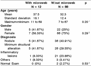
Initially, we distributed the patients in the study according to gender, age and diagnosis of laryngeal lesion. Next, the patients were divided in two groups: with microweb and without microweb. We compared the mean age, distribution of gender and frequency of concomitant laryngeal lesion in each group.
The statistical analysis was conducted through the exact Fischer test to compare proportions (gender and concomitant laryngeal lesion) and non-paired t Student test for comparisons of mean (age). We adopted the statistical significance level of p below or equal to 5% (p £ 0.05).
RESULTS
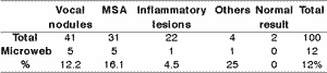
We included in the study 73 women and 27 men aged 7 to 69 years (mean age 32.95 years, median 33 years). We did not find statistically significant difference concerning age among male subjects (mean = 36.04 years; standard deviation = 17.11 years) and female patients (mean = 31.31 years, standard deviation - 10.38 years). The presence of nodules was the most frequent diagnosis among the patients (n=41), followed by minimum structural alteration (n=31), inflammatory lesions (n=22) and other diagnosis (N=4). Two patients did not present laryngeal affections at the exam (Table 1).
It was possible to identify the presence of microweb in the exam of 12 patients (12%; CI95% - 6.4% to 20%). Among them, microweb was concomitantly diagnosed with other laryngeal lesions, and it was not isolated in any case. The associated diagnoses with microweb were nodules (5 cases), cyst (3 cases), sulcus (2 cases), corditis (1 case) and neoplasm (1 case). The two patients with normal exam did not present microweb.
The patients with microweb did not differ from the others concerning gender, mean age and association with other laryngeal lesions (Table 2).Table 1. Laryngeal lesions identified at laryngovideostroboscopy and the association with microweb.
Fischer exact test (p = 0.53).
Table 2. Social-demographic characteristics and diagnosis at laryngovideostroboscopy of patients with and without microweb (n = 100).
1 value of p obtained with t Student test.
2 value of p obtained with Fischer exact test.
DISCUSSION
In our study, microweb was diagnosed in 12% of the cases, occurring in association with vocal nodules (5 cases), cyst (3 cases), sulcus (2 cases) corditis (1 case) and neoplasm (1 case). The association with microweb and other laryngeal lesions has been described in the literature2, 4, 7. Bouchayer (1990), reviewing 100 cases of microweb, confirmed that 78 presented an association with nodular lesion and 22 had other types of lesions such as polyps, cysts and fibrosis3. The prevalence of this association, however, is probably underestimated, since the laryngoscopic exams conducted in daily practice many times do not reach the region of the anterior commissure. In our study, more than 80% of the exams were excluded because they did not enable the appropriate assessment of the region. Thus, the diagnosis of microweb can not be conducted during the routine ambulatory exam, requiring proper attention and interest from the examiner to identify it.
Ford et al. (1994) identified 105 patients with vocal nodules and the presence of microweb in 11 cases (10.5%), in which 10 were diagnosed in the intra-operative stage and only one with the laryngovideostroboscopy exam2. In another study Benninger & Jacobson (1995) found microweb in 8 cases (7%) of the 115 patients with vocal nodules and of them, half were diagnosed through the exam4. Although our methodology was based on ambulatory findings and not on intra-operative data, we found a rate of 12.2% of microweb in the group of vocal nodules, data similar to those of other authors previously mentioned, which theoretically identified the lesion under better conditions, that is, microlaryngoscopy and palpation of the region. This similarity among the rates should be probably due to the fact that we have been very judicious in including only high quality exams in the study, that is, those that allowed good visualization of anterior commissure through complete glottic opening and absence of secretion in the region.
We did not find statistically significant differences concerning gender and age of patients with and without microweb, suggesting that these characteristics are not relevant for the identification of the lesion.
We observed that the lesions most associated with microweb were vocal nodules and minimum structural alterations, which together corresponded to more than 80% of the videolaryngostroboscopic lesions associated. However, in the group without microweb, these lesions were also very frequent, present in more than 70% of the cases. The comparison between the groups did not show statistically significant difference, suggesting that there is not a strong association between microweb and these lesions.
Conversely, owing to the relative prevalence of microweb in patients with vocal nodules, some authors believe that this association is not only a coincidence. Based on the site of the microweb, Bouchayer et al. (1988) suspected that there would be reduced vibration of the anterior third of the vocal folds, leading to mechanical trauma that favored the formation of vocal nodules3. Ford et al. (1994) referred that the presence of microweb would reduce the vibration segment of the vocal fold with consequent increase of subglottic pressure and vibration amplitude, leading to a greater contact of the vocal folds. In addition, its presence associated with vocal misuse would hinder the spontaneous absorption of the nodular lesion. However, according to the same authors, the abnormal vocal fold vibration was similar in patients with nodules associated or not with microweb 2. There are also some studies that related the presence of microweb associated with failure in voice therapy with patients that had vocal nodules. Ford et al. (1993) observed that 50% of the patients who failed in voice therapy had nodules associated with microweb. Therefore, this association would increase the incidence of surgical treatment need8. On the other hand, Benniger & Jacobson (1995), succeeded in voice therapy in 78% of the patients with vocal nodules and microweb, showing that the surgical indication depends on severity of the nodules, voice therapy results and size of the microweb 4.
In our study with dysphonic patients, microweb did not occur isolated in one single case. Thus, it is likely that the lesion responsible for the initial vocal complaint was not the microweb, but the associated lesion (nodule, minimum structural alteration, inflammatory lesion, or other). The role of microweb in the pathophysiology of dysphonia and in the origin of other secondary lesions still remains unclear and deserves further studies to help explain it.
CONCLUSION
Microweb was observed in 12% of the patients with vocal complaints and it was associated with nodules, minimum structural alterations, inflammatory lesions and neoplasms, and there were no significant correlations with age, gender or association with laryngeal lesions.
REFERENCES
1. Sataloff RT, Spiegel JR, Gold WJ. Endoscopic Microsurgery. In: Gold WJ, Sataloff RT, Spiegel JR. Voice Surgery. 1ª ed. St Louis, MO: Mosby; 1993. p. 227-267.
2. Ford NC, Bless DM, Campos G Leddy M. Anterior comissure microwebs associated with vocal nodules: detection, prevalence, and significance. Laryngoscope 1994; 104(11):1369-1375.
3. Bouchayer M, Cornut G. Microsurgery for benign lesions of the vocal folds. Ear Nose and Throat J 1988; 67(6):446-466.
4. Benninger MS, Jacobson B. Vocal nodules, microwebs and surgery. J Voice 1995;9(3):326-331.
5. Tsuji DH, Sennes LU. Videoquimografia de laringe: novo método de avaliação da vibração cordal. Arq Fund Otorrinolaringol 1998;2(4):136-140.
6. Rosen CA, Murry T. Diagnostic Laryngeal Endoscopy. Otolaryngologic Clinics of North America 2000;33(4): 751-757.
7. Bouchayer M. Congenital lesions of the vocal fold. Presented at the First Scandinavian Seminar on Phonosurgery, Stockholm, 1990.
8. Ford CN, Bless DM, Campos G, Leddy M. Anterior comissure microwebs associated with vocal nodules: detection, prevalence, and significance. NCVS Status Prog Rep 1993;4:85-92.
1 Post-graduation studies under course, Division of Clinical Otorhinolaryngology, Medical School, University of São Paulo.
2 Ph.D., Supporting Teacher, Discipline of Otorhinolaryngology, Hospital das Clínicas, Medical School, University of São Paulo.
3 Ph.D., Assistant Physician, Discipline of Otorhinolaryngology Hospital das Clínicas, Medical School, University of São Paulo.
4 Full Professor, Discipline Otorhinolaryngology, Hospital das Clínicas Medical School, University of São Paulo.
Study conducted at the Discipline of Otorhinolaryngology, Medical School, University of São Paulo -HCFMUSP
Address correspondence to: Drª Adriana S. Lima Av. Brigadeiro Faria Lima 1853, 1º andar
Jardins São Paulo SP 01452-912
Tel (55 11) 3813.0211 Fax (55 11) 3814.9491 E-mail: adri_lima@hotmail.com
Article submitted on May 23, 2002. Article accepted on July 11, 2002
Print: ![]()