

Year: 2001 Vol. 67 Ed. 4 - (12º)
Artigos Originais
Pages: 527 to 529
Results of the Endoscopic Endonasal Surgical Technique for Dacryocystorhinostomy.
Author(s):
Patsy P. V. Iturralde*,
Fábio O. Reis*,
Rigoberto A. Oliveira Michel Cahali**,
Richard L. Voegels***.
Keywords: endonasal surgery, dacryocystorrhinostomy, dacryocystitis
Abstract:
Introduction: The advent of endoscopic endonasal surgery provided a new alternative for the treatment of nasal lachrymal duct obstruction. Study design: Clinical retrospective. Aim: The objective of the present study was to report our experience with the approach in the treatment of dacryocystitis. Material and method: We conducted a retrospective study of 18 patients submitted to surgical treatment due to obstruction of the nasal lachrymal duct during the period of March 1998 to March 2000. Regarding the etiology of dacryocystitis, nine patients were classified as idiopathic, four as iatrogenic, four as recurrent after external dacryocystorhinostomy and one as posttraumatic. The symptoms included epiphora, purulent secretion and edema of the lachrymal sac. Dacryocystography and tomography of the paranasal sinuses were performed in all patients. Results: The patients were followed in average for one year, with resolution of the symptoms in 94.4% of the cases. In only one case there was persistent epiphora after surgery, although less intense than preoperatively. Conclusions: In our experience endoscopic endonasal dacryocystorhinostomy has high success rates associated with excellent aesthetic results, without any major complication. There is enough evidence that endoscopic endonasal dacryocystorhinostomy is effective and physiological and it should be considered the first option in the surgical treatment of these patients.
![]()
INTRODUCTION
As a result of the advent of endoscopic surgery, a great advance in endonasal techniques employed to disobstruct the lachrymal path system has been noticed.
The external approach described by Toti in 190412 is the most widely known and used technique by ophthalmologists.
Endonasal approach was proposed by Caldwell (1893)1 and developed by West (1910)8.
The development of endonasal surgical techniques (Rouviere et al., 1981, Kennedy, 1985, Mc Donogh, 1992, El Khoury and Rouvier, 1992)3 improved the knowledge of sinus-nasal-orbit anatomy, presenting endonasal dacryocystorhinostomy as the preferred choice for the cases of lachrymal-nasal duct obstruction, because it prevents external scars in the face.
The purpose of the present study was to present our experience with endonasal endoscopic dacryocystorhinostomy, showing the results we obtained with the use of the technique.
MATERIAL AND METHOD
We performed a retrospective study of 18 patients with diagnosis of nasal-lachrymal duct obstruction who were submitted to endoscopic dacryocystorhinostomy between March 1998 and March 2000.
There was predominance of female over male patients (13:5), and ages ranged from 3 to 69 years, mean age of 41.5 years.
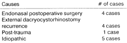
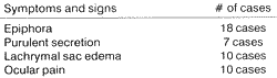
Etiologies of dacryocystitis are demonstrated in Table 1, and clinical picture in Table 2.
All patients were evaluated with nasal endoscopy, dacryocystography and paranasal sinuses CT scan.
The surgical procedure was performed with 4mm rigid endoscope at 0 degree, identifying the lachrymal sac by translumination after canalization with flexible optical fiber standard size 20, through the lower lachrymal canaliculus. We then proceeded an incision of the nasal mucosa, creating a posterior pediculated flap, exposing the bone that recovers the lachrymal sac, which is either drilled or removed with laser, followed by the removal of the mucous flap. We performed hemostasis control, blood aspiration and duct secretion and irrigation. As a routine, we placed a silicone probe (Crawford) through the upper and lower lachrymal canaliculi, fixed on the nasal septum mucosa with Nylon 5-0 suture. Normally, the tube is removed 4 to 8 weeks after the surgical act.
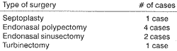
Patients who required other procedures besides dacryocystorhinostomy are described in Table 3.
As postoperative medication, we prescribed ocular corticoid eve drops for 10 days, nasal corticoid spray during the first month and nasal lavage with sterile solution for six months.TABLE 1 - Etiology of dacryocystitis.
TABLE 2 - Clinical picture of dacryocystitis.
RESULTS
Patients were followed up for one year, with endoscopic control during the first month, and we avoided removing intranasal crusts and synechia. We observed symptom improvement in 17 patients (94.4% of the cases) and one case (5.6%) presented only persistence of epiphora, but less severe than the preoperative condition. There were no intra-operative complications. The silicone tube was removed between 4 and 8 weeks after the surgery, depending on the tolerance level of each patient.
DISCUSSION
The purpose of dacryocystorhinostomy was to develop adequate drainage of lachrymal intranasal flow.
The causes of nasal-lachrymal duct obstruction may be infectious, post-surgical, traumatic or idiopathic. Preoperative assessment includes clinical picture characterized by epiphora, purulent secretion, lachrymal sac edema and ocular pain. Moreover, we conduct nasal endoscopic examination that should look for possible factors for surgical failure (maxillary-ethmoid sinus pathology, marked septal deviations, middle turbinate hyperplasia, or nasal fossa synechia); dacryocystography (showing the level of obstruction) and paranasal sinuses CT scan (which enables assessment of the general status of the nasal cavity and paranasal sinuses, as well as the thickness of lachrymal bone, in order to better plan the surgery).TABLE 3 - Complementary surgeries.
The obstruction of nasal-lachrymal duct may be effectively approached either externally, by the ophthalmologist, or endonasally, by the rhinologist.
The endoscopic technique has gathered more and more advocators, because it causes less surgical trauma, absence of skin incision, absence of scar retractions resulting from external dissections, and preservation of palpebral ligaments and medial canthus of the orbit, preserving the physiological mechanisms of the lachrymal pump3-10.
Toti's technique, in experienced hands, generates a success rate of 90%, whereas endonasal methods produce success rates-of 95%15, as demonstrated by our study.
The surgical technique we employed13 is based on the removal of the bone that recovers the lachrymal sac, using drilling or laser. It is also different from others concerning the mucous flap, because some authors prefer to preserve the flap to form the fistula with the mucosa of the lachrymal sac, by simply placing the mucosas close to each other.
As to recurrence cases, endonasal dacryocystorhinostomy is technically simpler, since osteotomy has already been performed and it consists of removing only the destroyed portion of the lachrymal sac.
CONCLUSIONS
The high success rate (94.4%) of endonasal endoscopic surgical technique by dacryocystorhinostomy demonstrates that it is a highly effective procedure. The fact that it provides an excellent visualization of the surgical field during the whole procedure characterizes it as a physiological technique, in addition to preventing the rupture of the internal canthal ligament and lesions of the internal canthus of the eye, as well as the antiesthetical scar resultant from the external approach. It also enables combined surgical procedures, such as septoplasty, sinusectomy or polypectomy, if required.
REFERENCES
1. CALDWELL, G. W - Two New Operations for Obstructions of the Nasal Duct With Preservation of the Cannaliculi. Am. J Ophthalmol., 10: 189, 1893.
2. ELIE E.; REBEIZ, M. D.; STANLEY, M. SHAPSHAY, M. D.; JOSEPH, H.; BOWLDS, M. D.; MICHAIL, M.; PANKRATOV M. S. - Anatomic guidelines for dacryocystorhinostomy. Laryngoscope, 102: (10), 1992.
3. ELOY E; BERTRAND, B.; MARTINEZ, M.; HOEBEKE, M.; WATELET, J. B.; JAMART, J. - Endonasal dacryocystorhinostomy: Indications, technique and results. Rhinology, 33 (4): 229-233, 1995.
4. H. HALIS UNLU, FIGEN GOVSA, CEMIL MUTLU, ALI VEFA YUCETURK, YILMAZ SENYILMAZ. - Anatomical guidelines for intranasal surgery of the lachrymal drainage system. Rhinology Mar, 35 (1): 11-5, 1997.
5. JAVATE, R. M.; CAMPOMANES, B. S. A. JR.; CO, N. D.; DINGLASAN, J. L. JR.; GO, C. G.; TAN, E. N.; TAN, F. E. The endoscope and the radiofrequency unit in DCR surgery. Ophthalmic Plastic and Reconstructive Surgery, 11 (1): 54-58, 1995.
6. MANNOR, G.; MILLMAN, A. L.- The prognostic value of preoperative dacryocystography in endoscopic intranasal dacryocystorhinostomy. American Journal of Opbtbalmology, 113(2): 134-137, 1992.
7. METSON R. - Endoscopic surgery for lachrymal obstruction. Otolaryngol Head and Neck Surgery, 104: 473-479, 1991.
8. RICE D. Endoscopic intranasal dacryocystorhinostomy results in four patients. Arch Otolaryngol Head and Neck Surgery, 116: 1061, 1990.
9. SCHAUSS, F.; WEBER, R.; DRAF, W; KEERL, R.- Surgery of the lachrymal system. Acta oto-rhino-laryngologica belg., 50(2): 143-146, 1996.
10. SERDAHL CL, CRAIG EB, CHOLE RA. - Nasolacrimal duct obstruction after endoscopic sinus surgery. Arch. Ophthalmol., 108:391-3, 1990.
11. STAMBERGER H. - Endoscopic endonasal surgery: concepts in treatment of recurring rhinosinusitis. II. Surgical technique. Otolaryngol. Head and Neck Surgery, 94: 147-56, 1986.
12. TOTI A. ZUM princip, zur techniq and zur deschicte der dacrocystorhinostomie. Z. Augenbeilkd, 23: 232-239, 1910.
13. VOEGELS L. RICHARD, CAHALI B. MICHEL, MATAYOSHI SUZANA, BUTUGAN OSSAMU, MINITI AROLDO. - Dacriocistorrinostomia endonasal, Rev. Bras. Otorrinolaringologia, 65(1): 44, 1999.
14. WEST J. A window resection of the nasal duct in cases of stenosis. Trans. Am. Opbtbalmol. Soc., 12: 659, 19.
15. WERNER HOSEMANN, MD; MANFRED WEIDENBECHER, MD; WOLFGANG BUHR, MD. Endoscopic endonasal dacryocystorhinostomy: Results in 56 patients. Ann. Otol. Rhinol. Laryngol., 103: 363-6, 1994.
* Resident Physician, Discipline of Otorhinolaryngology, Hospital das Clínicas da Faculdade de Medicina, Universidade de São Paulo.
* * Postgraduate Physician, Discipline of Otorhinolaryngology, Hospital das Clínicas da Faculdade de Medicina, Universidade de São Paulo.
*** Ph.D., Professor, Discipline of Otorhinolaryngology, Hospital das Clínicas da Faculdade de Medicina, Universidade de São Paulo.
Study conducted at the Division of Clinical Otorhinolaryngology, Hospital das Clínicas da Faculdade de Medicina, Universidade de São Paulo.
Address correspondence to: Priscilla Vasquez Iturralde - Rua Apeninos, 539 - Apto. 71-Aclimação - 01533-000 São Paulo/SP
Tel: (55 11) 284-6576-E-mail: ppvic@zipmail.com.br
Study presented as poster at 35° Congresso Brasileiro de Otorrinolaringologia, held on October 18, 2000, in Natal/RN.
Article submitted on March 7, 2001. Article accepted on April 16, 2001.
Print: ![]()