

Year: 2001 Vol. 67 Ed. 4 - (9º)
Artigos Originais
Pages: 507 to 510
Etiological Correlation Between Anatomical Variations in Computed Tomography and Chronic Rhinosinusitis.
Author(s):
Richard L. Voegels*,
Elder Y. Goto**,
Daniel Chung**,
Luciana M. Nita**,
Marcus M. Lessa***,
Ossamu Butugan****.
Keywords: chronic rhinosinusitis, computed tomography, anatomical variations
Abstract:
Introduction: Computed tomography (CM has been increasingly used both to diagnose and to evaluate nasal structure. Furthermore, CT scanning has become the evaluation of choice for patients with sinunasal diseases. Anatomical variations such as septal deviation, middle turbinate and anterior ethmoid cell anomalies may impair the flow of secretions through the ethmoid infundibulum, leading to the development of sinusitis. Nevertheless, these findings are not supported by the literature. Study design: Clinical retrospective. Purpose: The purpose of the present study was to analyze the anatomical variations found in CT scans of patients with chronic sinusitis treated surgically. Material and method: Paranasal sinus CT scans were obtained in 93 patients from December 1995 to December 1999. Results: There were 48 men (51.6%) and 45 women (49.4%). The mean age was 35.31 years. The most prominent findings were concha bullosa (12.89%), severe septal deviation (4.30%), hypertrophic inferior turbinate (3.220/0), paradoxical middle turbinate (2.15%), Haller cells (2.15%), and hypertrophic uncinate process (2.15%). The most affected paranasal sinuses were maxillary, posterior and anterior ethmoid. Conclusion: The number of CT scan abnormalities in our stud was not sufficient to allow assessment of the correlation wit chronic sinusitis. Further studies are necessary to establish the role of these anatomical variations.
![]()
INTRODUCTION
Chronic rhinosinusitis may be defined as a sinusal disease in which there is persistence of symptoms, such as nasal obstruction, cough, purulent rhinorrhea, headache and/or hyposmia for a minimal of three months9. As a result of the recent development of endoscopic approach of paranasal sinuses, introduced by Messerklingler and Stammberger8, together with the technical innovations in the radiological field, much has been studied about the anatomy of the ostiomeatal complex and the lateral nasal wall and the implications of anomalies in the genesis of the inflammatory paranasal sinuses affections.
Computed tomography (CT scan) has been the preferred method for diagnosis of paranasal sinuses affections, especially at coronal sections, in which we can clearly identify the most important structures of the nasal cavity and adjacent areas4,9. We should keep in mind that most of the times the examination is important not only to diagnose the inflammatory disease, but rather to better prepare the surgical plan, because CT scan reveals the structures very clearly, serving as an excellent intraoperative guide for the surgeon.
At first sight, it may seem obvious that abnormalities of middle concha, anterior ethmoid cells, nasal septum and other bone structures may prevent the physiological flow of secretions from the ethmoidal infundibulum, and lead to conditions that favor the development of infections and inflammation of paranasal sinuses. However, this suspicion has not been confirmed by the literature3,4,7,8,9.
The objective of the present study was to use CT scan to investigate the existing anatomical abnormalities of the nasal lateral wall, especially on the anterior ethmoidal region, in patients with diagnosis of chronic rhinosinusitis.
MATERIAL AND METHOD
We evaluated 93 patients with diagnosis of chronic rhinosinusitis, surgically treated between December 1995 and December 1999, at the Division of Clinical Otorhinolaryngology, Hospital das Clínicas, FMUSP.
Data were collected from the retrospective analysis of the medical files. CT scans were carried out with no previous preparation, such as use of topical vasoconstrictors or nasal lavage with sterile solution. We used devices CT PACE or PROSPEED, by General Electric (Milwaukee, WI, USA), 120kV 130mA and exposure of 2 seconds. Patients were positioned in supine posture and 5mm coronal sections were obtained through perpendicular positioning of the hard palate. In the ostiomeatal region, we carried out 2mm sections. Five-millimeter axial sections were also collected, with the patient in prone posture, analyzing from the frontal sinus to the floor of the maxillary sinus, in order to subside the identification of the disease and other anatomical variations, such as the presence of Onodi's cells. In order to assess the structures, we used soft part windows (approximate level of 50 and opening of 250H) and bone structure windows (level of 400 and opening of 4,000H). The contrast used was meglumine ioxithalamate (Telebrix 30® - Guerbet), at 30% iodine, dose of 2 mL/kg.
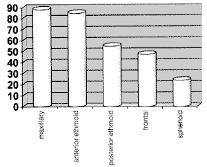
Graph 1. Distribution of affected paranasal sinuses (in %).
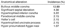
All CT scans were carried out and interpreted in preoperative meetings that gathered the team of assistant physicians of the Rhinology Group. The investigated anatomical abnormalities were: bullous middle concha, paradoxical middle concha, Hatter's cell and Agger nasi, hypertrophied uncinate process, significant septal deviation and hypertrophied inferior concha.
RESULTS
We studied 93 patients with diagnosis of chronic rhinosinusitis submitted to surgical treatment. There were 48 men and 45 women. Ages ranged from 3 to 76 years, mean age of 35.31 years (standard deviation: 17.85).
The distribution of affected paranasal sinuses is shown in Graph 1. The analysis of CT scan results (Table 1) detected that bullous concha was the most frequent anatomical abnormality. Other alterations were septal deviation, hypertrophied inferior concha, paradoxical middle concha, Haller's cell, hypertrophied uncinate process and hyper pneumatized Agger nasi.
DISCUSSION
During initial diagnostic approach of patients with chronic rhinosinusitis, it is important to determine which are the affected sinuses and what is the magnitude of the disease (partially or completely obstructed sinuses). This knowledge enables appropriate follow up of the progression of the case and the plan of a specific surgical intervention, if necessary.
TABLE 1 - Anatomical alterations that would favor the development of chronic rhinosinusitis (in %).
In our study, the most frequently affected paranasal sinuses in decreasing order were: maxillary, anterior ethmoid, posterior ethmoid, frontal and sphenoid (Graph 1). Our findings were similar to the ones reported by Willner9 concerning the three most affected sinuses (maxillary, 60%; anterior ethmoid, 51%, and posterior ethmoid, 35%); however, the data are different concerning the two least affected, because he found more cases of sphenoid sinusitis (32%) than frontal ones (24%). In their studies, Tonai and Baba8 and Bolger2 observed different incidences concerning the two most affected sinuses in chronic sinusitis, reporting, respectively the following findings: anterior ethmoid (86% and 84.3%) and maxillary (80.7% and 77.7%) sinuses, and equivalent incidences for the other sinuses.
A middle concha that is pneumatized is called bullous concha; it may lead to obstruction of middle meatus and infundibulum, through the compression of the uncinate process4. The incidence in the normal population ranges from 9% to 20%8. It was the anatomical alteration most commonly found (12.89%). Lusk4 described that bullous concha, together with septal deviation, were the most frequent alterations in children with chronic rhinosinusitis. Yousem10 and April1 reported exactly the same incidence (24%) among their patients and it was also their most frequent finding. Nadas6 investigated CT scans of 308 patients with chronic rhinosinusitis and concluded that there was no correlation between presence or size of bullous concha and increased risk of rhinosinusitis. Upon analyzing the fact that there is similar incidence between studied cases and the asymptomatic population, there seems to be no correlation between presence of bulldus concha and development of choronic rhinosinusitis; however, a study with a control group would be the correct way to study this aspect.
Hypertrophied inferior concha was observed in most patients, but it was considered positive in only 3.22% of the cases, because the criteria for inclusion of this category was need for inferior turbinectomy. Lusk4 reported hypertrophy in 6% of his sample, but criticized the validity of this piece of data, because there was no control of nasal cycle or use of topical vasoconstrictor in his study.
The cause of obstructive ostiomeatal complex disease may be attributed to septal deviation. There are different degrees of septal deviation, but in the present study we only included patients who necessarily needed corrective surgery (septoplasty), which was performed at the same time (4.30% of the cases). This figure is much lower than the incidences reported by Lusk4 (10.4%), April' (13%) and Willner9 (13%), but it is probably due to the strict criteria we followed - only septal deviations with surgical indication.
Middle concha may present a curvature contrary to the one normally seen and in such conditions it is named paradoxical concha. We found it in 2.15% of our cases and its was the fourth most frequent finding. In the literature, we found variable data in patients with chronic rhinosinusitis (4.4%5 to 29.8%8), suggesting that we still lack appropriate evidence to consider it a predisposing factor to chronic rhinosinusitis.
Infraorbital cell, also called Haller's cell, is part of the ethmoidal cells and it is differentiated from the others by the fact that it becomes pneumatized inferiorly to the ethmoidal bulla, and laterally, towards the roof of the maxillary sinus, interposing between the papyraceous lamina and the uncinate process. It may potentially obstruct the maxillary ostium and ethmoidal infundibulum, causing rhinosinusitis; however, no study in the literature has determined its role in the disease. It seems to be more common than the results from the present study suggested (2.15%), as shown in Milczuk5 study, who found the abnormality in 5.3 % of the patients, or Tonai and Baba8, who observed Haller's cells in 33.3% of the cases. A curious fact, reported by the latter, makes us wonder about the importance of the structure in maintaining the sinusal pathology. They observed these cells in 38.9% of the control group (no rhinosinusitis), which could inaccurately suggest that its presence is a protecting factor against chronic rhinosinusitis.
Deviation or enlargement of uncinate process may generate difficulties in drainage of the ostiomeatal complex. It is relatively rare and difficult to identify (2.5% of the cases reported by Bolger2 had pneumatized process), and we noticed enlargement in only 2.15% of our cases.
Agger Nasi cells are placed right above and anteriorly to the anterior insertion of the middle concha. Since its definition is poor, its incidence in the literature varied a lot, from 3% to 98.5%g. In our opinion, this factor does not seem to be implied in chronic rhinosinusitis, because it was considered obstructed in only 1.07 % of our patients. However, Brunner3 studied the role of Agger nasi cells in frontal sinusitis and concluded that they were a significant part of the etiological mechanism.
CONCLUSION
We did not collect enough CT scan findings to suggest a correlation between anatomical alterations and development of chronic rhinosinusitis. We hope that further studies be carried out so that we may understand the real importance of these anatomical variations.
REFERENCES
1. APRIL, M. M.; ZINREICH, S. J.; BAROODY, F. M. Coronal CT Scan Abnormalities in Children with Chronic Sinusitis. Laryngoscope, 103: 985-990, 1993.
2. BOLGER, W E.; BUTZIN, C. A.; PARSONS, D. S. Paranasal Sinus Bony Anatomic Variations and Mucosal Abnormalities: CT Analysis for Endoscopic Sinus Surgery. Laryngoscope, 101: 56-64, 1991.
3. BRUNNER, E.; JACOBS, J. B.; LEBOWITZ, R. A.; SHPIZNER, B. A.; HOLLIDAY, R. A. - Role of the Agger Nasi Cell in Chronic Frontal Sinusitis. Ann. Otol. Rhinol. Laryngol, 105: 694-700, 1996.
4. LUSK R. E; ALISTER, B. M.; FOULEY, A. - Anatomic Variations in Pediatric Chronic Sinusitis. Otolaryngol. Clin. North Am., 29: 75-91, 1996.
5. MILCZUK, H. A.; DALLEY, H. A.; WESSBACHER, R. W Nasal and Paranasal Sinus Anomalies in Children with Chronic Sinusitis. Laryngoscope, 103: 899-903, 1993.
6. NADAS, S.; DUVOISIN, B.; LANDRY, M.; SCHNYDER, E - Concha Bullosa: Frequency and Appearance on CT and Correlations with Sinus Disease in 308 Patients with Chronic Sinusitis. Neuroradiology, 37: 234-237, 1995.
7. SAUNDERS, N. C.; BIRCHALL, M. A.; ARMSTRONG, S. J.; KILLINGBACK, N.; SINGH, D. - Morphometry of Paranasal Sinus Anatomy in Chronic Rhinosinusitis. Arch. Otolaryngol. Head Neck Surg., 124: 656-658, 1998.
8. TONAI, A.; BABA, S. - Anatomic Variations of the Bone in Sinonasal CT. Acta Otolaryngol (Stockh), suppl 525: 913, 1996.
9. WILLNER, A.; CHOI, S. S.; VEZINA, G.; LAZAR, R. H. Intranasal Anatomic Variations in Pediatric Sinusitis. Am. J Rhinol., 11: 355-360, 1997.
10. YOUSEM, D. M.; KENNEDY, D. W; ROSENBERG, S. Ostiomeatal Complex Risk Factors for Sinusitis: CT Evaluation. J. Otolaryngol., 20: 419-424, 1991.
* Ph.D., Professor, Discipline of Otorhinolaryngology, Hospital das Clínicas, Faculdade de Medicina da Universidade de São Paulo.
** Resident Physician, Discipline of O0torhinolaryngology, Hospital das Clínicas, Faculdade de Medicina da Universidade de São Paulo.
*** Postgraduate Physician, Discipline of Otorhinolaryngology, Hospital das Clínicas, Faculdade de Medicina da Universidade de São Paulo.
**** Associate Professor, Discipline of Otorhinolaryngology, Faculdade de Medicina da Universidade de São Paulo.
Study presented as free paper at 35° Congresso Brasileiro de Otorrinolaringologia, held on October 17 - 20, 2000, in Natal/RN.
Study conducted at the Division of Clinical Otorhinolaryngology, Hospital das Clínicas, Faculdade de Medicina da Universidade de São Paulo.
Address correspondence to: Richard L. Voegels - Av. Dr. Enéas de Carvalho Aguiar, 255 - 6° andar-Sala 6021-05403-000 São Paulo/ SP
Tel: (55 11) 3069-6288 - Fax: (55 11) 270-0299.
Article submitted on February 2, 2001. Article accepted on March 19, 2001.
Print: ![]()