

Year: 2001 Vol. 67 Ed. 3 - (15º)
Artigo de Revisão
Pages: 394 to 402
Update of Grafts in Rhinoplasty.
Author(s):
Lucas G. Patrocínio*,
José A. Patrocínio**.
Keywords: rhinoplasty, grafts, implants
Abstract:
Several types of grafts and materials are used in rhinoplasty: implant (polyethylene, metilmetacrilate, silicon, Supramide, Proplast, politetrafluoretilene, Dacron, Gore-Tex), homologous and autologous cartilage, etc. We present a historical and updated revision of the grafts in the plastic surgery of the nose. An ideal graft, for all the areas of the nose, doesn't exist. The appropriate material is chosen in agreement with the surgeon's preference, based on the characteristic of that and on the surgical needs. It is in research the production, in laboratory, of cartilaginous tissue for transplants to be used in the completion of defects of the face. That is future.
![]()
INTRODUCTION
The search for beauty has been part of humanity throughout history. Differently from what might have been thought, nose plastic surgery is a very old specialty. Dated 7BC, there are perfect descriptions of nose reconstructions in Indian sacred books. According to the history, war prisoners, thieves and unfaithful women had their noses amputated as punishment. In order not to have that symbol of shame throughout their lives, these people searched for reconstructive surgeons whose techniques are used until today. There is no doubt, though, that after the first and second world wars, from when innumerous surgeons started to dedicate to nose reconstruction, that the plastic surgery had its main impulse.
Among all facial characteristics, many authors have advocated that the nose is responsible for conveying peculiarity to faces19,21.
Nose shape imperfections may be perceived differently, according to racial, cultural and ethnic perspectives of the observer. Studies have shown that there are patterns of social characteristics, such as nose size and shape, which are wished by most people.
In most cases, rhinoplasties require the use of some kind of material to complement the nose. The medical challenge is to find the best graft or implant to meet the needs of the surgery, trying to find methods and materials that are as similar as possible to the nasal material. The world trend is to choose autologous materials1,9. Different techniques have been abandoned, some remained and others are currently been experimented. As a result of new plastic medical materials, there is a new light in the horizon.
Classification
Terminology concerning different kinds of grafts has changed through the years, but the current terms are listed below:
Autografts: free grafts transferred from one region to the other in the same subject - there is no rejection.
Isografts: free grafts transferred from one subject to another genetically identical subjects (identical twins); rejection is rarely identified.
Homografts (allografts): grafts transferred from a subject to another genetically different subject, but within the same species; there is rejection at different levels, depending on the transferred tissue, preparation and receptor.
Heterograft (xenograft): grafts transferred from a representative of one species into a subject of another species; they are normally quickly rejected, depending on tissue preparation and receptor.
Alloplastic (implant): biologically inert foreign materials, used to augment or reconstruct parts of the body; there is rejection sometimes, depending on the degree of inflammatory reaction induced by the material used38.
Types of graft
There are different kinds of grafts and materials used in esthetic and reconstructive nasal surgery. The ideal graft should have shape, consistency and resistance of the defected nasal structure. It should be easily obtained and molded, at low cost. Moreover, it should cause minimal tissue reaction and resist to extrusion and body reabsorption.
Implants
First of all, an ideal implant should be nontoxic and non-allergenic, with minimal tissue reactivity, in addition to being easily sterilized, molded, non-absorbable, extrusion-resistant and enable removal, if necessary.
Stabilization of implants is obtained by the mechanical interaction of the adjacent tissue. Polymers, normally known as plastic, are chains of atoms formed by covalent bounds of molecules, through the division of electrons. During polymerization, the molecules, also called monomers, are combined in various chains to form the polymers. They have different physical characteristics depending on the size and the position of chains, similarly to intermolecular forces. Monomers may be of the same type - silastic (silicone), polymethylmetacrilate and polytetrafiuorethylene (Teflon, Fluon), or for polymers with the bound of two or more different monomers terephtalate polyethylene (Dacron or Terylene).
The chemical nature of an implant is what defines its effect on the body and its cytological near and far effects (the implant may be completely tolerated by the body but not work mechanically). If the surface is not porous, the process of reconstruction produces a membrane of non-adherent fibrous tissue. Under these circumstances, the stabilizing effect may derive only from the conformation of the membrane for micro irregularities of the surface, leading to a compressive force on the implant. The thicker the membrane, the more deformed and the less capable it is to limit the relative mobility of the implant. Microporosity is an attractive alternative, because it develops a much larger interactive surface as to macro perforations or micro irregularities which are obviously limited when it comes to fixating the implant12.
Different materials have been used throughout the years with different levels of success. In 1889, Gersuny in Vienna, published a study in which it used an injection of Vaseline to correct saddle noise deformity. Later, Gersuny, Delanger and Eckstein (1900) used paraffin that caused local inflammation on the nose and migrated. Buck and Brockaert (1903) denominated paraffinomas the tumors that arose from the proliferation of connective tissue in the area of nasal implant, because of the use of paraffin. Fink (1915) conducted 24 surgeries implanting stones from the Dead Sea in saddle noses. Other materials, such as gold, silver, porcelain, platinum, aluminum and celluloid, were experimented by researches. Rueda (1916) used catgut. Bogorodinski (1925) used silver. Eitner (1930) tried ivory. In 1930, paladone was introduced as a nasal implant, a material normally used to fix dental prostheses. The material was abandoned because after some time it broke up8.
Rollin, in 1937, used synthetic resins for the first time as nasal implants: polyethylene and methylmetacrilate high molecular weight polymers. In 1940, tantalum and vitallium (cobalt, copper and nickel), non-electrolytic metals were used for the first time, with poor results. Months or years after the implant, there was inflammation, ulceration and suppuration, spontaneously or after minor traumas. Some were rejected.
In 1943, polyethylene was introduced in surgeries. It is a hard polymer, thus more inert, flexible and well tolerated. Nevertheless, it tends to get harder and harder with time, in addition to the fact that it is not a good material to replace soft tissues. Rubin and Walden (1940) used it in 46 cases to perform nasal reconstruction, presenting 8 cases of failure. Rubin, Robertson and Shapiro (1948) published good results with the material.
Theissing, in 1949, in Germany, recommended the use of Supramid, a super polyamide based on phenol and chemically related with Dacron and Nylon to correct saddle nose deformity. Legler (1957) tried it in 70 cases of saddle nose and there were only two cases of late rejection. It is flexible, leads to low tissue reactivity and has high melting point29. A mesh of polyamide was developed in 1970 and it has been used with promising results. It is porous and allows growth of tissue through it, leading to stabilization, Beekhuis (1974)2 published 30 cases of operated saddle noses with Supramid and only three cases required removal of implant because of infection. However, in subsequent studies, Beekhuis et al.36 reported absorption of the material in the long run. Stucker (1982)37 obtained 99% of success in mesh polyamide implants for the correction of saddle nose, in a 9-year follow-up.
Grindlay and Wang (1951) experimented polyvinyl in animals' nostril. More (1952) showed the practicality of macro perforations to improve dental implant fixation. Cottle, Quilty and Buckingham, in 1953, published cases with Ivalon, a saturated polyvinyl alcohol in the shape of an sponge, similar to a loaf of bread, which may be easily sliced, modeled with a pair of scissors and implemented on the nose. Freman (1955) presented four cases of complications due to infection caused by Ivalon. Mirabet et al. (1964) used acrylic prosthesis with various perforations in all directions, which was invaded by the connective tissue and reduced its mobility. Chang Ti-Sheng and Ohmori27, in 1965, described 39 cases of saddle nose in which they used cartilage, bone or plastic (acrylic resin) with unsatisfactory results in five cases29.
Ohmori27, in 1965, described a technique indicated to correct saddle nose in syphilis patients, in which they used a removable wire prosthesis associated with a fixed prosthesis of dimethylpolysyloxane. Kent et al. (1972)15 presented a pilot study of implant of porous resin in oral surgery, replacing inert metal, silicone, acrylic resin and ceramics. Milward (1972)21, Rozner (1980), Marvin (1980) and Mackay (1983)20 published cases in which silicone was used (silastic). Some extruded, other infected or were unstable and had to be removed, in addition to not being able to support the tip of the nose. Silastic is currently the most widely used polymer in implants. It is inert but deficient in force and resistance. It has the consistency of rubber, it is easy to section and mold, but has the disadvantage of migrating, getting infected and bulge the skin. As a whole, silicone has more disadvantages than advantages3,9.
Janeke et al.13, in 1974, presented Proplast as a new alloplastic material for the use in reconstructive head and neck surgeries, which had been tested in cats and monkeys with excellent results, both for mastoid and frontal sinuses obliteration and nose, followed up for 9 months. Kent et al. (1975)16 had a 5-year follow-up of patients treated with Proplast for dental and facial reconstruction.
Polytetrafluorethylene (Teflon and Politel) is well tolerated and has good functional results. However, the need to anchor the graft for stabilization purposes results in limited used. Politef is used in the treatment of vocal fold paralysis.
Carbon polytetrafluorethylene (Proplast Plastipore) is a material manufactured specially for surgical purposes, which consists of Teflon and carbon fibers. It is biocompatible, black and has high chemical stability, easily adjusted to the desirable shape by using scissors, and it is 70 to 90% porous, enabling growth of adjacent connective tissue to stabilize it. The main failures with this material were caused by infections. After the removal of the implant, conventional antibiotic therapy was necessary. Some infections were solved without requiring removal of implant. Another cause of problem was its displacement. It has the same effect of a tattoo because of its black color which becomes more evident with time, and it may be avoided by recovering it with thick subcutaneous tissue29.
Cavalcanti (1983)6, in Curitiba, Brazil, used a mesh of marlex or prolene to smoothen the columella-labial angle and improve nasal profile. According to him, because of its early fixation, the material prevents unpleasant displacements caused by silicone. Niechajev (1999)26 implanted it in the nose dorsum of 23 patients, and Turegun et al. (1998)39 implanted in 36 cases a high-density porous polyethylene, Medpor, in difficult nasal reconstructions, specially in tertiary and quaternary rhinoplasties. It is a material used as a routine in facial skeletal reconstructions and mentoplasties.
Dacron (Debakey-Double Velour Dacron Fabric), polyethyleneglycol terephtalate, is used primarily in cardiovascular surgeries replacing vessel walls. It is soft, white and-easy to cut with the scissors. Arrais has been using it for 30 years, in layers that are sutured with chromed catgut certix 3-0 for the correction of saddle nose.
Implanted metals, due to ion discharge, cause reactions in the body forming a hard, dense and fibrous barrier that encapsulate them. If ions are cytotoxic and concentration is high, there may be inflammation and pain, triggering the process of rejection. Implanted metals also suffer damage, such as corrosion. Disadvantages are superior hardness compared to bone, leading to local osteoporosis and low biocompatibility, causing luxation, absorption and bone necrosis. Ceramics have the same disadvantages of metals, in addition to brittleness.
Implants of Supramid, Mercilene, Proplast, Silastic and Porex continue to be used but the latest news is the use of Gore Tex28-33. It is a synthetic material that presents good results. It is a polymer of expanded fibrillar polytetrafluorethylene with micro porous structure. After its application, there is a intraprosthetic cell colonization, with no formation of capsule or foreign body, and it is the less dangerous alloplastic implant known until today. The advantages are that it is porous, easy to mold, white and well tolerated. The disadvantages are considerably high price, softness that does not offer enough support, the fact that it is not pre-molded, palpable and unstable, leading to high rates of extrusion if placed on regions subject to mobility. Some complications from Gore Tex use have already been reported: infection, mobilization and need to remove it. Another material, the Softform, an expanded tubular polytetratfluorethylene is similar to Gore-Tex and has more stability.
Alloplastic implants are being extensively tested in rhinoplasty, because on the one hand they simplify the surgery, whereas on the other hand, there is a significant rate of rejection. Up to today, it has been concluded that implants cause poor long-term results. However, in some situations fresh autogenous does not solve the problem. There is the clear need for new materials with good biocompatibility that have appropriate efficacy, resistance to use and that may be easily molded into the desirable shapes. Currently, alloplastic materials are used in the dorsum, pre-maxilla and ala. They should not be placed in the tip nor in the medial crus. We suggest the use of external rhinoplasty, that is, away from the other incisions, it should be sterilized for 30 minutes and removed if exposed.
Heterograft
Cartilage heterograft was first used in 1947, from bovine manure, sterilized in boiling water and preserved in a solution of Merthiolate. This type of cartilage presented great percentage of absorption, according to the studies by Gibson and Davis10, in 1953.
Heterograft of bone and/or cartilage was obtained from different animals and used as implant. It was abandoned because of the high percentage of long term absorption.
Homograft
Nasal septum or rib grafts are about to be abandoned. Few centers still use them, but only when irradiated. It should be carefully collected concerning asepsis and anti-asepsis of previously selected cadavers and in hospitals in which there is all necessary infrastructure to preserve them.
The surgery with cartilage homograft was introduced in the 40's. The material used to be preserved in formaldehyde, Merthiolate or Cialit. It was used for a number of years. Easily moldable, it maintained nose's flexibility. It does not bind to the dorsum bone and tends to lateralize. In the long run, it is not always viable. There are cases of absorption, it may get infected and extrude. Cartilage homograft is normally like an autograft. We know that six weeks after the homograft, the chondrocytes are involved in a gelatin stabilizing avascular matrix. This quick barrier of mucopolysaccharides contributes to non-antigenic behaviors of the homograft, preventing the penetration of antibodies of the host connective tissue. As a result of time, the matrix is gradually absorbed and the chondrocytes are consequently destroyed, but the cells of the grafted cartilage survive for many years.
Homologous cartilage has advantages over the autologous cartilage because it does not require a donation surgery, it is less subject to shortening, easy to mold, well tolerated, flexible and it may be preserved for a long time. Some of the disadvantages are great tendency to long term absorption, rejection and the maintenance of a cartilage bank. In cases in which the cartilage may be submitted to tension, we should select some other material because it is more prone to reabsorption. Absorption signs are evidenced after 18 to 24 months and finally the implant may disappear completely11.
The meniscus cartilage was used in the 50's and abandoned due to its unsatisfactory results. Currently, some centers use homografts of irradiated coastal cartilage23, inserted via external rhinoplasty. However, it leads to chondrocyte destruction and there are different degrees of fibrosis and shortening of graft. According to Bujía et al.4 transplanted homologous cartilage suffer reabsorption because of immune reactions caused by class II antigens existent in the perichondrium, which destroy them completely by preserving in Merthiolate, inactivating immune reactivity.
Bone homograft was obtained from other healthy people, under ideal conditions of asepsis, hermetically kept in refrigerators under low temperatures or preserved in solution of Merthiolate or Cialit. Materials were periodically cultured to make sure that the material was still sterile. Homologous bones have a higher absorption rate than autologous ones. It is essential that there is good contact between the graft and the bone in the nose, so that the graft may catch. The main advantage of such a technique is how easy it is to obtain the necessary quantity of bone for the surgery, sparing the patient from undergoing another surgery. The main disadvantages are high rate of absorption and poor acceptability by the host. In general, surgeons prefer autologous to homologous bone.
Homograft is not longer frequently used. The impact that conventional microorganisms may have, in addition to the surge of unknown diseases, such as AIDS and the prion of BSE, affect negatively the frequency of use.
Autograft
Common materials for autograft are bone, cartilage31 and soft tissues. The advantages are absence of antigenicity or reaction to foreign body and the availability of head and neck sites. Disadvantages include limitation of material available in some cases and potential morbidity of the donating site.
Cartilage or bone from the nose itself If there is still nasal septum cartilage, it may be used. We proceed with a resection of submucous, it is piled up in two layers, if necessary (if three layers, the middle one is absorbed), molded as a saddle and fixed with catgut 3-034. We should preserve 1.5-2.0 cm anterior and superior of the nasal septum, in order to maintain the nasal support30. We may also use a portion of the inferior lateral cartilage. It is a simple method, in the same surgical area, with good results. There is not always enough cartilage and it may be used for minor defects only.
In the correction with superior lateral cartilage a paramedian incision is made 2-3 mm lateral and parallel to the nasal septum. The same may be done with the inferior lateral cartilage. They are rotated to the midline and sutured10. It is used for the correction of minor defects of the middle third and does not present satisfactory results.
Inferior lateral cartilage is normally used when the septum is insufficient. Some millimeters from the cephalic rim of the inferior lateral cartilage are bilaterally sectioned and they are rotated to the dorsum and fixed with a subcutaneous suture (Flying Wing Procedure)24. It is used for minimal saddle noses and has the same disadvantages of the previous techniques.
The nose bone may be removed from the vomer, perpendicular lamina of ethmoid, nasal concha and frontal process of maxilla. The amount is normally limited. The removal of hump with transposition is common in cases in which the hump is prominent. We remove it and apply to correct the saddle on the middle thirds17,20 due to its simple transposition. This technique is used to correct small defects. The advantage is to have easy access to material and the disadvantage is the limited quantity necessary for the surgery, in addition to the likelihood of long term absorption. It is more frequently indicated in women.
Cartilage, bone and soft tissues from other regions
The autograft of subcutaneous muscleaponeurotic system may be used to correct deformities of nasal dorsum during associated surgery of ritidectomy in which one of its portions is resected18. It should be used only for filling because it does not offer support.
Fat autograft should not be used because its rate of reabsorption is approximately of 50% and sometimes it is necessary to repeat the surgery within 6 months. It is not a good technique and it has been abandoned.
The autograft of bone may be harvested from the rib, calvarium, olecranon, iliac or tibia bones. Among bone grafts, most nose surgeons prefer bone from the iliac crest because it is more abundant. The removal of iliac crest is made with an incision on the margin of the pelvis and detachment of periosteum using a osteotome. The closure of periosteum is made by catgut 2-0 and the skin suture with mononylon 4-0. If hemostasis is correct, there is no need for drain. The removal of tibia is conducted by a longitudinal incision on the anterior portion of the leg. The medial portion of the bone is-exposed after detachment of the periosteum and the cortical bone is obtained with the use of an osteotome and a circular saw. The autograft of calvarial membranous bone of parietal-temporal region may be used to correct moderate or severe defects. It may be easily obtained with minimal scars1. Several authors32,35,40 described innumerous degrees of cerebral lesion and the inherent risks occurred when there was partial calvarial bone autograft. It is recommended to avoid midline and cranial sutures. They refer that membranous bones present less absorption than endochondral bone. Byrd (1994)5 confessed he had abandoned the use of calvarial bone after three years of experience because of the unacceptable number of irregularities and secondary deformities that became visible.
Iliac and coastal bones have been used with success, although they may be absorbed late, experience pain in the donating area, and sometimes secondary infection with loss of graft in the receptor region. The bone graft remains adhered to the nasal bone and immovable, projecting the tip of the nose. It has the advantage of not bending and gets to life when bound to the nasal bone. It should be fixed on the nose with steel wires, screws or mini plaques. The x-ray shows bone with survival of 20 years or more.
It has the following disadvantages: difficult to be molded, the inferior half of the nose is normally semi-mobile and it may be fractured by a severe trauma or suffer a luxation and protrusion, leading to an artificial appearance of the nose.
In all cases, the harvesting of grafts require a separate surgical procedure under general anesthesia, increasing the duration of rhinoplasty and making an additional scar that is sometimes not welcomed by patients, especially women. Excision of bone is normally followed by a certain degree of pain, limiting movements and walking for some days and extending the length of hospital stay.
In 1959, Converse and Campbell7 published the report of 189 operations in which they used bone homo and autograft from various facial sites. Out of 46 nose cases, five presented problems and overall, 12 cases experienced problems. The failure rate of nose grafts was higher than the other facial areas. The same figures are found in other grafts and implants. Rib bone is not a good graft because it tends to absorb and lead to fracture. Auricular cartilage may also be used. It has the advantage of having the second surgical field closely related with the first one, conducted under local anesthesia, with low morbidity and minimal bending, displacement or extrusion. The limited amount of cartilage, additional surgical time and different surgical field, requiring separate incisions, are some of the disadvantages of the technique, in addition to a possible esthetical deformity in the earl. It may also reconstruct ala cartilages (seagull wings). Auricular pinna is an almost ideal transplant because: 1) the harvesting of material is a low risk procedure, quick and may be conducted under local anesthesia; 2) pinna cartilage is stable enough to produce support and elastic enough to produce contours; 3) it is easy to model; 4) it has little tendency to get displaced; 5) there is no absorption; 6) there is no rejection or infection. The collection is made by anterior incision in the pinna, and since it is easier, on the right side (if necessary, cartilage may be removed from both ears), taking always autograft with posterior perichondrium14,22.
Autograft of coastal cartilage, obtained from the coastal-chondral junction of sixth, seventh, eighth or ninth ribs of the patients is the preferred material for nose surgeons. It should be removed following the balanced sections described by Gibson in order to prevent shortening. The sixth rib is the flattest one and it is normally preferred because it practically does not bend. In women the incision to remove grafts should be conducted on the inframammary sulcus. The advantages of this technique are that it is live tissue, catches well, has minima absorption, some flexibility, is easily molded and well tolerated. The disadvantages are: it requires additional surgical procedure, has the tendency of shortening or bending, it is difficult to obtain a piece of the necessary size and shape, in addition to being a different surgery it another area under general anesthesia, resulting in scar and potential risk for intercoastal vessels and pleura, and early postoperative pain and discomfort.
Implanted cartilages, by osmosis, absorb plasma and lymph and do not require blood supply to continue to live, reason why they are the ones with fewer absorption problems. Easily moldable, they maintain the flexibility o the nose. They do not bond to the dorsum bone, tends to lateralize, not all of them are viable in the long run, there are cases of absorption, and they may get infected and bi extruded. Its main disadvantage is the need for another surgical procedure in another area.
Complications
Potential complications of complex nasal reconstructions may be minimized by a planned and detailed technique. In addition to rejection, reabsorption and extrusion of graft already reported, infection is s threat, especially when there is blank space after closure. The use of grafts is another risk factor for infections. For these reasons, the use of preoperative antibiotics and local irrigation with antibiotics are recommended.
FINAL COMMENTS
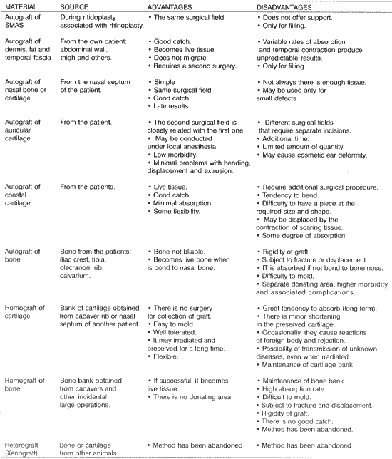
The appropriate material is chosen according to the preference of surgeon based on the characteristics of the graft and the surgical needs (Chart 1). The best results are obtained with autograft of septal cartilage when it is place in the tip, dorsum and/or columella. The ear cartilage is used for ala, dorsum and tip; the cartilage of the 6th rib and calvarial and/or iliac bone are used in the dorsum and/or columella. Up to 2mm increase of dorsum may be obtained with nasal septum autograft. From 2 to 5mn increase requires ear cartilage or calvarial and/or iliac bone. Beyond 5mm, coastal cartilage is necessary. There is no ideal autograft for all nose areas. Obviously, bone graft are more appropriate to the dorsum, whereas cartilaginous are more adjusted for the mobile-cartilaginous portion of the nose.CHART 1 - Characteristics of grafts.
The production of cartilaginous tissue for transplants has been studied and it is soon to be released for the use in facial defects correction24. Chondrocytes are obtained through nasal-septal biopsy, which are cultivated in a means of bioabsorbable polymers of polyglycolic and polylactic acids, forming a tridimensional matrix of the same shape of the defect to be corrected, from where the new cartilage will grow, and after six months, when mature, it will be used to correct the defect in question. This is what is ahead of us.
REFERENCES
1. BASER, B.; SHAHANI, R.; KHANNA, S.; GREWAL, D. S. Calvarial bone grafts for augmentation rhinoplasty. J. Laryngol. Otol., 105(12): 1018-20, 1991.
2. BEEKHUIS, G. J. - Saddle nose deformity: etiology, prevention, and treatment. Augmentation rhinoplasty with polyamide. Laryngoscope, 84: 2-42, 1974.
3. BERTILLON, G. J. - Use of silicone-rubber in nasal reconstructive surgery. Arch. Otolaryngol., 86 88-91, 1967.
4. BUJIA, J.; WILMES, E.; HAMMER, C.; KASTENBAUER, E. - Class II antigenicity of human cartilage: relevance to the use of homologous cartilage graft for reconstructive surgery. Ann. Plast. Surg., 26(6): 541-3, 1991.
5. BYRD, H. S. - Rhinoplasty and rib grafts: evolving a flexible operative technique. (Discussion) Plast. Recontr Surg., 94: 610, 1994.
-7. CONVERSE, J. M. et al. - Deformities of the nose. In:______. Reconstructive Plastic Surgery. 3.ed. Philadelphia: W B. Saunders, 1970. p.743-54.
8. DENECKE, H. J.; MEYER, R. - Plastic surgery of head and neck: corrective ang reconstructive rhinoplasty. N.Y: Sprengerverlag, 1967. vol. I.
9. DYER, W K. 2ND; BEATY, M. M.; PRABTHA, A. Architectural deficiencies of the nose: treatment of the saddle nose and short nose deformities. Otolaryngol. Clin. North Am., 32(1): 89-112, 1999.
10. GIBSON, T; DAVIS, WB. - The distortion of autogeneuos cartilage grafts: its cause and prevention. Brit. J Plast. Surg., 10: 257, 1958.
11. GIBSON, T. - Cartilage implants in rhinoplasty Problems and prospects. Rhinoplasty, 10: 1, 1972.
12. HOMSY, C. A. - Implant stabilization: chemical and biomechanical considerations. Orthopedic Clin. North Am., 4(2): 295-311, 1973.
13. JANEKE, J.B. et al. - Proplast in cavity obliteration and soft tissue augmentation. Arch. Otolaryngol, 100: 24-7, 1974.
14. JOVANOVIC, S.; BERGHAUS, A. - Autogenous auricular concha cartilage transplant in corrective rhinoplasty. Practical hints and critical remarks. Rhinology, 29(4): 273-9, 1991.
15. KENT, J.N. et al. - Pilot studies of a porous implant in dentistry and oral surgery. J Oral Surg., 30(8): 608-15, 1972.
16. KENT, J.N. et al. - Proplast in dental facial reconstruction. J. Oral Surg., 39(3): 347-55, 1975.
17. KOSTECKI, J.L. - Correction of saddle nose deformity with a turn-over hump segment procedure (case report). Plast. Reconstr Surg., 38(4):372-5, 1966.
18. LEAF, N. - SMAS autografts for the nasal dorsum. Plast. Recontr Surg., 97(6): 1249-52, 1996.
19. LUPO, G. - The history of aesthetic rhinoplasty: special emphasis on the saddle nose. Aesthetic Plast. Surg., 21(5): 390-327, 1977.
20. MACKAY, I. S. - The fate of silastic in the management of saddle deformity in the nose. J Laryngol Otol., 97(1): 43-7, 1983.
21. MILWARD, T.M. - The fate of Silastic and Vitrathene nasal implants. Br. J Plast. Surg., 25(3): 276-8, 1972.
22. MUENKER, R. - The bilateral conchal cartilage graft: a new technique in augmentation rhinoplasty. Aesthetic Plast. Surg., 8(1): 37-42, 1984.
23. MURAKAMI, C. S.; COOK, T A.; GUIDA, R. A. - Nasal reconstruction with articulated irradiated rib cartilage. Arch. Otolaryngol. Head Neck Surg., 117(3): 327 -30, 1991.
24. NAUMANN, A.; ROTTER, N. BUJIA, J.; AIGNER, J. Tissue engineering of autologous cartilage transplants for rhinology. Am. J. Rhinology, 12(1): 59-63, 1998.
25. NAUMANN, H.H.; FARRIOR, R.T - Correction of the saddle nose. Am. J Otolaryng., 1(5): 219-25, 1980.
26. NIECHAJEV I. - Porous polyethylene implants for nasal reconstruction: clinical and histologic studies. Aesthetic Plast. Surg., 23(6): 395-402, 1999.
27. OHMORI, S. - Surgical treatment of saddle nose. Plast. Reconstr Surg., 38: 477-8, 1966.
28. OWSLEY, T. G.; TAYLOR, C.O. - The use of Gore Tex for nasal augmentation: a retrospective analysis of 106 patients. Plast. Reconstr Surg., 94: 241-8, 1994.
29. PATROCÍNIO, J.A. - O use do implante de carbono na correção cirúrgica do nariz em sela. São Paulo, 1985. (Tese - Mestrado - Escola Paulista de Medicina).
30. PATROCÍNIO, J.A.; SOUZA, A.D.; ARRAIS, A. - Nariz em sela - Uma nova classificação. Acta AWHO, 5(2): 53-56, 1986.
31. PATROCINIO, J. A. Utilizarão de cartilagem na correção cirúrgica do nariz em sela. Acta AWHO, 8(3): 94-96, 1989.
32. POWELLI, N.B.; RILEY, R.W Cranial bone grafting in facial aesthetic and reconstructive contouring. Arch. Otolaryngol Head Neck Surg., 113: 713-9, 1987.
33. SABATOVICH, O.I.D.M.; RAMALHO, M.C.; CORREA, WE.; PITANGUY, I. - Uso de Gore-Tex SAM como implante facial na cirurgia plástica a reconstrutiva. Revisão da Literatura. Rev. Bras. Cir, 85: 241-4, 1995.
34. SHEEHAN, J. -Plastic surgery of the nose. 2nd ed. New York: Hoeber, 1936.
35. SHEEN, J. H.; SHEEN, A. E -Aesthetic Rhinoplasty. 2 ed. St Louis: Mosby, 1987. v 1.
36. SILVER, WE. - Augmentation of the nasal dorsum. In: KRAUSE, C. J. (ed.). Aesthetic facial surgery. Philadelphia: J. B. Lippincott, 1991. p. 321-327.
37. STUCKER, F.J. JR; HIROKAWA, R.H.; BRYARLY, R.C. JR. - Technical aspects of facial contouring using polyamide mesh Otolarvngol. Clin. North Am.. 15(1): 123-31. 1982.
38. SWENSON, R.W; KOOPMANN, C.F. JR. - Grafts and implants. Otolaryngol. Clin. North Am., 17(2): 413-28, 1984.
39. TUREGUN, M.; SENGEZER, M.; GULER, M. Reconstruction of saddle nose deformities using porous polyethylene implant. Aesthetic Plast. Surg., 22(1): 3841, 1998.
40. WILKINSON, H. A. - Autogeneic skull bone grafts (letter). Neurosurgery, 21: 760, 1987.
* Undergraduate, School of Medicine, Universidade Federal de Uberlândia.
** Faculty Professor and Head of the Service of Otorhinolaryngology, Universidade Federal de Uberlândia.
Affiliation: Serviço de Otorrinolaringologia da Universidade Federal de Uberlândia, Uberlândia /MG.
Address correspondence to: José Antônio Patrocínio - Rua XV de Novembro, 327 - Apt. 1600 - Centro - 38400-214 Uberlândia /MG -Tel/Fax: (55 34) 215-1143.
E-mail: lucaspatrocinio@triang.com.br
Article submitted on November 22, 2000. Article accepted on February 9, 2001.
Print: ![]()