

Year: 2001 Vol. 67 Ed. 2 - (14º)
Artigo de Revisão
Pages: 234 to 241
Hearing and Iatrogenesis.
Author(s):
César L. Terra Brito*,
Walmi B. Braga**,
Darcy R. Lima***.
Keywords: hearing, iatrogenesis, ototoxicity, deafness, aminoglycosides
Abstract:
Introduction: Medicines are therapeutic resources routinely prescribed by doctors. However, among expected actions, there are other events that aren't desired and may present during certain treatment. Everytime we find a non desired effect due to the use of certain drug, we call it iatrogenic effect or iatrogenic disease. Drugs, in general, may produce several undesired effects, among them, auditory and vestibular disturbances; as deafness and vertigo. Then, doctors, specialistic or not, must know about the main ototoxic drugs in order to reduce iatrogenic effects and avoid permanent defficiencies. Aim: This work aims to review the main ototoxic agents, present the clinical manifestations of ototoxicity and some situations that frequently predispose the patient to that.
![]()
INTRODUCTION
Is it common for physicians to prescribe medications they do not know about to treat diseases they barely know in patients they have never met? The wide and increasing number of medication available for clinical use has led to the development of iatrogenic medication diseases. Unfortunately, this negative aspect of clinical practice is sometimes overestimated, especially by lay people, as if iatrogenesis were something common place and highly dangerous. Similarly to electrical power, automobile, aviation, nuclear power and the press, medications are an integral and necessary part of human evolution.
All drugs pose risks, especially if used inadequately and unknowingly.
Although there are accidents with cars and planes, there is not a reductions of its use for the benefit of human beings. People who use modern technological devices are aware of its functioning. Owing to lack of knowledge or imprudence, serious accidents may happen. The same is true for medications: the physician should be updated and informed about new medications and current drugs and know their beneficial and harmful effects for the human body.
Drug-induced reactions are potentially important and common problems in medical practice because of the large number of medications available and the lack of constant update of medical doctors. The main world studies enabled detection and prevalence of problems, as follows10:
inpatients: about 10% to 20% of inpatients have undesirable effects to the use of drugs, and 0.3% to 3% of deaths are caused by iatrogenic problems;
hospital admissions: in general, about 3% to 5% of hospital admissions are caused by undesirable effects of medications.
Classification of undesirable effects of medications
No terminology or classification of undesirable or side effects and iatrogenic diseases is satisfactory. For practical purposes, undesirable factors may be classified in two main types:
A. Dose-dependent or type A
Undesirable dose-dependent effects (undesirable quantitative effect or type A) are normally caused by an abnormal pharmacodynamic or pharmacokinetic effect, excessively producing the known pharmacological effect of the medication. These problems are more common in low-therapeutic index drugs, such as anticoagulants (heparin, warfarin), antiarrhythmic (lidocaine, disopiramide), antihypertensive (beta-blockers, hydralazine), bronchodilators (theophylline, adrenaline) and cardiac glycosides, cases in which a small increase in dosage may result in undesirable effects such as toxicity. Such effects may result from variations of pharmaceutical formula (different doses in different tablets), pharmacokinetic variations (less metabolization in hepatology patients, or less excretion in renal patients), or variations in pharmacodynamics of products (digitalic intoxication in the presence of hypopotassemia).
Variations of pharmaceutical formulation may determine more availability of product, leading to intoxication even with therapeutic doses, which has been observed in a large number of countries with phenytoin and digoxin, in which different formulations determine higher levels of intoxication. In Brazil the same has been observed with therapeutic inefficiency of tetracycline tablets that did not contain the established quantity, because of frauds10. Therefore, whenever the physician tells the patient to change drugs from one manufacturer to another, he or she should monitor the patient during the first days of use to detect changes in bioavailability and undesirable effects as a result of use. The same drug from another manufacturer may provide different bioavailability and, therefore, they are non-bioequivalent; sometimes it is due to differences in manufacturing technique, and one product is more dissolved than another, resulting in higher plasma levels. They are medications with the same components and the same dosage, but not bioequivalent.
Pharmacokinetic alterations may be present in patients who have hepatic or congestive heart failure, in which hepatic perfusion is reduced, as well as metabolization of drugs, a process that may improve as a result of the correction of the hemodynamic disorder. This is the reason why the physician should know which drugs have primary passage effects, as well as those metabolized by the liver such as propanolol, lidocaine and morphine. Nephropathy may also interfere in pharmacokinetics, causing lower clearance and accumulation of drugs and metabolites, requiring calculation of doses based on the degree of renal dysfunction. Thyroid disorders, such as hypo or hyperthyroidism, may also modify the kinetics of drugs, changing the therapeutic response, what is a fact with the use of beta-blockers.
Modifications of pharmacodynamics may also lead to hepatic diseases, when the use of diuretics trigger the portal-systemic encephalopathy, and retention of sodium and water may be aggravated by the use of antacids and penicillin with sodium, in addition to the use of drug that retain water, such carbenicillin, indometacine and corticosteroids. Toxicity of cardiac glycoside is maximized by hypopotassemia and hypercalcemia, whereas efficacy of anti-arrhythmic such as lidocaine, procainamide and disopiramide, is reduced by hypopotassemia. Dehydration maximizes the hypotensive effect of anti-hypertensive medications.
B. Dose-dependent or type B
Undesirable dose-dependent effects (qualitative or type B undesirable effect) are a consequence of immune or pharmacogenetic mechanisms. The main characteristics of this kind of problem are absence of the relation with usual medication effects, existence of a time lapse between use and onset of problem, onset of reaction even with small amounts of drug and disappearance of the problem if medication is discontinued. The most widely known effects of this type are immune reactions such as fever, cutaneous rash, serum disease, asthma, hives, angioedema, and anaphylaxis. Some patients are more prone to develop allergic reactions than others. They are the so called atopic patients, who present disorders caused by type I hypersensitivity reactions, induced by specific antigens that result from release of vasoactive substances by mast cells and basophils previously sensitized by specific antibodies of immunoglobulin class IgE (reagins). Patients usually have history of atopic disease, such as asthma, allergic rhinitis, and previous medication-induced eczema or allergic reaction. In type I reactions (anaphylaxis or immediate hypersensitivity), the drug or the metabolite interacts with antibodies of IgE type fixed in the cells, especially tissue mast cells and blood basophils. It triggers a reaction that releases pharmacological mediators, such as histamine, kinin, serotonin and prostaglandin, which cause allergic response. Clinically, type I reactions are urticaria, asthma, angioneurotic edema, and anaphylaxis. The medications that normally cause these reactions are penicillin, local anesthetic and radiological contrast that contains iodine. In type II reactions (cytotoxic reactions), circulating antibodies type IgG, IgM or IgA interact with hapten - the medication combined with proteins of cell membrane to form antigenantibodies complexes. The complement is activated and there is cell lysis. Most of the reactions are hematological, such as thrombocytopenia associated with quinidine or digoxin; neutropenia due to phenylbutazone, anticonvulsant and metronidazol; or hemolytic anemia caused by the use of penicillin, cephalosporin and quinidine. In type III reactions (reactions due to immune complex), IgG antibodies bind to antigens in the circulation, occurring a deposition of the complex in tissues, with activation of the complement system and endothelial capillar damage. Serum disease is a typical example, characterized by fever, arthritis, lymphnodemegalia, urticaria and macular-papular rash. Medications that normally cause this problem are penicillin, sulfonamide and anti-thyroid medications. In type IV reactions (late or cell-mediated hypersensitivity), T lymphocytes are sensitized by an antigen complex (hapten - medication - bond to a protein). Reactions are caused when previously sensitized lymphocytes make contact with the antigen, through the direct toxic cell action or by release of lymphokine, which affects local activity and other cells. Clinically, these reactions manifest as contact dermatitis after use of local anesthetic, topical creams with antihistaminic, antibiotics and anti-fungal.
Some patients develop complex immune reactions, characterized by a syndrome in which connective tissue is impaired. One example is lupus syndrome mimicking systemic erythematous lupus with joint impairment, which maybe observed with the use of medication such as hydrazine, procainamide or phenytoin; or nodular poliarthritis, which may be observed after use of sulfonamide.
REVIEW OF LITERATURE
Anatomy and physiology of the auditory system
In order to better understand the auditory process and ototoxicity, we will conduct an objective review of anatomy and physiology of the auditory system.
Ears are vestibulocochlear organs due to its importance in the mechanisms of hearing and balance. They are divided into 3 portions: external ear, middle ear or tympanic cavity and inner ear or internal ear and labyrinth.
A. External ear
It consists of a) ear, also called pinna or auricular portion; and b) external acoustic canal. Functionally, the tympanic membrane is part of the middle ear, but it is normally studied as a part of the external ear.
The pinna is a skin cutaneous fold of the lateral face of the head, that recovers the elastic cartilage plaques and it is reinforced by it. It has margins, faces, elevations and depressions. The external acoustic canal is a sinuous structure that extends as far as the concha of the eardrums, and it is approximately 25mm long. The tympanic membrane is about l0mm in diameter and it forms a septum between the external acoustic canal and the tympanic cavity. We may divide the eardrum into four quadrants, which are very important parameters because of their relations with deep middle ear structures.
The external ear leads the sound to the middle and inner ears and protects it from external damage. Despite this function, the pinna barely increases hearing. This fact may be verified in audiometric findings of traumatized patients who had total amputation of ear13.
B. Middle ear
It consists of the tympanic cavity, an air space in the interior of the temporal bone that is lateral-medially crossed by three articulated ossicles - staples, incus and malleus. It communicates with the posterior mastoid air cells, through the aditus and anteriorly with the Eustachian tube, through the tympanic ostium of Eustachian tube. The mucosa lining of the tympanic cavity involves a columnar ciliated epithelium that is transformed into cuboidal in a transitional area localized posterior and superiorly; there is also simple squamous epithelium recovering the antrum and the mastoid air cells.
The tympanic cavity may be compared to a six-sided box that is larger vertically than horizontally and narrow in depth. It is responsible for transforming sound waves in mechanical force through the ossicle chain.
C. Inner ear
Also known as labyrinth, it is located inside the petrous part of the temporal bone. It consists of a series of complex spaces full of fluid - membranous labyrinth, that contains endolymph, and those located inside a similarly shaped cavity (bone labyrinth) that contains perilymph between it and the membranous labyrinth. Structurally, the bone labyrinth has a system of continuous cavities, with three distinguished cavities: one middle zone, the vestibule, a posterior-lateral zone that corresponds to the semicircular canals, and an anterior-medial zone, the cochlea. The bone labyrinth has a maximum length of 20mm. The labyrinth is functionally divided into two elements: a) cochlea, responsible for the auditory function, and b) vestibular system, that is involved in balance, as well as vision and proprioception.
Semicircular ducts are a set of three structures contained inside the semicircular canals, which have a disposition and denomination similar to them, but occupy only one quarter of the diameter of the canals. Each semicircular duct has a dilation called ampulla - and each ampulla has an ampullar crest, formed by neuroepithelial hair cells and supporting cells. Stereocilia of neuroepithelial sensorial cells are immersed into a conic jelly cupula that extends until the opposed wall of the ampulla, obstructing the dilation of the semicircular canals. Such neuroepithelial cells are intimately related to fibers of the vestibular nerve, and once stimulated by the oscillation of the cupula towards the endolymph, they carry vestibular impulses.
The three semicircular ducts are disposed in three spatial plans, and its vestibular activity responds to stimuli of angular acceleration, occasionally by rotation movement of the body.
Utriculus and sacculus are located in the vestibule and present inside them small regions (2-3mm) of thick epithelium differentiated into neuroepithelium, the so called maculae. Maculae share a similar structure with the ampullar crest, with supporting and neuroepithelial cells, whose steriocilia are embedded in a flat jelly layer. Differently from ampullar crests, the macular jelly membrane has crests of calcium carbonate crystals called otolytes, otoconia, or statoconia; in addition, the free margin of the macula does not obstruct the lumen of the utriculus and sacculus cavities.
Utriculus and sacculus maculae share the same structure and are perpendicularly placed. Both structures participate in balance in response to linear acceleration of the body - utriculus at horizontal acceleration and sacculus at vertical acceleration.
The cochlea is a helicoidal tube that has approximately 2.5 turns and about 35mm in length. It is formed by three elements: a) modiolus, nucleus or columella, a small cone of spongy bone tissue that occupies the axis of cochlea and whose base has a double row of orifices, crivus spiroid, through which the filaments of the cochlear nerve pass at the end of the internal ear canal. It is in the modiolus that we find the spiral ganglia; b) spiral cochlear canal, a bony tube that is rolled around the modiolus, describing two turns and a half and forming a spiral; c) bone spiral lamina, starting from the floor of the vestibule, projecting towards the modiolus as the margins of a screw, it is introduced into the cochlear canal and describes the same turns as the structure formerly described. It has posterior and anterior faces, an adherent internal rim and a free external rim.
Membranous cochlea, also known as cochlear duct or scala media, is located in the spiral cochlear canal, as from the free rim of the bony spiral lamina following its shape, starting from the inferior portion of the vestibule, where it connects the sacculus through the reuniens duct, and ending in a blind portion at the apex of the cochlea. The spiral cochlear canal is divided by the spiral lamina and cochlear duct in two portions: scala vestibuli starting from the vestibule and scala tympani, facing the tympanic cavity. Both scalae communicate with the vertex of the cochlea by an orifice named helicotrema.
At transversal sections, the cochlear duct has a triangular shape, both sides supported on the bony spiral lamina and the vertex pointing to the modiolus. Its inferior portion is anatomically located posteriorly and it corresponds to the basilar membrane, which has in its internal region a formation called Corti's organ, responsible for hearing and formed by a complex arrangement of supporting cells and neuroepithelial cells.
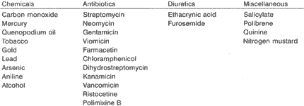
After activation of ossicle chain by sound waves captured by the external ear, the platinum of stapes (the last bone of the chain) promotes a propagation wave through the perilymph, from the oval window, a structure located between the middle and the inner ears. Such propagation reaches a portion of the cochlea - basilar membrane - causing its vibration and posterior activation of external hair neuroepithelial cells, leading to depolarization of internal hair cells. Next, signs are transmitted to the acoustic nerve, at the level of cochlea, reaching the spiral ganglion. From the spiral ganglion, impulses propagate to structures of the brainstem (cochlear nucleus, olivary complex, lateral leminiscus, inferior colliculus) and reach the medial geniculate nucleus, from which they head towards the cortical auditory region at the anterior transversal temporal gyros - and are finally recognized.TABLE 1 - Agents that cause ototoxicity.
Iatrogenesis and hearing
Drugs that affect the ear may produce vertigo or tinnitus and deafness, depending on the damage caused predominantly to the vestibular or hearing portion of the eighth cranial nerve. There are still some other otological problems induced by drugs: otorrhea may be caused by anticoagulant drugs, and external otitis has been produced by Pseudomonas or Proteus after the use of large spectrum antibiotics. In addition to drugs, other chemicals may damage the inner ear.
The following agents have been considered harmful and although this is not a complete list, it illustrates the wide variety of substances included in these cases (Table 1).
A. Pathophysiology of lesions
Among ototoxic drugs, aminoglycoside antibiotics are the most important and undoubtedly the most widely studied. Some authors concluded that these antibiotics bind to receptors of the membranes of hair cells of Corti's organ, sacculus and utriculus maculae and crests of the vestibular system. These receptors are the polyphosphoinositide, lypid components of cell membrane, which have an important role in bioelectrical events and in permeability of membrane by interaction with calcium ion. The formation of complexes among aminoglycoside complexes and polyphosphoinositide produces modifications of the physiology of the membrane and its permeability, and it affects first the structure and function of cilia and finally the membrane itself causing destruction of receptor cells12.
Segal et al. noted that aminoglycosides also have a neurotoxic action related to NMDA receptors, but it is probably not very relevant, because they are not capable of overcoming the hematoencephalic barrier (HEB) under normal conditions. However, this fact becomes important in those who have impairment of HEB14.
B. Ototoxicity and clinical manifestations
Vestibular damage
Streptomycin first affects the eighth cranial nerve by causing vestibular damage. The patients manifest dizziness and sudden movement vertigo. The potential of nerve damage is caused by high doses of the drug, deficient renal function and aging'. Deafness is rarely caused by streptomycin, except in cases of tuberculous meningitis, in which concentrations of streptomycin in the CSF will increases. It is important to monitor patients treated with streptomycin to cure pulmonary diseases, for example, to avoid toxicity.
Hearing damage
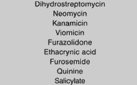
Tinnitus and deafness. Aminoglycoside antibiotics, except for streptomycin and gentamicin, cause primary tinnitus and deafness (Table 2). However, streptomycin may cross placenta barrier and lead to deafness without affecting the vestibular system of the fetus. Tinnitus is normally associated with deafness, and it is almost always presented before it. There are few medications, among which mandelarnine, that produce isolated tinnitus. These ototoxic medications should be used very carefully in patients with renal affections. Dihydrostreptomycin affects first the acoustic labyrinth, and later the vestibular system. It was withdrawn from use because it led to high incidence of deafness, even after discontinuation of treatment2.
Neomycin, kanamicin and viomicin may lead to deafness. Neomycin is not used for parenteral administration because of its renal and ototoxic effects. Insignificant amounts are absorbed by the intestine when the drug is administered as an intestinal disinfectant6.TABLE 2 - Drugs acknowledged as cause of deafness.
Ulcerative intestinal lesions, however, may result in higher absorption of the drug and in the presence of reduced renal function, it may produce high blood levels of the drug and result in deafness. The use of neomycin combined with polimixine B via plastic catheter for irrigation of osteomyelitic lesion has also resulted in hearing loss. Deafness caused by kanamicin is more probably found in high dose or prolonged treatment courses or in the presence of reduced renal function. Deafness may result or progress, but it is maintained after interruption of drug use3.
Some non-aminoglycoside antibiotics have been reported in the literature as ototoxic. They are: erythromycin, with reversible effects; chloramphenicol, especially with topic action; ampicillin and minocillin, derived from tetracycline, have vestibular toxic action; cephalosporin, counting on few citations; viomicin and capreomicin, have more vestibular toxicity; polimixine B and E; and cholistine. Viomicin, an anti-tuberculosis drug, produces mainly deafness, but it also affects the vestibular function. Furazolidone causes tinnitus and deafness considerably less frequently12. Ethacrynic acid, normally administered via endovenous route, but also prescribed as an oral therapy for uremic patients, has caused transient vertigo, ear fullness, tinnitus and deafness. Permanent deafness may follow both oral and intravenous treatment. Permanent moderate to severe hearing losses have been detected in uremic patients treated with ethacrynic acid and small doses of streptomycin. Synergic effect of these two drugs may be the cause of permanent hearing loss. Transient vertigo, tinnitus and hearing loss have also been noticed as a result of high doses of intravenous furosemide in patients with impaired renal function6.
Blood levels of quinine and salicylate may lead to cinchonism, characterized by headache, vertigo, tinnitus, hearing loss, blurred vision, nausea and vomiting. These symptoms disappear after discontinuation of the drugs'. Some degree of tinnitus and hearing loss are frequently present after use of high doses of salicylate acid to treat rheumatoid arthritis or acute rheumatic fever. Tinnitus normally precedes deafness and it is temporary, resulting in a hearing loss of 30 to 40 dB, especially in high frequencies2. When salicylate acid is used for the treatment of psoriasis, there may be absorption of small quantities of salicylate by the skin, enough to cause a transient hearing loss. Absorption of salicylate depends on the pathological status of the skin and the type of base in which the salicylic acid is dissolved. Small doses of quinine may cause tinnitus in sensitive subjects. Anti-neoplastic drugs may be ototoxic, such as cisplatin, nitrogen mustard and vincristine. The cochlear toxicity may be reversible or irreversible. In some cases, oral contraceptive therapy may cause unilateral or bilateral progressive or irreversible hearing loss3,12.
Local Otoxicity
A number of amynoglycoside antibiotics and other drugs are used to treat chronic otitis media and to manage otorrhea before the performance of myringotomy or tympanoplasty, if necessary. The toxic drug applied topically affects the middle ear through the perforation of the tympanic membrane, reaching the niche of the round window, on the medial wall of the middle ear, and it may contact the permeable wall of this window, the secondary tympanic membrane. Structural studies have shown that the membrane of the round window has three planes12,8,11:
a) the external epithelium, formed by a single layer of cuboidal cells with interdigitations on the lateral walls of cells;
b) intermediate connective tissue, with fibroblasts, collagen, elastic fibers, blood and lymphatic vessels, fibrocytes, myelinized and non-myelinized nervous fibers.
c) the internal epithelium, squamous cells with large lateral extensions, continuous basal membrane, cytoplasm with pinocytotic vesicles, large cell space and space among epithelial cells.
The membrane of the round window has a number of functions. It may play the role of release valve of energy that comes from cochlear scala tympani, through the perilymph and it is an alternative route of sound energy from the cochlea, from the middle ear. In addition, it may promote absorption of substances (endocytosis), secretion of substances (exocytosis) and cleaning of perilymph and defense (immunity). The membrane of round window is permeable, enabling transport by diffusion and through pinocytotic vesicles. Permeability has been shown in trials. A number of substances cross the membrane and reach scala tympani and perilymph, such as sodium, cationic ferritin, albumin, solvents, HRP (horseradish peroxidase), toxins (Staphylococcus), antibodies, products derived from hyaluronic acid, anti-mycotic, antiseptic, such as procaine, pantocaine, tetracaine, lipocaine. The anesthetic drugs applied in the ear with tympanic membrane perforation may cause severe vertigo, nausea and vomiting, because of the anesthesia of vestibular receptors on the side of the application12.
Some antibiotics, such as chloramphenicol, streptomycin, farmicetin, tetracycline, penicillin and erythromycin, have been proved to cross the round window. Among ototoxic antibiotics, the following have been proved to cross the round window and enter the cochlea: gentamicin, polimixine and neomycin. All these drugs go from the middle ear to inner ear via lymphatic route, through the membrane of round window or blood circulation. Since permeability of the membrane of the round window has been demonstrated by a number of studies, it is expected that the ear is susceptible to substances such as bacterial toxins or ototoxic drops. Therefore, these drugs would cause sensorineural hearing loss or internal ear alterations. In animal models, these cell alterations in Corti's organ have been demonstrated when the drugs listed above were used12.
Despite all animal studies leading to cochlear modifications, it has been clinically observed that in humans there is nearly no local toxicity. Few studies reported ototoxicity. How could we explain such controversy? In animal models, specially rodents, inner ear alterations caused by drugs are explained by the higher susceptibility these animals have because their round window membrane is delicate and directly exposed to the middle ear, without a niche. In humans, drugs as used in topic drops if there is chronic otitis media. This pathology causes modifications of the connective and epithelial external layers of the membrane, hyperplasia and metaplasia of external epithelium, and as far as connective tissue goes, the membrane is thicker and there is secretion and granulation tissue in the niche of the window. These and other factors could hinder the passage of ototoxic drugs to the human inner ear. The presence of a thicker membrane, with secretion and granulation tissue may transform it into an impermeable membrane, accounting for few cases of local ototoxicity in humans12.
Despite these clinical observations, we should be careful in order to prevent local ototoxicity: apply a- safe dose for a short period of time; discontinue the medication when the otitis is better and the membrane has restored its normal permeability; avoid application of ototoxic drops in cases of secretory otitis media with ventilation shunts; avoid the use of ototoxic drops in traumatic perforations. If the patient has risk factors, we should conduct careful monitoring with pure tone bone audiometry. If all these suggestions are followed, it is possible to apply these drugs locally with a good safety margin.
DISCUSSION
In face of the above description, we observed that iatrogenic auditory and vestibular diseases are clinical entities that should be known and remembered by the physician, regardless of his or her specialty. Aminoglycoside antibiotics should be prescribed with criteria, and its administration should be carefully performed, since they are daily use ototoxic drugs, especially in hospital settings.
It is important to identify drugs that have ototoxic action in order to follow up the patient. If possible, we should also monitor and prevent exposure that may result in otological damage, as well as understand the clinical manifestations of cochlear and/or vestibular toxicity that may surge during the course of treatment.
FINAL COMMENTS
Iatrogenesis plays an important role in current medial practice. Physicians should practice accordingly in order to reduce the side effects shown here. Therefore, we will provide better quality of life to patients. All in all, we should not only treat the manifested disorders but also prevent relief or solution interventions of the former from leading to the development of other complications.
REFERENCES
1. ABBRUZZESE, A. and SWANSON, J. - Jaundice after therapy with chlordiazepoxide hydrochloride. New Eng. J Med., 273:321, 1965.
2. ADAMS, G. L. et al. - Doenças do ouvido interno in: Adams, G. L.; Boies, L. R.; Paparella, M. M. Otorrinolaringologia. 5a ed. Rio de Janeiro: Interamericana, 1979. 142.
3. ADAMS, E W; WYNN, V; ROSE, D. E; et al. - Effect of pyridoxine hydrochloride (vitamin B6 upon depression associated with oral contraceptives. Lancet, 1: 897, 1973.
4. AGGELER, E M.; O'REILLY, R. A.; LEONG, L. AND KOWITZ, E E. - Potentiation of anticoagulant effect of warfarin by phenylbutazone. New Eng. J Med., 276. 496, 1967.
5. ALARCON-SEGOVIA, D. - Drug-induced lupus syndromes. Mayo Clin. Proc., 44: 664, 1969.
6. ALARCON-SEGOVIA, D.; WORTHINGTON, J. W; WARD, L. E. and WAKIM, K. G. - Lupus diathesis and the hydralazine syndrome. New Eng. J. Med., 272: 462, 1965.
7. DANGELO, J. G. & FATTINI, C. A. - Sistema sensorial In: Dangelo, J. G. & Fattini, C. A. Anatomia humana sistêmica e segmentar - para o estudante de medicina - 3a reimpressão da 2a edição - São Paulo, Rio de Janeiro, Belo Horizonte: Atheneu, 1998. 167-170.
8. GARDNER, E. - A orelha In: Gardner, E. et al. Anatomia Estudo regional do corpo humano - 4a ed. - Rio de Janeiro: Guanabara Koogan, 1988. 605-617.
9. HUNGRIA, H. - Anatomia In: Otorrinolaringologia/ Helio Hungria - 8ª ed. - Rio de Janeiro: Guanabara Koogan, 2000. 299-310.
10. LIMA, D. R. - Manual de farmacologia clinica, terapeutica e toxicologia. Rio de Janeiro: Guanabara Koogan, 1995.
11. MCLAY, K. - Anatomy and physiology In: Turner, A. L. Logan Turner's diseases of the nose throat and ear. - 9th ed. - Littleton: Wright PSG Inc, 1982. 286-304.
12. OLIVEIRA, J. A. - Ototoxicidade In: Costa, S. S. et al. Otorrinolaringologia: princípios e prática. Porto Alegre: Artes Médicas, 1994. 215-221.
13. SEBASTIAN, G. et al. - Fisiologia In: Sebastian, G. et al. - Audiologia prática - 3a ed. - Rio de Janeiro: Enelivros, 1986. 41-56.
14. SEGAL, J. A.; HARRIS, B. D.; KUSTOVA, Y; BASILE, A.; SKOLNICK, E - Aminoglycoside neurotoxicity involves NMDA receptor activation. Brain Research, 815 (1999) 270-277.
* Undergraduate, Medical School, Faculdade de Medicina de Petrópolis/Fundação Octacílio Gualberto (FMP/FOG).
** Master Degree in Otorhinolaryngology at Universidade Federal do Rio de Janeiro (UFRJ). Asosistant Professor of Otorhinolaryrgology at Universidade do Rio de Janeiro (UNI-RIO).
*** Ph.D. in Medicine at University of London, England. Professor of Clinical Pharmacology at INCD, Universidade Federal do Rio de Janeiro (UFRJ). Associate Director for Medicinal Plants Studies, Vanderbilt University Medical Center, Nashville, TN, USA.
Address for correspondence: César Leandro Terra Brito - Estrada dos Três Rios, 1416 - BL 5 - Apto. 401 - Freguesia - Jacarepaguá - Rio de Janeiro/RJ.
Tel: (55 21) 436-1458 - E-mail: terrabrito@usa.net
Article submitted on June 29. 2000. Article accepted on November 23. 2000.
Print: ![]()