

Year: 2001 Vol. 67 Ed. 1 - (13º)
Artigos Originais
Pages: 84 to 88
Paracoccidioidomycosis of the Larynx: A 21 Year Review.
Author(s):
Adriana G. D. Bastos*,
Andréa G. Martins**,
Fernando C. Cunha***,
Marise P. C. Marques****,
Paulo P. Melo*,
Shiro Tomita*****,
Valéria M. O. Alonso******.
Keywords: paracoccidioidomycosis, larynx, mucosae lesions
Abstract:
Introduction: A retrospective study of 17 patients with laryngeal paracoccidioidomycosis (PCM) was performed, from June 1978 to May 1999. Method: Aspects related to epidemiology, clinical, endoscopic findings, diagnosis and treatment were analyzed. Results: There were more cases in men. The medium age was 54,7 years. The vocal folds were injured in 64,7% of the cases and the epiglottis in 47%.These lesions had a vegetation appearance in 58,8% of the cases. All the patients had diagnosis by biopsy of lesions of the larynx, being in 16 patients (94,1%) histologically. Sixteen patients were submitted to treatment, with an index of cure about 23,5%. Conclusion: We concluded that in cases of PCM an otorhinolaryngologic evaluation is fundamental. Suspect lesions of the larynx need biopsy, with accomplishment of direct mycological and histopatological examination and, when available, sorology.
![]()
INTRODUCTION
Paracoccidioidomycosis (PCM) is the most frequent systemic mycosis of Latin America caused by a dimorphic fungus - Paracoccidioides brasilienses. It is normally geographically limited to the Americas, from Mexico to Argentina. As to occupational distribution, it prefers to affect rural workers 4,6.
The disseminated chronic form is the most common one, amounting to 60 to 70% of the cases in adults, attacking especially adult males over 30 years. Although the lungs are most frequently affected, in the chronic form the extrapulmonary lesions with affection of mucosa, skin and cervical ganglions is the most frequently seen by the physicans 1,4,6.
The most common lesions of PCM are located in the lungs and oral mucosa. Mucosal lesions are more frequent in the lips, gums, tongue, jugal mucosa, palate, uvula, tonsil pillars, floor of the mouth, nose and larynx. If there is laryngeal affection, it affects mainly the vocal folds and dysphonia is the main complaint³.
The purpose of the present study was to conduct a retrospective study of the cases of laryngeal PCM diagnosed at SME/ENT of HUCFF/UFRJ.
MATERIAL AND METHOD
We reviewed the medical files of patients with confirmed diagnosis of laryngeal PCM in the period between May 1978 and May 1999. We analyzed epidemiological history and clinical picture, endoscopic findings, as well as the method used to define the diagnosis and the treatment approach.
Out of a total number of 27 treated patients at SME/ ENT with diagnosis of laryngeal PCM, 10 were excluded because there was no medical file at HUCFF. Out of the 17 patients, 10 (58.85°/a) had been referred with complaints of dysphonia and the seven remaining cases (41.2%) had suspicion and confirmation of PCM in another organ. About 50% of the patients had mucosal lesions also on the palate, nose, oral cavity, lips and/or pharynx.
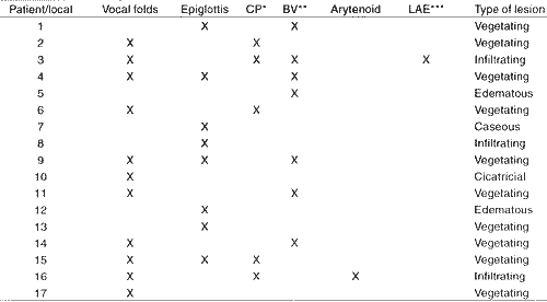
Laryngeal lesions were analyzed as to nature and location, according to the description provided by the direct and/or indirect laryngoscopic, exam. Lesions were classified as vegetating, infiltrating, edematous, caseous or cicatricial. As to location, we considered epiglottis, vocals folds, ventricular folds, arytenoid cartilage, aryepiglottic ligament and posterior commissure.
We designed a protocol to collect the data, which was later analyzed.
RESULTS
Out of 17 patients, 16 were male (94.1 %) and one was female (5.9%), at a ratio of 16:1. The mean age of patients was 54.7 years, ranging from 37 to 72 years. A positive epidemiological history confirming the rural origin of patients was found in 13 patients (76.5%).
The most common presentation was the chronic form (82.4%, 14 patients), and the remaining cases were ignored. In addition to laryngeal affection, 14 patients (82.4%) also had respiratory manifestations. Seven patients (41.2%) had mucosal lesions on the palate, oral cavity, nose, pharynx and/or lips. One patient also had a nodular lesion in the central nervous system, and 9 (52.9°/0), had cervical and/or submandibular adenomegalia in the physical exam.
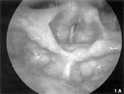
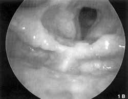
In our study, vocal folds were affected in 11 cases (64.7%) - two isolated cases and nine associated with other laryngeal segments (Chart 1). In eight cases (47%), lesions were described as vegetating (Figures 1a and lb), in two as infiltrating, and in one as cicatricial. Epiglottis was the second most affected site, with eight cases (47%), four of them with isolated lesions. Five lesions were vegetating, one was infiltrating, one was edematous and one was caseous. Ventricular folds were affected in seven cases (41.2%), with only one isolated lesion - five vegetating lesions, one infiltrating and one edematous. Posterior commissure was affected in 5 cases (29.4%) - three were vegetating and two were infiltrating. Arytenoids and aryepiglottic ligaments were affected in one situation, both with infiltrating lesions.
All patients had the diagnosis confirmed by biopsy of laryngeal region, and in 16 patients (94.1%) the diagnosis was confirmed by histopathological exam and in one patient by direct mycological exam. Three patients (17.6% of the cases) also had the diagnosis confirmed by material collected from the airways, two from sputum exam with direct mycological exam and positive culture and one from pulmonary aspirated material, whose cytopathological exam was positive.
Chemotherapy treatment was conducted in 16 patients (94.1%) and 11 (64.7%) used only sulfonamides, five (29.4%) used sulfonamides and amphotericin B, and in one case treatment was unknown. Seven patients (41.1%) started treatment with sulphadiazin, one gram PO QID for two weeks. If the patient did not manifest intolerance or hypersensitivity to the medication, the antimicrobial agent was replaced by sulphadoxin at one gram PO every five days. Four patients started treatment with the association sulphamethoxazole/ trimethoprim (SMX/TMP) at the attack dose of PO 10mg/kg/ day BID. If the patient responded well to the drug within the period of 6 weeks, the dose was cut in half and maintained for 24 months. In five patients (29.4%) who had severe forms of PCM, treatment was started with endovenous amphotericin B in the hospital, and the advocated initial dose was 0.25mg/Kg and 0.5mg/Kg, in alternated days, until it induced the tolerance to a maintenance dose of l mg/Kg/day up to a maximum of 50mg per application. The total dose varied according to the case, but it was in average 1.5 to 3.0 grams. After hospital discharge, patients were maintained with sulfonamides for 24 months, as recommended. Only 7 patients (41.1%) concluded the 2 expected years of treatment, with a cure rate of 23.5% (four patients). Seven patients (41.1%) abandoned treatment and four (23.5%) lost communication with the hospital. One patient is still under treatment. We detected one case of recurrence after two years of treatment.Chart 1 - Distribution of patients according to location and type of laryngeal lesion..
Key: * CP = posterior commissure; ** BV = ventricular bands; *** LAE = aryepiglottic ligament
DISCUSSION
According to the literature, the general epidemiological data we collected fell within the expected criteria, that is, prevalence in males, old minimum and medium ages, positive epidemiological history and chronic diseases 3,4.
The lungs were the primary most affected organ. In 82.4% of the patients with laryngeal PCM we detected lung affection. Dysphonia was the symptom most frequently related to laryngeal compromise, present in 10 cases (58.8%). Dysphonia was related to the affection of fungi on the vocal folds. Do Valle et al., in 1995, demonstrated the presence of permanent dysphonia in 50% of the cases of 30 studied patients with laryngeal PCM. Machado Filho and Miranda, in 1961, in a study of 313 cases of PCM, demonstrated the presence of dysphonia in 84.6% of the 26 cases of primary laryngeal PCM. Santanna et al., in 1999, demonstrated in a study of 7 patients that dysphonia was the most frequent symptom, present in 86% of the cases.
Vocal folds and epiglottis were the most frequently affected laryngeal sites, and vegetating lesions were the most frequent manifestation. Machado Filho et al, in 1960, in a study of 104 patients without previous treatment for PCM (35 with laryngeal lesion), demonstrated that the most affected laryngeal anatomical elements were the vocal folds (68.5%), and epiglottis (62.8%). In both cases, ulceration was the predominant type of lesion (74.3%), and the other types also described were hyperemia, edema, granulation and destruction. Santanna et al., in 1999, demonstrated that in seven studied cases most of the lesions affected multiple laryngeal segments, and four of them were vegetating and three were ulcerative.
Figures l A and 1B. Lesions of laryngeal PCM with vegetating aspect on the right ventricular fold, laryngeal face of epiglottis. Destruction of epiglottis. 1A in adduction and 1B in abduction.
Definite diagnosis of the pathology may be confirmed by direct mycological exam, culture, histopathological exam or serology. Direct exam is definite and simple. The gold standard is to identify the fungus in the lesion, although this method has only been used for diagnosis of 3 cases (one of larynx, one of sputum and another of oral cavity), because of few specialized staff. Culture usually takes long and it is less sensitive than direct exam. The former was used in only one case, in which sputum was the collected material. Histopathological exam is a diagnostic option, especially when the material is obtained by biopsy - the presence of yeast with thick walls and multiple gemmation identifies Paracoccidioides brasilienses. In the biopsy laryngeal material of all patients, histopathological exam was positive in 16 cases (94.1%), showing that it is a good method to diagnose laryngeal mycosis. Fixation serologic reactions of complement and double agar gel immunodiffusion were not used in any situation, because of poor availability of the exam. Serologic reactions are useful both as a diagnostic element and as a parameter for the assessment of the activity of the disease 4,6.
Drugs currently available for treatment are sulfonamides, amphotericin B and derivatives of imidazol (cetoconazol and itraconazol), and when appropriately employed they all have similar efficacy. Sulfa is the first choice because in general patients come from less privileged social strata. Amphotericin B is very toxic and requires hospitalization, and it is indicated in very severe cases, when the patient develops resistance or intolerance to sulfa. Imidazol derivatives, when compared to sulfa, lead to faster resolution of lesions, but do not reduce the rates of recurrence, and they are indicated in the case of chronic affections in adults. Treatment lasts 2 years in average and it should only be suspended when the disease is inactive. In our study, we observed that the advocated treatment was in accordance with the literature; 94.1% of the patients were treated with sulfonamides, and five patients (29.4%) needed hospitalization for the treatment of severe PCM with amphotericin B6.
Rates of interruption of treatment were higher than the acceptable level (41.1%), and many cases remained with an unknown evolution (23.5%). These data showed the lack of infrastructure of the health care system in Brazil, taking into consideration that the long duration of treatment is another key element. Do Valle et al., in 1995, demonstrated that the main factors for irregular treatments were distance from home to medical care setting, duration of treatment and low social-economic background¹.
CONCLUSIONS
All patients with suspicion or diagnosis of PCM should be submitted to an otorhinolaryngological evaluation and indirect laryngoscopy.
Dysphonia is the most frequent symptom of patients with laryngeal PCM. Suspected laryngeal lesions should undergo biopsy and the collected material should be analyzed by direct mycological and histopathological exam, in addition to serology, if possible.
Advocated treatment was employed in most of the cases but reached low cure rates, because of the high rates of interruption of treatment.
The high rates of treatment interruption showed the lack of infrastructure of the public health care system in Brazil, associated with factors such as prolonged duration of treatment, low social-economic background and distance from home to medical care setting.
REFERENCES
1. DO VALLE, A. C.; APRIGLIANO FILHO, F.; MOREIRA, J S.; WANKE, B. - Clinical and endoscopic finding in the mucosae of the upper respiratory and digestive tracts in post treatment follow-up of Paracoccidioidomycosis patients. Rev Inst. Med. Trop. São Paulo, 37: (05). N° 05, 407-13, 1995.
2. MACHADO FILHO, J. & MIRANDA, J. L. - Considerações relativas à Blastomicose Sul-Americana. Evolução, resultados terapêuticos, moléstias associadas em 394 casos consecutivos. Hospital (RJ), 60, 375-412, 1961.
3. MACHADO FILHO, J.; REGO, A. P.; CHAVES, A. L. F.; MIRANDA, J. L.; SILVA, C. C. - Considerações relativas à Blastomicose Sul-Americana. Da participação laríngea e brônquica em 104 resultados endoscópicos. Hospital (RJ), 58, 19-34, 1960.
4. RESTREPO, A. M. -Paracoccidioides brasilienses. In: Mandell GL : Principles and practice of infectious diseases. 4th ed., New York, Churchill Lingstone,1995, v-2, pg. 2386-9.
5. SANTANNA, G. D.; MAURI, M.; ARRARTE, J. L. & CAMARGO, H. JR.- Laryngeal manifestations of paracoccidioidomycosis (South American blastomycosis). Arch. Otolaryngol .Head Neck Surg., 125: 12, 137-8,1999.
6. WANKE, B. - Micoses Profundas. In : Schester M & Marangoni DV, Doengas infecciosas conduta diagnóstica e terapêutica, 2ª ed., Rio de Janeiro, Guanabara Koogan, 1998, pg. 318-23.
* Resident Physician of the Service of Otorhinolaryngology at Hospital Clementino Fraga Filho (HUCFF), Universidade Federal do Rio de Janeiro (UFRJ).
** Master degree in Otorhinolaryngology under study at the Service of Otorhinolaryngology, HUCFF/UFRJ.,
*** Faculty Professor of the Service of Otorhinolaryngology at HUCFF/UFRJ.
**** Physician and Preceptor of Residents of the Service of Otorhinolaryngology at HUCFF/UFRJ.
***** Assistant Professor and Head of the Service of Otorhinolaryngology at HUCFF/UFRJ
****** Assistant Professor of the Service of Otorhinolaryngology at HUCFF/UFRJ.
Study developed at the Service of Special Methods - Serviço de Metódos Especiais (SME) of the Service of Otorhinolaryngology at HUCFF/UFRJ.
Study presented at I Congresso Triológico de Otorrinolaringologia, held on November 13 - 18, 1999, in São Paulo /SP.
Address for correspondence: Adriana Geórgia Davim Bastos - Rua Alberto Maranhao, 387 - Bloco 2 - Apt° 303 - Jardim Guanabara - Ilha do Governador
21940-490 Rio de Janeiro /RJ - Tel: (55 21) 466-2211 - E-mail: sbtadr@domain.com.br
Article submitted on April 25. 2000. Article accepted on November 1, 2000.
Print: ![]()