

Year: 2001 Vol. 67 Ed. 1 - (11º)
Artigos Originais
Pages: 73 to 76
Is Tongue Base Hypertrophy Related to Gastroesophageal Reflux?
Author(s):
Rui C. M. Mamede*,
Gustavo C. D. Elchenberger**,
Daniel Garcia Jr.***,
Roberto O. Dantas****,
Gabriel J. Leite*****.
Keywords: limphatic ring, gastroesophageal reflux, postreflux pharingitis
Abstract:
Introduction: Patients have hypertrophied lymphoid follicles without apparent cause. Aim: The objective of the present study was to determine the possible association between gastroesophageal reflux (GER) and the presence of hypertrophied lymphoid follicles at the base of the tongue (HBT). Material and method: Prospective study was realized at University Hospital. Twenty eight patients submitted to 24 hour pH measurement were examined by videolaparoscopy in order to identify lymphoid follicles at the base of the tongue, which were classified according to size. HBT were considered to be present when the follicles prevented a view of the valecula (++) and they were considered to be severe when they prevented a view of the epiglottis. The patients were divided into groups according to the presence of signs of GER detected by pH measurement, and an attempt was made to relate these signs to the prece of HBT. Results: One case of severe HBT (+++) was identified among the patients with GER, corresponding to a little more than 3% (1/28) of the total sample studied. HBT was detected in 66,7% (10/15) of the patients with signs of GER and in 38,5% (5/15) of those with no signs of GER. The result was not statistically significant, perhaps due to the reduced sample size, but showed a higher incidence of HBT among patients with positive pH monitoring. Conclusion: We believe that HBT represents additional evidence of reflux in the pharynx and further studies are necessary to relate otolaryngologic signs and symptoms to GER.
![]()
INTRODUCTION
The function of lymphoid follicles located in the pharynx is still a mystery; however, it is known that during childhood immune reactions triggered by them are very meaningful. It is also known that in free-of-inflammation adult and elderly subjects there is reduction of the production of immunoglobulin³. In these situations, lymphoid follicles practically disappear from rhinopharynx and oropharynx. Therefore, Waldeyer's ring, consisting of lymphoid tissue in the rhinopharynx and oropharynx, is exuberant at birth and tends to disappear in adults, and palatine tonsil is the only portion left. However, there are situations, without an apparent cause, in which lymphoid follicles of the base of the tongue are enlarged, promoting bulging of oropharynx and unpleasant symptoms, posing the patient's life at risk.
Exuberant lymphoid tissue, when present in the base of tongue, has a smooth pink-whitish surface, with poorly defined margins that tend to prevent the view of vallecula and sometimes even of the epiglottis. At palpation, we noticed bulging of the base of the tongue, more consistent but without hardened local or general portions. The presence of hypertrophied lymphoid tissue on the base of the tongue may cause cough, nasal voice and the sensation of oropharyngeal globus, as if there were something stuck there. This feeling is increased during swallowing of liquids and it is sometimes gone for some minutes during swallowing of solids. In addition to these complications, the presence of hypertrophied tissue on the base of the tongue may result in significant consequences to patients, especially to those that have to undergo general anesthesia. It may be difficult to localize the larynx during intubation, because of the large bowing of the base of the tongue. Exuberant lymphoid tissue of the base of tongue should be differentiated from ectopic thyroid, carcinoma, sarcoma and infectious pharyngitis.
In recent years, many problems of airways without apparent cause have been attributed to esophagopharyngeal reflux (EPR). The ones related with the upper airways are otitis media, sinusitis, laryngitis, laryngospasm, stridor, recurrent cough. Among those related with the lower airways are chronic bronchitis, asthma, recurrent pneumonia and broncho-pulmonary dysplasia. Burton¹ pointed out that in the presence of atypical symptoms of airways or abnormal therapeutic responses, we should associate the possibility of having the gastroesophageal reflux as the main factor responsible for these atypical behaviors or anomalies. Globus histericus, a symptom normally associated with psychiatric disorders, was suggested by Malcomsen9 in 1968 to be related with GER. Other pharyngolaryngeal symptoms associated with GER are hoarseness, vocal fatigue, chronic throat clearing, chronic cough, dysphagia and presence of mucus in the throat¹².
It is also interesting to notice that sometimes pharyngolaryngeal complaints do not present the typical GER symptoms, such as regurgitation, emesis, dysphagia, choking or heartburn. Kennedy7, in 1962, called these cases "silent reflux" cases; but Koufman8, in 1991, referred to them as hidden reflux cases.
Since there seems to be a relation between GER and airway affections, we tried to identify in the present study the association between hypertrophy of lymphoid tissue on the base of the tongue and gastroesophageal reflux in adults, by assessing the presence of hypertrophy in patients with positive and negative pH monitoring.
MATERIAL AND METHOD
We invited all patients from the Gastroenterology clinic treated in the previous two years 1997-1999 who had symptomatic diagnosis of GER to undergo videolaryngoscopy under local anesthesia in order to try to identify the presence of lymphoid follicles on the base of tongue. Twenty-eight patients came and signed the informed consent that included the purpose of the research. In addition to videolaryngoscopy, they were also submitted to 24-hour esophageal pH monitoring (Bernstein's test), which consisted of the continuous monitoring of esophageal pH for 24 hours. The result of pH monitoring was not known when the videolaryngoscopy was conducted.
Analyzing videotapes and photos from the laryngoscopy, lymphoid follicles were classified according to the criteria described by Mamede14, as follows:
+ presence of follicles on the base of tongue; ++ lymphoid follicles preventing the visualization of vallecula; +++ lymphoid follicles preventing the visualization of epiglottis or spread on the pharynx and larynx.
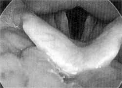
We defined that patients who had lymphoid follicles classified as ++ or +++ (Figure 1) would be considered as having hypertrophy of the base of the tongue. We conducted a statistical analysis of the data collected at the level of 5%, applying the exact Fisher's test if the groups had samples smaller than 10. The project was approved by the committee of ethics of the institution, complying with the Internal Act 196-96 by the National Council on Health.
RESULTS
The 28 patients who were included in the sample had a mean age of 34 years, ranging from 25 to 52 years, divided as 16 female and 12 male subjects. Out of the total, 15 had hypertrophy of the base of tongue (HBT) and in 13 cases there were lymphoid follicles but they were very small (+). Among those classified as HBT, 14 lymphoid follicles prevented visualization of vallecula (++) and only one patient had lymphoid follicles that prevented visualization of epiglottis (+++):
Figure 1. Hypertrophy of the base of tongue obstructing the view of epiglottis (HBT +++).
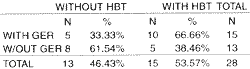
In the sample of 15 patients with GER confirmed by 24-hour esophageal pH monitoring, we observed 10 cases of HBT, 9 ++ and 1 +++. In the sample with 13 patients whose 24-hour esophageal pH monitoring was negative we observed 5 cases of HBT, all of them ++ (Table 1). In the control group formed by patients with negative 24-hour esophageal pH monitoring, results did not show statistically significant difference.
DISCUSSION
It is widely known that at birth lymphoid tissues are abundant and that they reduce with age. Zharikova, in 1984 14, and Kamata, in 1992 6, noticed that lingual tonsil starts to act later if compared with other lymphoid follicles, that is, in the first or second decades of life, reaching its peak of immunological activity during the forth and fifth decades of life. Age was irrelevant in our study, because the groups consisted of adults who were all very close in age.
Compensatory hyperplasia of lymphoid follicles as a response to adenotonsillectomy 4,13, 5, allergic processes, chronic infections and alcohol abuse and smoking5, have been suggested as etiological factors of HBT. However, there are no scientific studies with samples large enough to confirm the hypothesis, leading us to believe that a number of factors contribute to the surge of HBT. However, some of our patients had been submitted to adenotonsillectomy, both in the group with reflux (4 cases), and in the group that had no reflux detected by the 24-hour esophageal pH-monitoring (2 cases); moreover, none of our patients had signs of inflammation of lymphoid tissue (edema or hyperemia).TABLE 1 - Presence of hypertrophy of the base of tongue and gastroesophageal reflux.
Fisher's exact test: not significant at 5%.
In previous studied, results have shown increased incidence of GER in patients with HBT, confirmed by biopsy of esophagus, compared to patients submitted to videolaryngoscopy in the service of Head and Neck Surgery at Faculdade de Medicina de Ribeirão Preto, who had no complaints of GER and were the control group ¹¹ ¹°. In the present study, in which we used 24-hour esophageal pH monitoring as a parameter to define GER, there were no statistically significant differences, probably because of the limited size of the sample. We all know that 24-hour esophageal pH monitoring has limitations when it comes to show the presence of GER in patients who have typical symptoms. Therefore, it is possible that among the patients in the control group, some cases of GER have been left undiagnosed. Nevertheless, we observed a higher incidence of HBT in patients with positive 24-hour esophageal pH monitoring. This may indicate one further possible etiological agent of HBT.
Apparently, lymphoid follicle hypertrophy depends on the contact between the gastric juice and the lymphoid tissue. When we use two electrodes pH monitoring we notice that pharyngolaryngeal symptoms are present only in the presence of gastric juice on the proximal electrode, that is, at the level of pharynx. Currently, a new term has been created to denominate GER that causes manifestations in respiratory and upper digestive tracts - pharyngolaryngeal reflux, responsible for chronic hoarseness, aphonia, pharyngeal globus, chronic cough, asthma, sore throat, sinusitis, pharyngitis and laryngitis.
Another piece of data that suggests that GER has an important role in HBT is the fact that GER is capable of damaging the tissues located in the esophagus, larynx, pharynx and even trachea. How come these alterations are not present also in the lymphoid tissue? There are reports in the literature that state that GER would be responsible for recurrent hypertrophy of adenoids². If lymphoid follicles of the base of tongue are similar to those of oropharynx, they are expected to be subject to the action of GER as well. Cruz and Costa, in 1994 3, stated that the role of palatine tonsil against exogenous pathogens is important, because when they penetrate its crypts, the production of immunoglobulins in the germinative centers is stimulated. Histological and histopathological studies showed the presence of micro crypts that seemed to be enlarged in repetitive tonsillitis. Could the same phenomenon take place when the lymphoid tissues of the base of tongue get in contact with the gastric juice? We believe that HBT is a evidence of the presence of reflux in the pharynx. However, we encourage the performance of other investigations to relate otorhinolaryngological signs and symptoms of GER.
CONCLUSION
Based on our study, we concluded that the incidence of hypertrophy of the base of the tongue is higher in the presence of gastroesophageal reflux; however, there was no statistically significant difference.
REFERENCES
1. BURTON, D. M. - Pediatric Airway Manifestations of Gastroesophageal Reflux. Ann. Otol. Rhinol. Laryngol., 101: 7429, 1992.
2. CONTENCIM, P.; NARCY, P. - Nasopharyngeal pH monitoring in infants and children with chronic rhinopharyngites. Int J Pediatr. Otorrinolaryngol., 22: 249-56, 1991.
3. CRUZ, O. L. M.; & COSTA, S. S. - Imunofisiologia do anel linfático de Waldeyer. In: COSTA, S. S.; CRUZ, O. L. M.; & OLIVEIRA, J. A. A. Otorrinolaringologia. Princípios a Prática. Editora Artes Médicas, Porto Alegre: 381-3, 1994.
4. ELIA J. C. - Lingual tonsillitis. Ann NY Acad Sci, 82: 52-4,1959.
5. JOSEPH, M.; REARDON, E.; GOODMAN, M. - Lingual tonsillectomy, a treatment for inflammatory lesions of the lingual tonsil. Laryngoscope, 94: 179-84, 1984.
6. KAMATA T. - Histological study of human lingual tonsil, especially changes with aging. Nippon Jibiinkoka Gakkai Kaibo, 95: 825-43, 1992.
7. KENNEDY, J. H. - "Silent" Gastroesophageal Reflux: an Important but Little Known Cause of Pulmonary Complications. Dis. Chest., 42: 42-5, 1962.
8. KOUFMAN, J. A. - The Otolaryngologic Manifestations of Gastroesophageal Reflux Disease (GERD): A Clinical Investigation of 225 Patients Using Ambulatory 24- Hour pH Monitoring and an Experimental Investigation of the Role of Acid and Pepsin in the Development of Laryngeal Injury. Laryngoscope, 101: (4Pt 2 Suppl. 53), 1-64, 1991.
9. MALCOMSEN, K. G: - Globus Histericus del Pharyngis (a reconsideration of proximal vagal modalities). J. Laryngol. Otol., 82: 219-30, 1968.
10. MAMEDE, R. C. M.; MELLO-FILHO, F. V.; VIGÁRIO, L. C.; DANTAS, R. O. - Effect of gastroesophageal reflux on hypertrophy of the base tongue. Otolaryngol & Head and Neck Surg. 122: 607-610, 2000.
11. MAMEDE, R. C. M.; MELLO-FILHO, F. V.; VIGÁRIO, L, C.; PETRUCCI, A. J.; MOTONAGA, S. M. - Classificação dos tumores da Boca a Orofaringe. Rev. Bras. Cir. Cabeça e Pescoço, 19: 3-10, 1995.
12. ORMSETH, E. J.; WONG, R. K. H. - Reflux laryngitis: pathophysiology, diagnosis and management. Am J. Gastroenterology, 94: 2812-17, 1999.
13. SCHANTZ, A.; GOODMAN, M.; MILLER, D. - Papillary hypertroplasia of the lingual tonsil. Arch. Otolaryngol., 95: 272-3, 1972.
14. ZHARIKOVA O. L. - Development and structure of the lymphoepithelial pharyngeal ring in macacus rhesus. Arkh Anat. Gistol. Embriol., 84: 44-52, 1983.
* Associate Professor of the Service of Head and Neck Surgery of the Department of Surgery, Orthopedics and Traumatoloy at Faculdade de Medicina de Ribeirão Preto, Universidade de São Paulo (FMRP-USP).
** Resident Physician of the Service of Head and Neck Surgery at FMRP-USP.
*** Former Undergraduate of FMRP-USP.
**** Associate Professor of the Division of Gastroenterology of the Department of General Medicine at Faculdade de Medicina de Ribeirão Preto, Universidade de São Paulo.
***** Foreign Visit Physician of the Service of Head and Neck Surgery at FMRP-USP.
Study conducted at the Service of Head and Neck Surgery of the Department of Surgery, Orthopedics and Traumatology at Faculdade de Medicina de Ribeirão Preto, Universidade de São Paulo.
Paper presented at I Congresso Triológico de Otorrinolaringologia, held in São Paulo/SP.
Address for correspondence: Rui Celso Martins Mamede - Rua Nélio Guimarães, 170 - Alto da Boa Vista - 14025-290 Ribeirão Preto /SP.
Tel: (55 16) 623-1350 - Fax: (55 16) 623-1350 - E-mail: rcmmamed@rgm.fmrp.usp.br
Article submitted on April 3, 2000. Article accepted on November 1, 2000.
Print: ![]()