

Year: 2001 Vol. 67 Ed. 1 - (9º)
Artigos Originais
Pages: 61 to 65
Proposed Protocol of Video Fiberoptic Evaluation of Swallowing Disorders.
Author(s):
Claudia A. Eckley*,
Otávio Blain**,
André Fraga**,
André C. Duprat***,
Henrique O. Costa****.
Keywords: dysphagia, diagnosis, flexible fiber optic laryngoscopy
Abstract:
Introduction: The great morbidity and mortality encountered in patients with dysphagia, especially those with neurological disorders and/or head and neck surgery warrants adetailed and accurate evaluation of swallowing. Material and method: The authors report their experience using flexible endoscopy in the diagnosis of 27 patients with swallowing disorders and propose a protocol for this exam. Results: A detailed explanation of the examination is given, showing its diagnostic and prognostic importance in evaluating sensory and motor components of swallowing in patients with a history of dysphagia and aspiration. They also compare this method to videofluoroscopy swallowing studies, their risks and benefits. Conclusions: They conclude that the dynamic swallowing study through flexible endoscopy is safer and easier to carry out, besides being able to determine laryngeal sensitivity and mobility, as well as salivary aspiration, which are the main causes of morbidity and mortality in the dysphagic population.
![]()
INTRODUCTION
Swallowing disorders are very frequent in neurological patients and in patients with diseases or sequelae of head and neck surgery, and they are a very important cause of morbidity and mortality2,6,7. Motor and sensitive laryngeal abnormalities, especially those of neurological origin, are not very well known3,7. As a result of aging, there is a gradual reduction in oropharyngeal and laryngeal sensitivity, which may be maximized by other neurological affections4,¹°. Aspiration pneumonia may have different causes; however, its very strong correlation with dysphagia makes the study of the swallowing and phonation system essential for diagnosis and treatment of this and more severe respiratory complications. Therefore, the more data obtained about larynx functioning and swallowing system, the higher the likelihood of understanding the mechanisms involved in aspiration.
Patients affected by aspiration pneumonia are usually at extremes of age, and they are very fragile and susceptible to complications1,2,6,7,8. In many occasions, the patients have already had, clinical manifestation of dysphagia for a long time, and they have nutritional and immunological deficits, which worsen the prognosis of aspiration.
For many years now, swallowgram with videofluoroscopy has been considered the preferred choice to assess swallowing disorders8,12,14. However, there are limitations in some clinical situations, in addition, to exposing the patient to radiation and the constant risk of aspiration. In recent years, the use of flexible fiberoptic evaluation has been advocated for a dynamic and functional evaluation of patients with dysphagia and other swallowing-related complaints6,9,11,13,17. This exam provides many advantages when compared to videofluoroscopy: it may be conducted either in outpatient or inpatient setting, it is less invasive, does not use contrast nor expose the patient to radiation; and, above all, enables assessment not only of mobility, but also of sensitivity of larynx, pharynx and soft palate5.
The authors of the present study proposed a protocol for the performance of a comprehensive dynamic study of swallowing with nasolaryngeal fiberoptic evaluation, and they showed their experience with this kind, of diagnosis and follow-up of patients with swallowing disorders,
MATERIAL AND METHOD
We describe the protocol we used in our service to evaluate patients with swallowing disorders, emphasizing the importance of each step of the exam.
After a thorough clinical history addressing the symptoms of the swallowing and phonation system, head and neck, systemic and neurological sites, we started the exam. Whenever possible, we avoided the use of anesthesia, either nasal or oral, considering the risks of reducing the local sensitivity and in order to avoid compromising the data concerning the sensorial picture. If there is congestion of the nasal mucosa through which the endoscope is introduced, we may apply nasal vasoconstrictor, provided that there are no other clinical contraindications.
Although it is not possible to assess the complete oral preparatory phase using this exam, we may observe the tone and synchrony of oral orbicular muscle and passive and active movements of the tongue before the introduction of the flexible endoscope through the nasal fossa. These data will be later added to the others in other to complement the swallowing evaluation.
Next, the device is introduced through the most pervious nasal fossa and the following events are studied:
Mobility of the soft palate and efficiency of Passavant's ring. The fiberendoscope is placed right after the choanae to enable complete visualization of the ring. The patient is asked to repeat palatal phonemes and sentences that have palatal phonemes in order to visualize anterior-posterior and lateral-lateral mobility of the ring. Next, the patient is asked to swallow saliva and we observe palatal occlusion and, the presence or not of reflux of oropharyngeal content to the rhinopharynx and nasal fossae.
Static aspect of oropharynx, hypopharynx and larynx. Next, the fiberoptic device is taken to the rhinopharynx and twisted downwards, right below the uvula, in order to visualize the base of tongue, hypopharynx and larynx. The presence of salivary stasis on the vallecula, or on both pyriform sinuses and on the retrocricoid region on laryngeal paralysis is frequently observed. The passive observation of this region enables visualization of saliva aspiration and salivary stasis during inspiration, tremors and involuntary movement of pharynx and larynx, as well as tumors, asymmetries and bulging of the region.
Laryngeal and pharyngeal mobility. Vocal fold mobility is tested asking the patient to produce a sound vowel, modulation and connected speech. The patient should swallow saliva, and we may observe the presence of cough and the capacity to clean the material of stasis, if present. We also observe the synchronic movement of palate and tongue during swallowing.
Laryngeal and pharyngeal sensitivity. The device is introduced further, until it touches the posterior wall of pharynx, base of the tongue, laryngeal face of epiglottis, aryepiglottic folds, and arytenoids cartilages on both sides, in order to observe gag reflex or swallow reflex in each tested site.
Dynamic study of swallowing. We tested swallowing of liquid, paste and solid food, all of them stained with some drops of methylene blue to enable better visualization. Oral phase: The exam does not enable visualization of the oral phase per se, but only the control of the food bolus and/or liquid in the mouth before the pharyngeal phase is triggered. Regardless of the consistency of the food, the patient is asked to hold an amount in the mouth during some seconds and to swallow only when told to. In this stage, we indirectly observe the oral phase by checking if the patient is able to control the food in the mouth and observing if there is food leak to the vallecula and pyriform sinuses before swallowing (early leak) with or without aspiration. Pharyngeal phase: Next, the patient is asked to swallow the oral content. Although it is not possible to visualize the whole pharyngeal phase with a flexible endoscope, minutes before swallowing we may observe the elevation of the larynx and infer how the coordination of the beginning of the swallowing reflex will be. During swallowing, we lose direct visualization because of the closing movement of soft palate and all rhino, oral and hypopharyngeal muscles, as well as posterior placement of the tongue. After this seconds of blackout, it is possible to visualize again the studied area. We may observe if the swallowing was effective in cleaning entirely the pharynx and larynx or check if there were remains of food in the region. If there are remains of food, it is important to notice on each side they lie and if there is proper cleaning after repetitive swallowing movements (without food). These data are extremely important for therapeutic intervention. We may also observe late leak of oral content, which is normally present when the patient is unable to form the food bolus in the mouth, leaving remains of food in the mouth, which may accidentally go into the larynx (if there is associated sensitivity deficit), leading to aspiration. The process of swallowing should be repeated for liquids, paste and solid food in order to determine specific deficits and also to help, guiding the best food consistency for the patient, aiming at avoiding aspiration. Obviously, the exam will be interrupted if we detect sensorial deficit associated with significant aspiration during the exam.
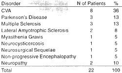
We reported our experience with 27 patients who had swallowing disorders caused by different pathologies, who were followed up in our ambulatory between January 1999 and July 2000. Twenty-two patients were adults and five were children. The mean age of adults was 57 years, ranging from 33 to 87 years. The mean age of children was 5.3 years, ranging from 4 months to 11 years. As to gender, we had 19 female patients and 8 male patients. All patients had been referred to the department because of complaints of dysphagia and/or aspiration. In the pediatric population, 2 of the 5 patients also had occasional reflux of liquid through the nose. They were all submitted to videoendoscopic evaluation with dynamic study of swallowing, as described above.TABLE 1 - Diagnosis of the neurological disorders of 22 adult patients with dysphagia.
RESULTS
All studied adult patients had neurological disorders, the most common one the cerebral vascular accident found in 8 out of 22 adults (36%), followed by Parkinson's disease and multiple sclerosis, affecting 3 patients each (13%), lateral amyotrophic sclerosis (8%) [Table 1]. Out of the 5 studied children only 2 had established diagnosis - one had Edward's syndrome and the other had a non-progressive encephalopathy. The other 3 studied children had dysphagia or aspiration of unknown etiology when they were referred to our ambulatory.
In the pediatric population, the dynamic videoendoscopic study of swallowing resulted in exclusion of laryngeal sensitivity and mobility deficits in 3 out of 5 patients. As to the 2 abnormal exams, one was of a 9-year old boy with non-progressive encephalopathy who presented left vocal fold paralysis in abduction and laryngeal global hyposensitivity with difficult to form the food bolus and oral phase leak, both early and late, of liquids with aspiration, although the pharyngeal phase seemed to be rather normal.The patient was instructed to thicken food and was submitted to speech therapy; the aspiration was controlled and it was possible to maintain oral feeding. The second child with abnormal exam was a 2-year-old girl who also had non-progressive encephalopathy, manifesting dysphagia and lack of coordination of the whole swallow and phonation system, with pharyngeal and laryngeal global hyposensitivity
and aspiration of liquid, paste and solid foods; the patient was instructed to have a gastrostomy due to the risk of developing aspiration pneumonia.
In the adult population, the disorder most frequently found in the dynamic study of swallowing was reduction of laryngeal sensitivity, leading to aspiration of liquids (30%), specially in cases with early leak of food to the oral cavity (24%) or alterations in the pharyngeal phase with remains of food on pyriform sinuses or vallecula after swallowing (20%). The aspiration reduced with more consistent foods such as paste and solid, especially after multiple swallows of the same food bolus associated with posture maneuvers. None of the 3 patients with Parkinson's disease had significant alterations of laryngeal sensitivity.
Laryngeal paralysis was found in 4 patients, one child reported above and three other adults - two with left vocal fold paralysis associated with CVA and one with bilateral paralysis after an episode of myasthenia gravis. This third patient had dysphagia and rapid progression dyspnea, requiring emergency tracheostomy; he also presented marked reduction of laryngeal sensitivity and significant aspiration of foods and maintained a nasoenteral tube for 3 weeks. Dynamic video fiberoptic study of swallowing was repeated after this interval, and we observed improvement of vocal fold mobility and laryngeal sensitivity, enabling indication of reintroduction of oral feeding.
DISCUSSION
Swallowing disorders may result in significant morbidity and mortality because they cause malnutrition and aspiration of foods and, consequently, aspiration pneumonia. In the United States it is estimated that about 40,000 deaths per year are caused by aspiration after cerebral vascular accidents². Unfortunately, we do not have the same statistics in Brazil; however, our empirical experience has shown that aspiration is the potential cause of severe respiratory complications. In view of that, it is essential that the we conduct diagnosis of aspiration episodes in risk patients, especially neurological patients with dysphagia or dysphonia, or even pediatric patients with repetitive pneumonia.
Swallowgram via videofluoroscopy provides significant data on swallowing, especially about the oral preparatory phase and the esophageal phase, and it is possible to check the alterations of peristalsis and food reflux, among other things7, 9. However, the exam is unable to assess sensitivity, which is the key factor in the pathogenicity of aspiration pneumonia.
Dynamic video fiberoptic study of swallowing, as described before, is less invasive than a swallowgram and may provide data on laryngopharyngeal sensitivity and mobility, in addition to the fact that it is easy to perform and may be conducted either in the hospital bed or in the ambulatory5,6. Since it is possible to couple a micro video camera to the fiberoptic, it enables recording for future analysis and com parison with other exams, allowing assessment of the evolution of dysphagic patients. The exam is well tolerated by patients, causing little discomfort, it permits swallowing and may be performed in all age ranges, provided that there is some collaboration. We avoid the use of topic anesthesia in order to prevent the results from being modified, since one of the main objectives of the study is to assess local sensitivity.
There are many studies comparing videofluoroscopy and flexible nasolaryngoendoscopy for the study of swallowing, and it has been agreed that both exams are capable of defining aspiration and its causes with the same sensitivity. However, videofluoroscopy evaluates well the oral phase and flexible nasofibrolaryngoscopy assesses laryngeal and pharyngeal sensitivity5,7,9,12,15,17. It is currently considered that these two exams complete each other, but the latter may be conducted at the bedside and enables early diagnosis of swallowing disorders, which is essential for prognosis. It is also essential to have a flexible nasoendoscopy before the swallowgram, because in cases in which there are alterations of pharyngeal and laryngeal mobility, the risks of massive aspiration of contrast during swallowgram are huge.
We know that the early the reintroduction of oral feeding in patients with neurological sequelae, the better the prognosis; nevertheless, it should be done safely. As we have seen in our small sample, video nasolaryngofibroscopy enabled determination not only of aspiration, but also of what was the most appropriate consistency for safer and effective swallowing. In many occasions, the exam contributed to the diagnosis in patients with neurological disorders that had not been previously defined.
CONCLUSION
Dynamic flexible nasolaryngoendoscopy is a diagnostic and rehabilitation method important for swallowing disorders in pediatric and adult populations. By means of an easy to learn and apply protocol it is possible to perform the exam in the ambulatory and at the bed side, with very little discomfort for the patient.
REFERENCES
1. ANGELARD, B.; FEVE, A.; MOINE, A.; FICHAUX, P.; GUILLARD, A.; LACAU, S.,T.; GUILY, J. - Abnormal Movements of the Larynx. Diagnostic Approach and Therapeutic. Ann. Otolaryngol. Chir. Cervicofac. 110(3): 125-128, 1993.
2. AVIV, J. E.; MARTIN, J. H.; SACCO, R. L.; ZAGAR, D.; DIAMOND. B.: KEEN. M. S.: BLITZER. A. - Supraglottic and Pharyngeal Sensory Abnormalities in Stroke Patients with Dysphagia. Ann. Otol. Rhinol. laryngol., 105(2): 92-97, 1996.
3. AVIV, J. E.; SACCO, R. L.; THOMSON, J.; TANDON, R.; DIAMOND, B.; MARTIN, J. H.; CLOSE, L. G. - Silent Laryngopharyngeal Sensory Deficits after Stroke. Ann. Otol. Rhinol. Laryngol., 106(2): 87-93, 1997.
4. AVIV, J. E. - Effects of Aging on Sensitivity of the Pharyngeal and Supraglottic Areas. Am. J. Med., 103(5A): 74S-76S, 1997.
5. AVIV, J.E.; KAPLAN, S. T.; THOMSON, J. E.; SPITZER, J.; DIAMOND, B.; CLOSE, L. G. - The Safety of Flexible Endoscopic Evaluation of Swallowing with Sensory Testing (FEESST): an Analysis of 500 Consecutive Evaluations. Dysphagia, 15(1): 39-44, 2000.
6. AVIV, J. E. - Clinical Assessment of Pharyngolaryngeal Sensitivity. Am. J. Med., 108(Suppl. 4a): 68S-72S, 2000.
7. AVIV, J. E. - Prospective, Randomized Outcome Study of Endoscopy Versus Modified Barium Swallow in Patients with Dysphagia. Laryngoscope, 110(4): 563-574, 2000.
8. BASTIAN, R. W. - Videoendoscopic Evaluation of Patients with Dysphagia: An Adjunct to the Modified barium Swallow. Otolaryngol. Head Neck Surg., 104(3): 339-350, 1991.
9. COSTA, H. O.; DUPRAT, A. C.; ECKLEY, C. A. Distúrbios da Deglutição na Criança. Em: Laringologia Pediátrica. Ed Roca Ltda., São Paulo, SP, 1999, pp.235-252.
10. DE PAULA, A.; FERNANDES, J. D.; FORTINGUERRA, M. B. - Estudo da Fase Faríngea da Deglutição em Voluntários Sadios através da Fibronasoscopia. Rev. Bras. ORL., 66(5): 434-38, 2000.
11. KAYE, G. M.; ZOROWITZ, R. D.; BAREDES, S. - Role of Flexible Laryngoscopy in Evaluating Aspiration. Ann. Otol. Rhinol. laryngol., 106(8): 705-709, 1997.
12. LANGMORE, S. E. - Laryngeal Sensation: A Touchy Subject. Dysphagia, 13(2): 93-94, 1998.
13. LEDER, S. B.; KARAS, D. E. - Fiberoptic Endoscopic Evaluation of Swallowing in the Pediatric Population. Laryngoscope, 110(7): 1132-1136, 2000.
14. LORENZ, R.; JORYSZ, G.; TORNIEPORTH, N.; CLASSEN, M. - The Gastroenterologist's Approach to Dysphagia. Dysphagia, 8(2): 79-82, 1993.
15. WILLGING, J. P. - Endoscopic Evaluation of Swallowing in Children. Int. J. Pediatr. Otorhinolaryngol., 32(Suppl.): S107-S108, 1995.
16. WU, C. H.; HSIAO; T. Y:; CHEN, J. C.; CHANG, Y. C.; LEE, S. Y. - Evaluation of Swallowing Safety with Fiberoptic Endoscope: Comparison, with Videofluoroscopic Technique. Laryngoscope, 107(3): 396-401, 1997.
17. YANAGISAWA, E.; OWENS, T. W.; STROTHERS, G.; HONDA, K. - Videolaryngoscopy. A Comparison of Fiberoptic and Telescopic Documentation. Ann. Otol. Rhinol. laryngol., 92 (5 Pt 1): 430-436, 1983.
* Assistant Professor of the Department of Otorhinolaryngology at Santa Casa, São Paulo. Master Degree in Medicine at FCMSCSP.
** Resident Physician of the Department of Otorhinolaryngology at Santa Casa, São Paulo.
*** Instructor of the Department of Otorhinolaryngology at Santa Casa, São Paulo. Master Degree in Medicine at FCMSCSP.
**** Joint Professor of the Department of Otorhinolaryngology at Santa Casa, São Paulo.
Study presented at 35° Congresso Brasileiro de Otorrinolaringologia, which was awarded a special citation.
Address for correspondence: Dra. Claudia Eckley - Rua Sabará 566, cjto. 23 - Higienópolis - 01239-011 São Paulo /SP - E-mail: cekley@unysis.com.br
Tel/Fax: (55 11) 257-2686.
Article submitted on August 15, 2000. Article accepted on October 20, 2000.
Print: ![]()