

Year: 2001 Vol. 67 Ed. 1 - (2º)
Artigos Originais
Pages: 16 to 21
Noise Exposure: Functional and Morphological Aspects of the Auditory System in Human and Experimental Animal. Model.
Author(s):
Laís P. Bonaldi*,
Ana P. Q. Olivares**,
Claudia Silva**,
Márcia L. Domênico**,
Cristiani Bartta**,
Liliane D. Pereira***,
Marco A. De Angelis****,
Silvia L. Floriano*****.
Keywords: noise, audition, cochlea
Abstract:
Introduction: Auditory thresholds may be temporary changing previously an occupational hearing loss and, even before that the organ's Corti, hair cells may be damage. Aim: The aim of this study was the noise exposure auditory system characterization. Material and method: Comparing auditory configuration changes in 203 normal hearing workers and outer hair cells guinea pig morphology (scanning electron microscopy), after temporary absence of the distortion product otoacoustic emissions. Results: The audiogram configuration was changed in 98 cases (48%) attesting the existence of a period before the temporary threshold changes. The otoacoustic emissions showed temporary functional injury of the outer hair cells and behavioral disturbances. The microscopical data indicates that the outer hair cells structure remains organized, no injury was observed. Conclusion: Analysis of the data suggests that temporary hearing loss could be present without microscopical damage. Additional methods and animal models could provide information to understanding noise as a cause of permanent occupational hearing loss.
![]()
INTRODUCTIONS
Harmful effects generated by noise exposure, regardless of the specific characteristics, vary according to level of exposure (frequency, sound pressure level and duration) and individual susceptibility. However, in occupation audiological assessment other associated factors may influence the results, such as the presence of physical or chemical factors, subjectivity of the method employed and criteria for normal results. Depending on the interaction of these factors, findings of individual assessments may vary from normal auditory thresholds to temporary threshold deviation and detection of noise induced hearing loss (NIHL). In both cases of altered thresholds, they are sensorineural :hearing losses in which even before affection of pure tone thresholds (functional), there may be alterations of hair cells of Corti's organ (morphological). Since they are reversible lesions, recovering the responses within normal auditory thresholds, the presence of damaged cochlear cells is not generally detected in pure tone audiometry7. A number of methods that involve objective assessment of hearing or observation of morphological modifications at different cochlear levels, such as otoacoustic emissions, electrophysiological tests, central auditory processing tests, histological studies and electronic microscopy, may complement audiometry by providing clinical and preventive applications to the process of hearing conservation. Among the methods, we point out the investigation of cochlear damage with otoacoustic emissions, because they are only present if conditions are close to normal and if there are no middle ear affections5. The emissions are generated by the biomechanical activities of outer hair cells that contract, acting as active cochlear emitters ² ¹¹. Otoacoustic emissions are divided into two main types - spontaneous and evoked, subdivided into transient, not very applicable for clinical diagnostic purposes7, and distortion product, which enables objective assessment of specific frequencies6. Therefore, when preventing temporary and permanent hearing losses caused by exposure to noise, otoacoustic emissions are an alternative to assess and monitor the cochlea, and the advantages are quick response and objectivity4. The objective of the present study was to characterize the auditory system exposed to noise, before the onset of NIHL, by correlating alterations in audiometric configuration of workers with normal auditory thresholds with morphology of outer hair cells of guinea pig after the temporary absence of distortion product otoacoustic emissions.
MATERIAL AND METHOD
In the first stage of the study, we selected 203 audiometries of workers exposed to noise in a food manufacturing plant, who used inserted individual hearing protecting equipment. All workers were submitted to otoscopy and 111 (55%) were female and 92 (45%) were male subjects, in the age range of 20 to 70 years. They were classified as normal hearing subjects according to the criteria of auditory thresholds lower or equal to 25dBHL. The exams were revalued according to the following criteria of configuration of curve: descending curve - fall in higher frequencies (3.000 Hz, 4.000 Hz or 6.000 Hz) with a 10dB difference compared to the previous frequency, and the posterior frequency had similar or higher threshold; and acoustic notch - fall in high frequencies with a 10dB difference between the worst frequency and the two subjacent ones.
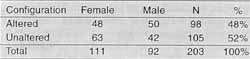
The second stage involved the study of the cochlea of guinea pig at adult development stage. It was assessed using otoacoustic emissions pre and post-noise exposure of 80 to 90dB in a graphic plant for 8 hours for two consecutive days, after a 16-hour rest interval and after a seven-day period. We carried out otoscopic investigation and tympanometry using portable equipment Hand-tymp before the conduction of distortion product otoacoustic emissions (ILO 92DP OAE System - Otodynamics, with standard probe). The test was conducted in the frequency range of 1.000 Hz to 6.000 Hz, because of the limitations of the device, and we used the software DP-Gram, maintaining the sound pressure at 70 dB SPL, F2/F1 ratio of 1.22 and three points per octave. The criteria used to consider the presence of emissions were to have results louder than the background noise, considering as reference the values previously found in 11 adult healthy guinea pigs. After functional assessment of the hearing system, according to the criteria listed above, tympanic bulla were detached from the skull and dissected in order to expose the cochlea and the hair cells from the cochlear spiral. The cochlea was fixed at 20% glutaldehyde with sodium cacodylate buffer 0,1M, pH 7.2 for 24 hours. The material was then washed with the same buffer solution overnight, following by two 10-minute washings and fixation with osmium tetroxide at 1%, buffered for 1 hour. After another washing with buffer solution, the material,was submitted to treatment with acid tannic at 1% in distilled water for 30 minutes; it was washed again with distilled water and submitted to dehydration in ethanol increasing series. Next, the cochlea was submitted to drying using the method of CO² critic point (Balzers-CPD 030), prepared in an appropriate support with colloidal silver glue (Silver Print) and recovered with gold (Spriettering -Balzers-SCD 050), to be later observed by the microscope JEOL JSM 5300 (CEMO/UNIFESP-EPM). The final sample of scanning electron microscopy was obtained in order to try to find evidence of cochlear alterations after exposure to noise, supporting the inferences made about the pathophysiology of the studied subjects.TABLE 1 - Altered x unaltered configuration in male and female subjects whose audiometry was initially classified as normal.
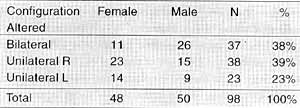
TABLE 2 - Altered configuration in subjects whose audiometry was initially classified as normal according to gender and side.
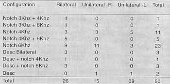
TABLE 3 - Types of altered configuration in male subjects whose audiometry was initially classified as normal.
RESULTS
Part 1 - Functional characterization of normal auditory system exposed to noise - altered configurations in normal audiometry.
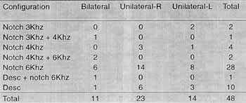
After the analysis of the configurations in our sample of 203 cases initially classified as normal, we noticed the presence of altered configurations in 98 cases (48%), as shown in Table 1. Alterations of configuration were mainly bilateral and unilateral on the right, amounting to 75 cases (77%) compared to 23 cases (23%) of unilateral left affections (Table 2). As to the characteristics of alterations, we pointed out the existence of notches in 6.000Hz, both in male and female patients (Tables 3 and 4), and in male there was also a percentage of 4.000Hz notches but in fewer cases.
Part 2 -Morphological characterization of auditory system - screening electron microscopy in guinea pig exposed to noise, assessed by distortion product otoacoustic emissions.
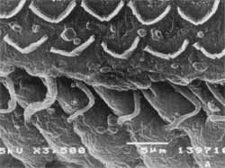
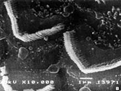
The values of amplitude of responses of distortion product otoacoustic emissions after 8 hours of exposure to 80-90dB were reduced considerably in all assessed frequencies, and we also noticed absence of response. However, in the two following assessments, after intervals of 16 hours and seven days, under the same conditions of noise exposure, results did not show pre and post-exposure significant changes, keeping the amplitude values within the normal range for healthy guinea pigs (Tables 5 and 6). In the second and third evaluations. behavioral disorders that are normally associated with noise exposure were noticed (agitation, hypersensitivity to touch, light and sound). The results at screening electron microscopy showed that there was no damage to cochlear outer hair cells and ciliary and structural organization were maintained intact (Figures 1 A, and 1B).Table 4 - Types of altered configuration in female subjects whose initial audiometry was considered normal.
Key: R = right; L = left; Desc = descending.
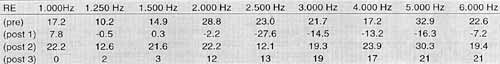
TABLE 5 - Values of amplitude of responses of otoacoustic emissions on the right ear (RE) of an adult guinea pig, before exposure (pre) and after the first (post 1) second (post 2) and third (post 3) exposure to noise.
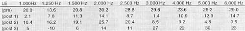
TABLE 6 - Values of amplitude of responses of otoacoustic emissions on the left ear (LE) of an adult guinea pig, before exposure (pre) and after the first (post 1), second (post 2) and third
DISCUSSION
Before the onset of NIHL, whose characteristics are irreversibility and slow progression as a result of systematic noise exposure³, we may detect temporary auditory deviations. The typical NIHL configuration at audiometry is deviation in the frequency of 6.000HZ, extending the process into 4.000Hz and possibly 3.000HZ; later, the frequency of 4.000Hz becomes worse and only at advanced stages of the condition the other areas are equally affected. As to temporary deviations of thresholds, they are due to post-stimulation auditory fatigue, that is, there may be changes in thresholds after exposure to noise, but they are reversible. These changes may be considered as the first manifestation of NIHL; if so, they may present characteristics of temporary deviation similar to NIHL configuration. In the present study, we analyzed audiometries of workers exposed to noise before any threshold deviation could be noticed, either permanent or temporary. However, findings were compatible with the characteristics of NIHL, showing that there is possibly a previous phase before deviation of temporary thresholds. The analysis o1 data from 203 audiometries classified as normal showed 98 cases (48%) with descending configuration. There was prevalence of bilateral alteration, especially notches in 6.000Hz extending into 4.000 and 3.000Hz. Based on these finding: and the fact that even without alterations of pure tone thresholds there could be alterations of hair cells from Corti's organ, we decided to conduct a complementary study with an experimental animal model. The study was carried out in a guinea pig because it is easy to perform the anatomical dissection and there are similarities with the human ear 9,10. Moreover, it is considered a model animal for the study of properties of evoked otoacoustic emissions 1,4 - an objective method that allows functional analysis of cochlea by specific spectrum of frequencies. Findings have shown that there was functional damage of outer hair cells after the first exposure to noise, which could be compared to a temporary deviation of threshold. However, the damage was not noticed after the second and third exposures, and we noticed the presence of emissions within the normal frequency range of healthy guinea pigs, representing normal cochlear function and suggesting normal auditory sensitivity. Behavioral variations observed after the exposure were compatible with those referred by workers, such as hypersensitivity to stimuli and marked irritability, among others. The observation of outer hair cells at screening electron microscopy showed findings compatible with those defined by distortion product otoacoustic emissions. The recovery of thresholds showed by the outer hair cells, detected by the presence of otoacoustic emissions, was confirmed by the absence of damage and structural disorganization at microscopy.
Figures 1 A and B -Scanning electron microscopy of guinea pig after noise exposure
CONCLUSION
The auditory system exposed to noise may have reversible auditory alterations that are undetected by the normally employed methods. These previous alterations without apparent damage at electronic microscopy may be associated with behavioral changes. The use of complementary methods and animal models collaborates to the expansion of knowledge about noise as a cause of hearing loss, stressing the importance of conducting further studies with more precise methods (histological and others).
REFERENCES
1. AVAN, P.; LOTH, P. A, D.; MENGUY, C.; TEYSSOU, M. Evoked otoacoustic emissions in guinea pig: basic characteristics. Hear Res, 44 (2-3): 151-60, 1990.
2. BROWNELL, W. E.; BADER, C. R.; BERTRAND, D.; DE RIBAI PIERRE, Y. - Evoked mechanical responses of isolated cochlear outer hair cells. Science, 227 (4683): 194-6, 1985.
3. COMITÊ NACIONAL DE RUÍDO E CONSERVAÇÃO AUDITIVA (CNRCA) - Características da PAIR. JAMB, 9, 1994.
4. HOTZ, M. A.; PROBST, R.; HARRIS, F. P.; HAUSER, R. -
Monitoring the effects of noise exposure using transiently evoked otoacoustic emissions. Acta Otolaryngol, (Stockh), 113 (4): 478-82, 1993.
5. KEMP, D. T. - Otoacoustic emissions in perspective. In Robinette, M. S. & Glattke, T. J. - Otoacoustic Emissions: Clinical Applicalions. Thieme, New York, 1-21, 1997.
6. KEMP, D.T. -Stimulated acoustic emissions from within the human auditory system. J. Acoaust. Soc. Am., 64(5): 1386-91, 1978.
7. LOPES FILHO, O. & CARLOS, R. - Produtos de Distorção das Emissões Otoacústicas. RBM Rev. Bras. filed., 3 (5): 224-36, 1996.
8. OLIVEIRA, J. A. A. - Fisiologia clínica da audição - cóclea ativa. In Nudelmann, A.A.; Costa, E.A., Seligman, J., Ibañez, R. N. - PAIR: Perda Auditiva Induzida por Ruído. Editora Bagaggem Comunicação Ltda., Porto Alegre, 101-42, 1997.
9. OLSZEWSKI, J. - The method of electron microscopic study of Corti Organ in cobaia. Folia Morphol" 48 (1-4): 137-41, 1989.
10. SEHITO GLU, M.A.; UNERI, C.; CELIKOYAR, M.M.; UNERI, A.U. - Surgical anatomy of the guinea pig middle ear. Ear Nose AND TROATJ, 69 (2): 91-97, 1990.
11. ZENNER H. P.; ZIMMERMANN, U.; SCHMITT, U. -Reversible contraction of isolated mammalian cochlear hair cells. Hear Res, 18 (2): 127-33, 1985.
* Doctorate studies in Morphology under course at UNIFESP/EPM.
** Speech and Hearing Pathologist.
*** Ph.D. in Human Communication Disorders at UNIFESP/EPM.
**** Joint Professor of the Discipline of Descriptive and Topographic Anatomy.
***** Otorhinolaryngologist.
Study presented at 35° Congresso Brasileiro de Otorrinolaringologia, which received a special citation. Study conducted in the Departments of Descriptive and Topographic Anatomy and Hearing Disorders at UNIFESP-EPM.
Address for correspondence: Laís Vieira Bonaldi - Disciplina de Anatomia Descritiva a Topográfica.
UNIFESP/EPM - Rua Botucatu, 740 - 04023-900 São Paulo /SP - Tel: (55 11) 579-6700 FAX: (55 11) 571-7597-E-mail: lais.morf@epm.br
Article submitted on August 15, 2000. Article accepted on October 20, 2000.
Print: ![]()