

Year: 2004 Vol. 70 Ed. 1 - (20º)
Relato de Caso
Pages: 124 to 128
 PDF PT
 Severe complication in the treatment of epistaxis: a case report
Author(s):
Carlos Z. Arbulú 1,
Robinson K. Tsuji 1,
Marcus M. Lessa 2,
Richard L. Voegels 3,
Ossamu Butugan 4
Keywords: epistaxis, treatment, embolization, complications
Abstract:
Epistaxis is a very usual disorder, it is usually self-restricted or controlled with conservative measures as local compression, cold gauze, arterial pressure control, cauterization under local anesthesia (chemical or thermoelectric) or anterior nasal packing. However, it could be presented as severe cases, and more aggressive measures could be necessary, like posterior nasal packing, arterial ligation or embolization. We present one case of a forty-nine-year-old patient with epistaxis who developed a severe treatment complication from another department.
![]()
INTRODUCTION
Epistaxis is bleeding of the nasal fossa and represents an affection of regular nasal hemostasis. The hemostasis can be impaired owing to vascular integrity compromise, nasal mucosa abnormalities or coagulation disorders. It is a very common affection in medical practice, being that approximately 60% of the population has already have or will have at least one episode of epistaxis in life 1, 2. It is normally self-limited, but there are about 6% of the cases in which medical intervention is required 3. Nasal hemorrhage, if recurrent, may lead to morbid consequences or even fatal consequences, such as aspiration, hypotension, anemia, hypoxia and acute myocardial infarction.
The most common site of bleeding is the anterior nasal region, corresponding to 80% of the cases 4. Anterior bleeding is also easily controlled and normally can be solved by more conservative measures, such as chemical or electrical cauterization of nasal septum, or anterior packing. Posterior bleedings, even though less frequent, are more difficult to treat and normally require more invasive measures, such as anterior-posterior packing or arterial ligation 5.
Arterial embolization in the treatment of epistaxis was described for the first time by Sokoloff et al.6 in 1974 and it has been increasingly used as complementary or alternative management for posterior epistaxis. In the literature, we can find successful rates that range from 79% to 100% 7-13, but this procedure is not completely safe and some severe complications can derive from it.
The authors report a case of epistaxis and severe complication resultant from arterial embolization associated with sphenopalatine artery ligation and anterior-posterior nasal packing.
CASE REPORT
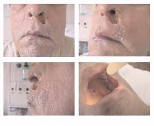
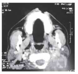
AJFT, 49-yearold male subject, presented the first episode of epistaxis on the left in January 2000, when he was treated with anterior ethmoidal ligation in another center. At the time, he was diagnosed with systemic blood hypertension, which was treated with atenolol 25mg a day. The patient remained asymptomatic up to 17 July 2001, when he presented a new episode of left nasal fossa epistaxis and was submitted to ipsilateral sphenopalatine artery ligation on the next day. One day after the procedure, the patient presented a new episode of epistaxis, and an arteriogram and embolization were then performed. The arteriogram showed an area of suspected bleeding in one of the branches of the left maxillary internal artery. Embolization was performed on the left facial and internal maxillary arteries, being that they were embolized using microspheres. The next day, the patient had a new episode of bleeding and was once again submitted to arteriogram and embolization of facial and internal maxillary arteries on the left (Figures 1, 2 and 3), in addition to embolization of the contralateral facial and internal maxillary arteries (Figures 4 and 5), using Gelfoam®. Anterior-posterior packing was prescribed for 3 days. On July 23, after removal of nasal packing, the attending physician detected the presence of a necrotic area on the left nasal ala, leading to skin necrosis on the nasolabial region. On July 31, he was referred to our center with complaint of new self-limited episodes of bleeding on the left and extensive areas of necrosis. As to personal history, the patient smoked 30 cigarettes a day and drank alcohol only socially, with no other associated diseases. Upon admission, the patient had no active bleeding and complained of facial pain on the left maxillary region. The physical examination revealed good overall health status, no fever, normal blood pressure, eupneic, with pale skin and mucosa. He had necrosis on the nasal ala on the left extending to the surrounding nasolabial skin area, with fistulation of the gingival-labial groove and crusts on the rims of the lesion. At oroscopy, he presented palate ulcers and oronasal fistula in the hard palate region (Figure 6). Lab tests showed hemoglobin 10mg/dl, hematocrit 31% and absence of coagulation disorders. We conducted computed tomography (CT) that showed lesion on the soft parts of the left nasal vestibule region (Figure 7), without any other affections. He was treated with local applications, nasal lavage (sterile solution), topical use of gentian violet and symptomatic drugs and progressed without recurrence of epistaxis. He was discharged 10 days after admission without progression of tissue necrosis and there was already some granulation tissue on the margins of the lesion.
DISCUSSION
The treatment of epistaxis normally follows a scale of management approaches that is followed by otorhinolaryngologists. We should start by taking more conservative measures such as local compression, arterial blood pressure control, sedation, cold dressings, chemical or thermal-electrical cauterization, anterior nasal packing or even anterior-posterior nasal packing. In case of refractory epistaxis, we can use more invasive measures such as surgical arterial ligation of sphenopalatine, internal maxillary, ethmoidal arteries or embolization of internal maxillary and/or facial arteries. Treatment modality depends on level and type of bleeding (anterior, posterior, septal) as well as clinical conditions of the patients. In our center, we prefer surgical ligation of ipsilateral sphenopalatine artery in case of refractory epistaxis, without the need for nasal packing at early postoperative conditions We reserve anterior ethmoid ligation for cases of failure in the previous procedure, a fact that is not frequent in our experience.
Arterial embolization, if properly indicated, yields high success rates. The advantages of this therapy are: location of bleeding, region, diagnosis of associated diseases such as tumors or vascular lesions, and no need for general anesthesia. Some authors suggest that the high cost of the procedure would be compensated by the reduction in length of hospital stay. The disadvantages are the need for specific equipment and specialized team. The method is limited for bleedings that arise from the ethmoid arteries and in patients with carotid artery atherosclerotic disease 9, 12, 13.
Complications of arterial embolization are classified as minor and major, occurring in 17% to 25% of the cases 9, 12. Minor complications are pain and facial paresthesis, headache and facial edema, normally solved within the first week. Facial pain was also referred in our case and it could have been present because of tissue necrosis or ischemic damage to the trigeminal nerve. The major complications are cerebral vascular accident, amaurosis, facial palsy, septal perforation, skin necrosis and palatine ulcer, presenting incidences below 2%.
In our study, the patient presented a massive lesion on the nasal ala with fistulation of gingival-labial groove and oronasal fistula in the hard palate. Tseng et al.12 and Elden et al.9 published the largest samples of cases including complications resultant from arterial embolization. According to the authors, the incidence of skin lesions was below 1%, and in no case there was extensive mucosa skin lesion similar to that reported in our case.
Facial arterial irrigation is made by the branches of maxillary and facial arteries. Facial artery produces branches to the gums (superior alveolar artery) and to the skin on the nasolabial region (superior labial artery). Maxillary artery produces branches to the palate (major palatine artery) and to the septum and mucosa of the nasal fossa (sphenopalatine artery). The vestibule is irrigated by the subseptal artery, which is a branch of the superior labial artery and also includes branches of the internal maxillary artery. In general, complications related in the literature are due to over-embolization, use of very small particles or use of long permanence particles such as Gelfoam®9. In our case, the patient was submitted to bilateral embolization of both internal maxillary and facial arteries, being that the left side was embolized twice with Inalom® and Gelfoam®. Epistaxis was controlled but there was severe ischemia in the areas vascularized by these arteries, leading to the lesions described above. Moreover, the patient received anterior-posterior packing that has probably contributed to ischemia. Another important factor is that the patient was a smoker and had arterial hypertension, which could have contributed to the development of ischemic lesions by atherosclerotic abnormalities.
Comparing surgical arterial ligation and arterial embolization, we can observe that both techniques have similar success rates. Embolization has the advantage of being performed under local anesthesia, but the major complications are more severe 11. In our center, we count on a highly competent and experienced team of interventionist radiologists and we reserve embolization only to the cases in which arterial ligation has not been satisfactory to treat severe and refractory epistaxis.
CLOSING REMARKS
The approach to epistaxis management should be carefully analyzed, taking into consideration type and severity of bleeding and the clinical conditions of the patients. Treatment should be as conservative as possible, following a scale of procedures, in order to prevent severe complications.
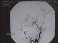
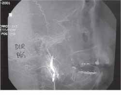
Figure 1. Arteriogram showing left external carotid artery before embolization.
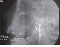
Figure 2. Selective embolization of left maxillary artery.
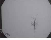
Figure 3. Left external carotid artery after embolization of maxillary and facial arteries.
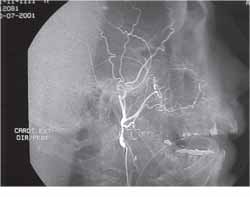
Figure 4. Arteriogram showing right external carotid artery before embolization.
Figure 5. Right external carotid artery after embolization of maxillary and facial arteries.
Figure 6. Necrotic lesions on the skin of the left nasal vestibule and surrounding nasolabial region, with fistulation to the gingival-labial groove. Note oronasal fistula on the hard palate region (arrow).
Figure 7. Paranasal sinuses computed tomography, at axial section, showing loss of continuity of the skin that corresponded to nasolabial ulcer.
REFERENCES
1. Petruson B. Epistaxis: a clinical study with special reference to fibrinolysis. Acta Otolaryngol 1974; 3:1-73.
2. Shaw C. Epistaxis: a comparison of treatment. Otolaryngol Head Neck Surg 1993; 109 (1): 60-5.
3. Small M. A study of patients with epistaxis requiring admission to hospital. Health Bull (Edinb) 1982; 40:24-9.
4. Schaitkin B. Epistaxis: medical vs. Surgical therapy: a comparison of efficacy, complications and economic considerations. Laryngoscope 1987; 97:1392-6.
5. Voegels RL. Endoscopic ligature of the Sphenopalatine artery for severe posterior epistaxis. Otolaryngol Head Neck Surg 2001; 124(4): 464:7.
6. Sokoloff J. Therapeutic percutaneous embolization in intractable epistaxis. Radiology 1974; 111:285-7.
7. Almeida R. Tratamento endovascular de epistaxe por microcatéter super-seletivo. Revista Brasilera de Otorrinolaringologia 2000; 66:30-6.
8. Cullen M. Comparison of internal maxillary artery ligation versus embolization for refractory posterior epistaxis. Otolaryngol Head Neck Surg 1998; 118:636-42.
9. Elden L. Angiographic embolization for the treatment of epistaxis: a review of 108 cases. Otolaryngolog Head Neck Surg 1993; 111:44-50.
10. Moreau S. Supraseletive embolization in intractable epistaxis: review of 45 cases. Laryngoscope 1998; 108:887-8.
11. Strong B. Intractable epistaxis: transantral ligation versus embolization. Otolaryngol Head Neck Surg 1995; 113:674-8.
12. Tseng E. Angiographic embolization for epistaxis: a review of 114 cases. Laryngoscope 1998; 108:615-19.
13. Vitek J. Idiopathic intractable epistaxis: endovascular therapy. Radiology. 1991; 181:113-6.
14. Bent J. Complications resulting from treatment of severe posterior epistaxis. Journal of Laryngology and Otology 1999; 113:252-4.
1 Residents physicians, Division of Clinical Otorhinolaryngology, Hospital das Clínicas, Medical School, University of Sao Paulo.
2 Post-graduate studies under course, Discipline Otorhinolaryngology, Medical School, University of Sao Paulo.
3 Ph.D., Professor, Discipline Otorhinolaryngology, Medical School, University of Sao Paulo.
4 Associated Professor, Discipline Otorhinolaryngology, Medical School, University of Sao Paulo.
Study conducted at the Discipline of Otorhinolaryngology, Hospital das Clínicas, Medical School, University of Sao Paulo.
Address correspondence to: Carlos Zevallos Arbulú - Av. Dr. Enéas de Carvalho Aguiar, 255 6°andar sala 6021 Sao Paulo SP 05403-000.
Tel: (55 11) 3069-6288 - Fax: (55 11) 270-0299 - E-mail: carzevarb@hotmail.com
Study presented at 2° Congresso de Otorrinolaringologia da Universidade de Sao Paulo, held on November 22 - 24, 2001, in Sao Paulo-SP.
Print: ![]()