
Year: 1994 Vol. 60 Ed. 4 - (8º)
Artigos Originais
Pages: 287 to 296
A New Method For Sensory Organization tests: The Foam-Laser Dynamic Postugraphy
Author(s): Lucio A. Castagno, MD (*)
Keywords: Disequilibrium; dynamic posturography; sensorial organization tests; ele tronystagmography
Abstract:
Balance is dependent ou three main systems: somatosensory, visual, and vestibular system. Two sophisticated vestibulomotor reflexes developed to maintain equilibrium. The vestíbulo-ocular reflex (VOR) connections distribute inputs mainly from semicircular canals to the brainstem for stabilization of retinal image during movement. The vestibulospinal reflex (VSR) distributes information mostly from utriculus and sacculus to CNS and muscles in the legs, allowing synchronized reciprocal stabilization of the body required for locomotion or standing. The VOR has been usually evaluated through ELECTRONYSTAGMOGRAPHY, while only in recent years, VSR has been scientifically assessed and quantified through DYNAMIC POSTUROGRAPHY A commercially available for computerized dynamic posturography (CDP) opparatus was introduced in 1986 (EquiTest) and is gaining popularity. However, its cost preevents world wide use. We present an inexpensive and reliable technique for performing sensory organization tests with the following "equipment": a one square meter cabinet as "visual surround" (2 m height) , a laser pen (as for slides presentation), anda 10 cm thick medium density furniture foam (0.5x0.5 m). Mathematical formulations used to calculate the angle of oscillation, and to plot the graphics of "equilibrium score" and " sensory analysis", are presented. This new technique of foam-laser dynamic posturography (FLP) has been used for one year and the results are encouraging as they do relate very well to those obtained with CDP. Three representative cases of the FLP usefulness are presented. Dynamic posturography is an important quantitative measure of posture control mechanisms, and especially useful for follow-up control, recording the course of disease and the effect of therapy.
![]()
INTRODUCTION
Maintaining static balance is dependent on three main systems: somatosensory (muscles and tendons, joints, and cutaneous receptors), visual, and vestibular system. Disturbances in these complex functions are often manifested as dizziness or vertigo, which are some of the most common symptoms encountered in otology or neurology. Motor processes in balance control coordinate the action of trunk and leg muscles into discrete synergies that minimize sway and maintain the body's center of mass within its base of support. Two highly sophisticated vestibulomotor reflexes developed for this purpose. The vestibulo-ocular (VOR) reflex connections are distributed conjugately to eye muscles for bilaterally synchronized stabilization of the eyes. Without this retina stabilization, clear vision during active or passive movement is impossible and accounts for the oscillopsia that may occur in severe vestibular ototoxicity 2. The vestibulospinal reflex (VSR), on the other hand, distributes information about the earth vertical to the legs allowing synchronized reciprocal stabilization of the body required for locomotion. Partially redundant orientation information can be provided simultaneously by the vestibular, visual and proprioceptive sensory systems to the central nervous system (CNS) causing symptoms. The relative role of these multisensory inputs to the VOR and VSR must, therefore, be determined in order to draw accurate conclusions about normal and abnormal vestibular communication systems 3.
During the last decades the emphasis on the vestibular investigation of patients with dizziness or vertigo was placed almost exclusively in electronystagmograpliy and VOR analysis 4,5,6. The vestibulospinal reflex (VSR) was mostly neglected, even though experiences with cephalography have been done in the 20s-30s 7,8, and with static posturography sinee the 50s 9,10. It was Nashner who initially described the principles of dynamic posturography in a doctoral thesis in 1970 11, and later introduced the concept of computerized dynamic posturography (CDP) 12. These early works were based on the use of a moving platform that became commercially available in 1986 EquiTest; Neurocom International, Inc., Clackamas, Ore.), and since then several papers have been published as its popularity increases. During computerized dynamic posturography the patient is standing with feet parallel, comfortably spaced apart, on a dual forceplate enclosed by a visual surround. Both the forceplate and the surround may move with the personis antero -posterior sway or independent of the sway. This dual forceplate records the vertical forces between feet and ground as well as horizontal anteroposterior shear forces, thereby allowing estimation of the position of the swaying body, and the pattern of sway (hip or ankle strategy). CDP performs two main analysis: the sensory organization tests (SOT), designed to assess the sensory contro) of postura (figure 1), and the movement coordination test (MCT) for appraisal of motor responses used to maintain balance. The sensory organization tests (SOT) seem the most important part of CDP and are divided into six separate tests, lasting 20 seconds each, with tests III to VI repeated to get more stable (mean) values: SOT 1 is a quantified version of the Romberg test; SOT II and V are done with eyes closed; SOT III and VI are performed with eyes open and the visual surround moves in response of the body sway; SOT I to III are done on a stable ground, while in SOT IV to VI the platform is moving. From each test an equilibrium score is considered: 100% for absolutely no sway, decreasing with the increasing sway range, and 0% in case of falling or greater than 12.5§ degrees sway (maximum possible sway without falling). Also the SOT protocol considers the amount of shear force exerted, expressing the degree of ankle or hip movements. The movement coordination test (MCT) evaluates latency and amplitude of reflex muscle contractions during sudden unpredietable platform translation or rotation, through electromyography of tire gastrocnemius (backward movement) and the tibialis anterior muscle (forward movement).
It is certainly important in t11e investigation of patients with equilibrium disorders to evaluate both the vestibulo-oculomotor and vestibulospinal pathways. Also the amount of information obtained with computerized dynamic posturography is indeed remarkable la. However, the equipment costs prevent its worldwide use. A clinical office vestibulospinal evaluation that could be easily and inexpensively done would be very useful. Recently Weber and Cass 14, in a very interesting article, compared foam posturography with Nashneris moving platform posturography, and demonstrated a significant correlation (p less than 0.005) with a 95% sensitivity and 90% specificity. Although this paper considers only normal or abnormal results (been abnormal those patients who fell within 15 seconds in the foam posturography and within 20 seconds in CDP) it proved that ir was clinically possible to assess pathological sway in sensory organization tests with the foam technique `s. Having this in mind we decided to develop an inexpensive procedure that could quantify the sway.
Figure 1 - Scoring Sensory Organization Tests (SOT).
SOT I, II and III are performed on a fixcd platform with eycs open, eyes closed, and vision sway- referenced. SOT IV, V, and VI are similar, but performed on a sway-referenced platform. Trials are 20 seconds each, and equilibrium is scored by retating thc patient's actual sway to theoretical limitis (100%= no sway; 0%= fall or grcater than 12.5° sway).
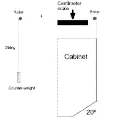
Figure 2 - Fiam-laser dynamic posturography.
The patient is placed inside aia one square-meter cabinet,
two meters height, with a visual surround consisting of 10 cens horizontal black-and-white stripes. SOT I, II, and III are performed-on the floor, while test IV, V, and VI ate done on a 10 cm thick medium density forniture foam. The cabinet is manually moved (with a simple mechanical counter-weight system) 20° forward and backward during SOT III and VI. Besides the patient, at the right hip levei (human center of gravíty), a laser peia is verticaly fixcd aiming a centimeter scale held in the ceiling.
MATERIAL AND METHODS
Our technique for foam -laser posturography is very simple. The patient is placed inside a one square meter cabinet, two meter height, with a visual surround consisting of 10 cm. horizontal black-and-white stripes (figure 2). Besides the patient, at the right hip level (center of human gravity), we place an ordinary laser pen (used for slide! presentation), turned on, and aiming a centimeter scale held on the ceiling (in the earth vertical plane). Sensor organization tests I, II, and III are done with the patients standing on the floor, both feet together, the right arm turned to the back and the left arm flexed anteriorly. SOT IV, V, and VI are done with the patient standing over a 10cm thick medium density furniture foam. During each 2 seconds test the examiner regards the red-laser dot on the ceiling as it moves, memorizing the maximum point anterior and posterior displacement. This anterior-posterior displacement (in centimeters) is entered in sevaral mathematical formulas to calculate the oscillation and (figure 3). During SOT III and VI the cabinet is alo moved approximately 20§ forward and then backward; the axis o£ this rotation is at the patients ankle level. Usually there is a small displacement of the red-laser dot in the same direction as the visual surround moves, even in normal patients. After all, the SOT analysis are done and the graphics for equilibrium score and sensory analysis prepared. Sensory analysis is presented ín accordance to several physiological concepts: somatosensory (SOT II / SOT I); visual (SOT IV / SOT I); vestibular (SOT V / SOT I); and visual preference ((SOT III + SOT VI) / (SOT II + SOT V)). It seems that sensory analysis detects the abílity for use of somatosensorial (SOM), visual (VIS), and vestibular (VEST) inputs to maintain equílibrium. It also indicates the level of visual preference (PREF) needed in balance control le. An ordinary microcomputer with any spreadsheet software may be used for calculations and graphics. We use MS Works for Windows 2.0 (Microsoft Corporation, Bellevue; Wa.) for calculations and to print a two-pages standardized report (figure 4).
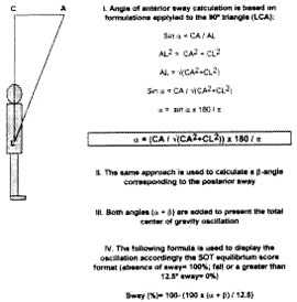
Figure 3 - Mathematical formulation for the proposed technique of foam-laser dynamic posturography
Thc anterior oscillation forma a triangle (LCA), where is the angle of sway, L is the laser pen at the hip Ievel (center of gravity), C is thc center of a centimeter scale, and A is the point of maximum anterior displacement. Distance CA is measured observing the movement of the red laser dot ; distance CL may be measured with an ordinary metric ruler or with any ultrasonic measuring tool.
Table 1 - Mean and fifth percentile in a normal sample of subjects used to define "normal values" for Sensory Organization Tests.
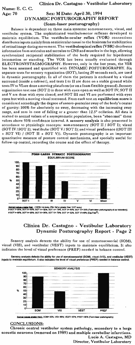
Figura 4
RESULTS
The foam-laser dynamic posturography has been used for ore year and the results are certainly encouraging as they do relate very well to those obtained with the EquiTest (Table I). Clinically normal performances are defined to include 95% of the asymptomatic sample (i.e., 2 standard deviations below the mean). Diseased patients are identified, as pathological sway is quantified, and progression with treatment is easíly assessed. Three illustrative cases are presented.
Case reports
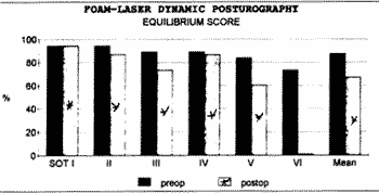
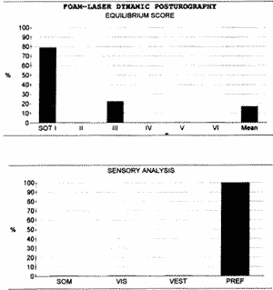
Patient 1: Clínica Dr. Castagno # 73709P, 32 years old, mate, presented with left hearing loss and tinnitus for ore year, without vestibular symptoms. Audiometry detected a profound left hearing loss (PTA= 95dB) with normal hearing in the right ear. BERA revealed a prolonged I-V period (6.2 ms. At 90 dB) on the left, and computerized electronystagmography showed a total absence of response on the left. MRI was done and a 2.8 cm. left acoustic neuroma was found. A subocciptal approach was used and the acoustic neuroma was totally removed, sparing the facial nerve. Preop foam-laser posturography was essentially normal, and at ore month postop presented only minor vestibular disfunction that most probably will improve with time (figure 5).
Patient 2: Clínica Dr. Castagno # 74662P, 70 years old, male, retired physician, was submitted to a left posterior fossa subocciptal approach for removal of a large acoustíc neuroma in 1989. The surgery required prolonged displacement of the cerebellum and sacrifice of the facial nerve. During the last four years the patient has suffered with severe incapacitating disequilibrium without any improvement. A computerized tomography was done and reveled several areas of cerebellar infarction. Foam-laser posturography presented a chronic vestibular dysfunction (figure 4).
Patient 3: Clínica Dr. Castagno # 74809G, 53 years old, female, had a severe prosthetic valve endocarditis treated heavily with aminoglycosides and laser was submitted to a valve replacement surgery. For the last five months, since therapy with aminoglycosides, the patient complains of severe incapacitating disequilibrium with mild hearing loss. Computerized electronystagmography revealed a total bilateral vestibular paresis (caloric and rotatory testing). Foam-laser posturography presented a complete vestibular dysfunction with visual preference, and total inability to maintain equilibrium without appropriate visual and somatosensory clues (figure 5).
DISCUSSION
Human balance and posture depend on coordinated integration of multiple sensory inputs (somatosensory, visual, and vestibular). These afferent informations are processed in the brainstem and cerebellum, and motor commands are initiated. Dysfunction in any part of this complex system may cause disequilibrium, dizziness or vertigo. Diagnosis o€ disorders of balance is often dífficult in clinical practice 17,18. Until recently most of the vestibular investigation was essentially based on electronystagmography data. Posturography is a much needed method for quantítating the vestibulospinal component of equilibrium, which cannot be assessed with conventional oculographic tests such as ENG: as for maintaining static equilibrium, the semicircular canals probably have less contribution than the utriculus and the sacculus 19.
The quantification of postural control has been studied for a number of years by various investigators since the Romberg test was described in 1853. Several techniques have been reported. Static posturography or stabilometry was first introduced and essentially reproduced the Romberg test. Earlier procedures required creative devices to measure sway (cephalographes, ataxiometers, potentiometers, etc.), but nowadays a vertical force platform with strain gauges is used; signals are sent to a computer for real-time calculations of the center of gravity displacement (antero-posterior and lateral), and graphicaly-oriented reports 20,21. Cranio-corpography has been used by Claussen since 1968 and reproduces the Unterbergefis test done in the dark. During the 1-3 minutes test the patient wears an interesting helmet with an anterior and posterior lights, while a Polaroid camera records all displacement of the head az. Computerized dynamic posturography was introduced by Nashner and colleagues in the early 1980s with a prototype platform that measured sway using a hip-mounted potentiometer, even though the concepts of dynamic posturography (and sensory organization tests) were presented several years before 8. From these initial experiments, a commercially available apparatus was developed (EquíTest) and is gaining popularity in several centers. Any of these posturography procedures is a complementary examination, adding information to the patient evaluation. Its valve is apparent when monitoring the progress of the vestibulospinal recovery following peripheral lesion, especially in a physical therapy program for vestibular rehabilitation 23,24.
Of course, our technique of foam-laser dynamic posturography (FLP) cannot replace completely computerized dynamic posturography (CDP). FLP cannot perform movement coordination test and detect latencies that may he useful in neurological diagnosis 25; neither performs hip or ankle strategy analysis; nor plots the displacement of center of gravity. However it ís a remarkable simple, inexpensive, and useful technique that produce analysis of sensory organization tests very much comparab1 to these obtained with the EquíTest. Indeed, our technique may be easily used to also report the lateral sway. If cost are considered, a few hundred dollars may be needed t build a visual surround cabinet, buy a piece of medium density furniture foam and a laser pen. On the other hand computerized dynamic posturography costs approximated US$ 80,000.
Dynamic posturography is a valuable further development in clinical equílibrium estimation and research Although clinically versatile, it must be noted that it is adjunctive measure of posture control mechanisms and n a site-of-lesion test. Indeed, any pattern observed in isolation could be misinterpreted without knowledge of pertinent physical findings (medications, deformities, patient compliance), electronystagmography and related tests results, and phychological faetors (emotional status, secondary gain) 26,27,28,29. All considered, however, dynamic posturography allows a more global view of the patient balance disorder and also help in the follow-up, recording the course of disease and the effect of possible treatment.
Figure 5 - Foam-laser dynamic posturography in a patient with a 2.8 cm acoustic neuroma before and after surgery (1 month postop).
Figure 6 - Complete vestibular loss secondary to ototoxicity (patient 3).
REFERENCES
1. Nashner, L.: Adapting reflexes controlling the human posture. Exp Braio Res, 26: 59-72, 1976.
2. HAWKINS, J.; PRESTON, R.: Vestibular Ototoxicity. In: The vestibular system. R F Naunton (Ed.), Academic Press, New York, pp. 321-349, 1975.
3. Black, F.: Vestibula function assessment in patients with Meniere's disease. Laryngoscope, 92: 1419-1436, 1982.
4. Henriksson, N.: An electrical method for registration and analysis of electionystagmographic data. Acta Otolaryngol (Stockh), 65: 200, 1955.
5. Mangabeira-Albernaz, P.; Ganança, M.; Caovilla, H.: Critérios em vestibulometria. Acta AWHO (São Paulo), suppl. 2, 1982.
6. CASTAGNO, L. et ai: Eletronistagmografia computadorizada: O novo sistema de aquisição de dados "ENGc UCPEL/CASTAGNO". Rev Bras Otorrinolaringol, 59: 263-268, 1993.
7. ROSEMBLUM, D.; MANDUKE, K.: Cephalography and methods of its evaluation. LaborHygiene, 3: 16-20, 1928.
8. SAVCHENCO, N.: Rationalization o€ cephalography. J Physiol, 7: 174-176, 1936.
9. BABSKIY, Y. et ai: A new method for studying the standing man. Phyziol Zhurnal, 3, 1955.
10. TEREKHOV, Y.: A system for study man's postural equilibrium. Bio-medicai Engineering, 10: 478-480, 1974.
11. NASHNER, L.: Sensory feedback in human posture eontrol. Thesis, Massachuseas Jnstitute of Technology, Gambridge, Mass., 1970.
12. NASHNER, L.; BLACK, F.; WALL, C. III: Adaptation to aitered support and visual conflitions during stance: patients with vestibular deficits. J Neurosci 2: 536-544, 1982.
13. LEDIN, T.; ODKVIST, L.:Dynamic posturography. Acta AWHO (São Paulo), 12: 116-120, 1993.
14. WEBER, P.; CASS, S.: Clinicai assessment of postural stability. Am J Otol 14: 566-569, 1993.
15. SHUMWAY-000K, A.; HORAK, F.: Assessing the influence fo sensory interaction on balance. Phys Ther, 66: 1548-1550, 1986.
16. NASHNER, L.: Computerized dynamic posturgraphy. In: The handbook of balance function testing. Mosby-Year Book Inc., St. Louis, pp. 280-333, 1993.
17. STRINGER, S.; MEYERHOFF, W.: Diagnosis, causes, and management of vertigo. Gomprehensive Therapy, 16: 3441, 1990.
18. CASTAGNO, L.: Distúrbios do equilíbrio: Um protocolo de investigação racional. Rev Bras Otorrinolaringol, na prensa, 1994.
19. OJALA, M.; MATIKAINEN, E.; JUNTUNEN, J.: Posturography and the dizzy patient: a neurologieai study of 133 patients. Acta Neurol Scand, 80: 118-122, 1989.
20. BIZZO, G. et ai: Speci€ications for building a vertical force platform designed for clinicai stabílometry. Med Biol Eng & Gomp, 23: 474-476, 1985.
21. OLIVEIRA, L.: Estudo de revisão sobre a utilização da estabilometria como método díagn6stíco clínico. RBE, 9: 36-55, 1993.
22. CLAUSSEN, C.: Cranío-corpo-graphy: A simple objective and quantitative whole-body as well as intracorporal posturography. Agmsologie, 24: 97-98, 1983.
23. NORRE, M.; FORREI, G.; BECKERs, A.: Clinicai application of vestibular reflex tests in peripheral vestibular disorders. Acta Otolaryngol (Stockh), suppl. 468: 337-339, 1989.
24. SMITH-WHEELOCK, M.; SHEPARD, N.; TELIAN, S.: Physical therapy program for vestibular rehabilitation. Am J Otol, 12: 218-225, 1991.
25. VOORHEES, R.: The role of dynamic posturography in
26. GOEBEL, J.: Contemporary diagnostic update: Clinicai utility of computerized oculomotor and posture testing. Am J Otol, 13: 591-597, 1992.
25. VOORHEES; R.: The role of dynamic posturography in neurotologie diagnosis. Latyngoscope, 10: 995-1001, 1989.
27. KEIM, R.: Clinicai comparisons of posturography and eleetrony stamography. Laryngoscope, 103: 713-716, 1993.
28. HAMID, M.; HUGHES, G.; KINNEY, S.: Specificity and sensitivity of dynamic posturography. Acta Otolaryngol (Stockh), suppl. 481: 596-600, 1991.
29. ASAI, M. et a1: Evaluation of vestibular function by dynamic posturography and other equilibrium examinations. Acta Otolaryngol (Stockh), suppl. 504: 120-124, 1993.
(*) Otolaryngologist. Director, Vestibular Laboratory, Clinica Dr. Castagno. Address: Clínica Dr. Castagno - Rua General Osório, 1585 - Pelotas, RS, 96020-000, BRAZIL - Phone: + 55 532 230555 - FAX: + 55 532 230308
Artigo recebido em 20 de junho de 1994.
artigo aceito em 07 de julho de 1994.

