

Image Details
V.77 Ed.4  |
V.77 Ed.4  |
V.77 Ed.4  |
V.77 Ed.4  |
V.77 Ed.4  |
V.77 Ed.4  |
Image Code: 3684
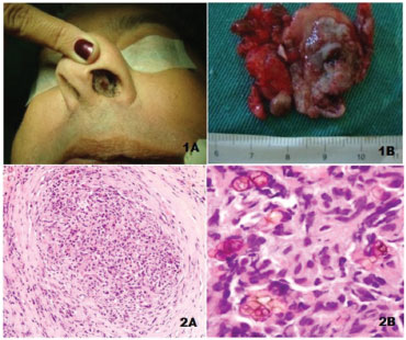
Figure 1 Clinical image of the septal lesion (1A).
Image published on/in:2014 Vol.: 80 Ed.: 1 - 16º
1
Description: Figure 1 Clinical image of the septal lesion (1A). Lesion removed with mucosal segment (1B). HE, small increase (2A). Chronic granulomatous inflammatory process with rounded and brownish fungi, some with septa. HE, large increase (2B).
Author(s) of the original article: Lídio Granato1; Ísis Rocha Dias Gonçalves1; Tomás Zecchini Barrese2; Carlos Kayoshi Takara1
Title and link to the article:
Primary chromohifomycosis of the nasal septum
oldfiles.bjorl.org/conteudo/acervo/acervo_english.asp?id=4555
Print: 
All rights reserved - 1933 /
2026
© - Associação Brasileira de Otorrinolaringologia e Cirurgia Cérvico Facial