

Image Details
V.77 Ed.4  |
V.77 Ed.4  |
V.77 Ed.4  |
V.77 Ed.4 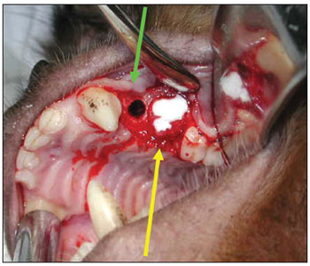 |
V.77 Ed.4  |
V.77 Ed.4  |
Image Code: 3532
Picture of a left nasal fossa
Image published on/in:2012 Vol.: 78 Ed.: 4 - 9º
4
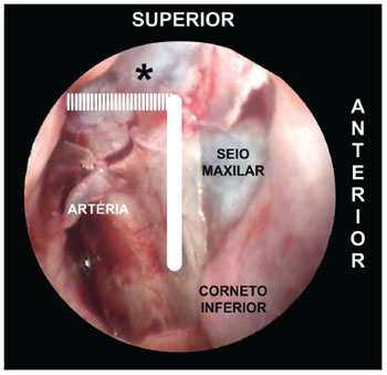
Description: Figure 4. Picture of a left nasal fossa showing details of the "sphenopalatine quadrangle." In the superior border, the tip to find the accessory foramens is the fat tissue from the pterygopalatine fossa (*). The perpendicular portion of the palatine bone is the anterior border and the starting point from which the mucoperiosteal flap is produced to find the sphenopalatine foramen.
Author(s) of the original article: Gustavo Lara Rezende1; Vitor Yamashiro Rocha Soares2; Waldete Cabral Moraes3; Carlos Augusto Costa Pires de Oliveira4; Márcio Nakanishi5
Title and link to the article:
The sphenopalatine artery: a surgical challenge in epistaxis
oldfiles.bjorl.org/conteudo/acervo/acervo_english.asp?id=4322