
Image Details
![]() Home ImageBank
Home ImageBank
![]() Search Images
Search Images
![]() New Images
New Images
![]() Home BJORL
Home BJORL
![]()
Image Code: 3674
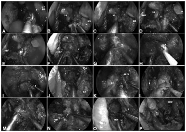
Figure 4. A: Malignant clival tumor extending laterally beyond the paraclival ICA; therefore, requiring endoscopic transpterygoid.
Image published on/in: 2013 Vol.: 79 Ed.: 6 - 18º
Description: Figure 4. A: Malignant clival tumor extending laterally beyond the paraclival ICA; therefore, requiring endoscopic transpterygoid. After a total ethmoidectomy and bilateral sphenoidotomies, a medial maxillectomy is completed to access the pterygoid process; B: The sphenopalatine artery is coagulated before removing the posterior wall of maxillary sinus; C: The greater palatine canal opened and the descending palatine artery and the greater palatine nerve are freed. Sequentially, the vidian nerve is transected to allow the lateral displacement of the soft tissue contents of the pterygopalatine fossa; D: After adequate exposure of anterior aspect of the pterygoid process; E-F: The floor of the sphenoid sinus is drilled flush with the level of the clival recess; G-H: Subsequently, the vidian canal is dissected and the ICA position is confirmed with nasal acoustic Doppler sonography; I-J: Removal bony of the pterygoid base enhances the exposure for tumor removal from the clivus; K-M: The medial pterygoid plate and Eustachian tube are removed; N-O: Allowing a total resection of the clival tumor with adequate margins; P: Finally, the HBF is placed to cover the defect and ICA. MS: Maxillary sinus; SS: Sphenoid sinus; HBF: Hadad-Bassagaisteguy nasoseptal flap; V: Vidian nerve; ICA: Internal carotid artery; M. Pt: Medial pterygoid artery; T: Tumor; ET: Eustachian tube.
Author(s) of the original article: Pornthep Kasemsiri; Daniel Monte Serrat Prevedello; Bradley Alan Otto; Matthew Old; Leo Ditzel Filho; Amin Bardai Kassam; Ricardo Luis Carrau
Title and link to the article: Endoscopic Endonasal endonasal technique: treatment of paranasal and anterior skull base malignancies
oldfiles.bjorl.org/conteudo/acervo/acervo_english.asp?id=4531
