

Image Details
![]() Home ImageBank
Home ImageBank
![]() Search Images
Search Images
![]() New Images
New Images
![]() Home BJORL
Home BJORL
![]()
Image Code: 3673
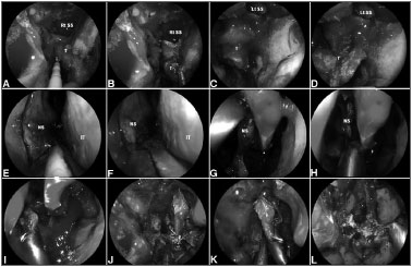
Figure 3. A-D: Intraoperative photograph demonstrating tumor at superior wall of nasopharynx.
Image published on/in: 2013 Vol.: 79 Ed.: 6 - 18º
3
Description: Figure 3. A-D: Intraoperative photograph demonstrating tumor at superior wall of nasopharynx. An incision is made just above the torus tubarius A allowing a subperiosteal dissection of the basisphenoid and clivus; E-K: A posterior septectomy and sphenoidotomies are performed; and the sphenoid sinus intersinus septa are removed; L: Finally, histological analysis is required to confirm the negative surgical margins. Rt. SS: Right sphenoid sinus; Lt. SS: Left sphenoid sinus; NS: Nasal septum; IT: Inferior turbinate.
Author(s) of the original article: Pornthep Kasemsiri; Daniel Monte Serrat Prevedello; Bradley Alan Otto; Matthew Old; Leo Ditzel Filho; Amin Bardai Kassam; Ricardo Luis Carrau
Title and link to the article: Endoscopic Endonasal endonasal technique: treatment of paranasal and anterior skull base malignancies
oldfiles.bjorl.org/conteudo/acervo/acervo_english.asp?id=4531