

Image Details
![]() Home ImageBank
Home ImageBank
![]() Search Images
Search Images
![]() New Images
New Images
![]() Home BJORL
Home BJORL
![]()
Image Code: 3584
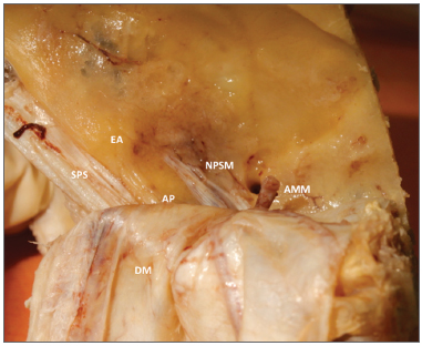
Figure 2. Anatomy of the middle cranial fossa viewed perpendicularly from the petrous.
Image published on/in: 2013 Vol.: 79 Ed.: 2 - 5º
2
Description: Figure 2. Anatomy of the middle cranial fossa viewed perpendicularly from the petrous. AE: Arcuate eminence; SPS: Superior petrosal sinus; GSPN: Greater superficial petrosal nerve; PA: Petrous apex. DM: Dura mater of the middle cranial fossa; MMA: Middle meningeal artery.
Author(s) of the original article: Aline Gomes Bittencourt1; Robinson Koji Tsuji2; João Paulo Ratto Tempestini3; Alfredo Luiz Jacomo4; Ricardo Ferreira Bento5; Rubens de Brito6
Title and link to the article: Cochlear implantation through the middle cranial fossa: a novel approach to access the basal turn of the cochlea
oldfiles.bjorl.org/conteudo/acervo/acervo_english.asp?id=4419