

Image Details
![]() Home ImageBank
Home ImageBank
![]() Search Images
Search Images
![]() New Images
New Images
![]() Home BJORL
Home BJORL
![]()
Image Code: 3574
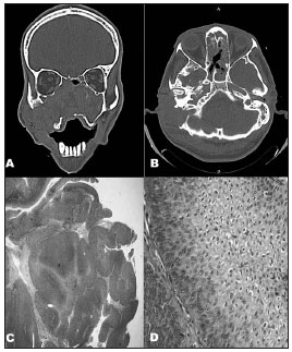
A and B: CT scans of the nose, paranasal sinuses, and mastoid
Image published on/in: 2012 Vol.: 78 Ed.: 6 - 20º
1
Description: Figure 1. A and B: CT scans of the nose, paranasal sinuses, and mastoid: tumor-like lesion with soft tissue attenuation invading the nasal fossae, the maxillary, ethmoid, and sphenoid sinuses, the rhinopharynx, the right medial orbit wall, and the ipsilateral mastoid, accompanied by massive lysis of the adjacent bone structures. C: Microscope image showing an IP: papillomatous proliferation of the squamous epithelium with endophytic growth pattern (40x - HE). D: Microscope image revealing endophytic projection of the squamous epithelium with preserved cell architecture containing various koilocytes (400x - HE).
Author(s) of the original article: Jônatas Lopes Barbosa1; Sebastião Diógenes Pinheiro2; Marcos Rabelo de Freitas3; André Alencar Araripe Nunes4; Elias Bezerra Leite5
Title and link to the article: Sinonasal inverted papilloma involving the middle ear and the mastoid
oldfiles.bjorl.org/conteudo/acervo/acervo_english.asp?id=4388