
Year: 2003 Vol. 69 Ed. 6 - (14º)
Artigo Original
Pages: 819 to 823
Pulmonary hipertension in patients with adenotonsillar hypertrophy
Author(s):
Bruno Bartolomei Sebusiani1,
Shirley Pignatari2,
Giuseppe Armínio3,
Levon Mekhitarian Neto4,
Aldo E. Cassol Stamm5
Keywords: pulmonary hypertension, adenotonsillar hypertrophy, cor pulmonale
Abstract:
Although adenotonsillar hypertrophy is a well-recognized disease since the beginning, it was not described to be cause of pulmonary hypertension until 1965 by Menashe and Ferrehi. There are only few records in the literature, analyzing the existence of pulmonary hypertension in children with obstructive adenotonsillar hypertrophy. Aim: The aim of this study was to evaluate the prevalence of pulmonary hypertension in children with obstructive adenotonsillar hypertrophy. Study design: Transversal cohorte. Material and Method: Fourteen patients with indication for adenotonsillectomy, from both sexs, with age ranging from O to 15 years, were analyzed in this study. AlI patients underwent to a complete pre-surgery evaluation, with questionary, complete ENT evaluation, lateral X ray films, nasofibrolaryngoscopy and echocardiography. Results: From all fourteen patients analyzed, one (7,14%) had pulmonary hypertension in the echocardiography examination. Conclusion: Adenotonsillar hypertrophy is a cause of pulmonary hypertension, and echocardiography examination is a very usefull exam to determine to presence of the pathology. Adenotonsillectomy may revert pulmonary hypertension in children with adenotonsillar hypertrophy.
![]()
Introduction
All physicians who dedicate to seeing children have already wondered about the reasons for indicating of adenotonsillectomy. Many complications are known to be the direct result of obstruction caused by adenotonsillar hypertrophy, such as for example oral breathing, cranial-facial developmental disorders, sleep obstructive apnea, difficulty to eat, among others, being all of them possible conditions for surgical indication. Among the most severe complications resultant from the chronic obstructive process caused by exaggerated increase in size of adenoid vegetation and palatine tonsils, we can include pulmonary hypertension and cor pulmonale. Even though we are not absolutely sure about prevalence, this clinical condition is considered an undisputable condition for performance of adenotonsillectomy.
Hindi records from about 3,000 years ago reported a surgical act that would have been described as a tonsillectomy. The procedure involved detachment of the mucosa capsule with the finger and enucleation of the palatine tonsil. Celsius and Paulo de Algena also used to perform a similar procedure approximately 2,000 years ago. It is believed that the first adenoidectomy was performed at the end of the 19th century, when Wilhem Meyer, in Copenhagen, suggested that adenoid vegetations would be responsible for hearing loss as well as other nasal symptoms in many patients.
Since adenotonsillectomy is a procedure with low morbidity/mortality, there was much enthusiasm about the performance of the surgery, especially in the first half of the 20th century. It used to be indicated in many patients with minimum symptomatology or those who had diseases not related to palatine tonsils or adenoid vegetation. In the 70's, in the USA, there were proportionally 1,200 adenotonsillectomy for each 100,000 general surgical interventions 1. The next decades, however, were marked by skepticism concerning the indication of adenotonsillectomy and we observed a reduction of approximately 30% in the number of surgeries performed 1.
Despite the significant reduction, adenotonsillectomy is still the most frequently performed surgical procedure in the pediatric age group 1. Chronic obstruction of the airways, followed by pulmonary hypertension and cor pulmonale is an undisputable indication for the performance of the surgical procedure.
The purpose of the present study was to assess the prevalence of pulmonary hypertension in patients with adenotonsillar hypertrophy.
Material AND METHOD
This prospective study was conducted at the Department of Otorhinolaryngology (Pediatric Otorhinolaryngology) and Echocardiography, Hospital Professor Edmundo Vasconcelos, in the period between March and October 2002. It included 14 patients aged below 15 years, both genders, with formal indication of adenotonsillectomy by adenotonsillar hypertrophy. It excluded patients with cranial-encephalic malformations, genetic syndromes, blood dyscrasias, and other adenotonsillar indications rather than adenotonsillar hypertrophy. All patients had legal representatives to sign the informed consent term including clinical-surgical procedures.
They all underwent preoperative assessment including questionnaire, clinical ENT assessment, paranasal sinuses simple x-ray, videonasofibroscopy and echocardiography.
A questionnaire containing related questions to the obstruction complaints presented by the patient was answered by the legal representative of the patients before the surgery, aiming at estimating the impact of adenotonsillar hypertrophy on the patients' quality of life. In case of positive complaint, it was later classified concerning severity (mild, moderate or severe).
During ENT examination, we could assess the size of palatine tonsils. The tonsils were then classified in 4 different grades, depending on the degree of obstruction proportional to the level of the oropharynx. We defined grade I obstruction as tonsil obstruction of up to 25% of oropharynx; grade II, as obstruction of 25-50%; grade III, obstruction of 50-75%, and grade IV, obstruction greater than 75% of oropharynx lumen.
Adenoid hypertrophy was also assessed by simple paranasal sinuses x-ray with videonasofibroscopy. These variables were also classified into four different levels, depending on the obstruction shown by the examination. The same criteria used for assessing the degree of obstruction of the tonsillar hypertrophy were employed in the assessment of adenoid increase. The degree of adenoid hypertrophy evidenced at videonasofibroscopy was later correlated with the obstruction provided by the adenoid vegetation at the simple paranasal sinuses x-ray. Videonasofibroscopy was conducted with flexible endoscope (Machida 3.2 mm®), video camera (Toshiba®), videocassette (Sony®), video monitor, light source (Storz®) and image capture system (Laudo & Imagem); after use of nasal topical vasoconstrictor (nafazoline chloridrate) and anesthetic spray (neotutocaine 5%).
Echocardiography was performed by the division of Echocardiography of the hospital with echocardiograph model HP ImagePoint HX®. During the procedure, we could assess the presence of suggestive signs of pulmonary hypertension, through the estimate of pulmonary artery systolic pressure (tricuspid reflux and/or pulmonary regurgitation). Patients with pulmonary hypertension evidenced by preoperative echocardiography were submitted to a new exam on the 7th postoperative day. All tests were conducted by the same examiner.
All patients were submitted to adenotonsillectomy in horizontal supine position under general anesthesia and orotracheal intubation. Adenoidectomy was conducted by conventional technique, with Beckman curettes of three different sizes. We used the simple technique of submucous dissection to conduct tonsillectomy. All patients were operated on by the same surgeon.
As to statistical analysis, the calculation of correlation coefficient between both variables was selected.
Results
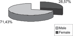
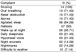
There were ten (71.43%) female patients and 4 (28.57%) male patients (Graph 1). The mean age of patients was 6.78 years, being that the youngest was 2 years and the oldest, 14 years. Snoring was the most common complaint, being reported by all accompanying people (100%) and considered severe for 64.28% of them. Oral breathing (92.86%) was the second symptoms in frequency observed in the questionnaire. Apnea and nasal obstruction were reported in ten patients (71.43%). The results obtained from the questionnaire are reported in Table 1.
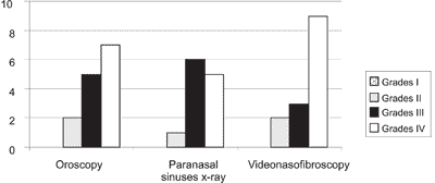
The analysis of grade of palatine tonsil through the ENT examination demonstrated seven (50%) children with tonsil hypertrophy grade IV; five (35.71%) grade III, and two (14.29%) grade II. There were no patients with obstruction below 25% at the oropharynx level.
Out of 14 patients included in the study, 12 (85.71%) were submitted to paranasal sinuses x-rays. Five patients (41.67%) presented adenoid obstruction grade IV, six (50%) had grade III and one patient (8.33%) had grade II. None of the patients were classified as having adenoid hypertrophy grade I, that is, obstruction below 25% of rhinopharynx lumen.
All 14 patients were submitted to assessment of the size of adenoid vegetation with videonasofibroscopy. Nine (64.28%) presented obstruction provided by adenoid vegetation greater than 75% of the lumen at the rhinopharynx level; three (21.43%) had obstruction between 50-70% of the lumen and two (14.92%) had obstruction between 25-50% (Graph 2).
One case (7.14%) had affection in the echocardiography evidencing signs of pulmonary hypertension. It was detected grade IV obstruction in this patient, both of palatine tonsils and adenoids. After seven days postoperatively, we detected regression of echocardiography signs suggestive of pulmonary hypertension.
Graph 1. Distribution of patients by gender.
Graph 2. Distribution of patients by degree of tonsil obstruction evidenced at oroscopy and degree of adenoid obstruction observed in simple paranasal sinuses Rx and videonasofibroscopy.
Table 1. Frequency of symptoms observed in the preoperative questionnaire answered by the legal representatives of the patients.
Discussion
Despite the fact that adenotonsillar hypertrophy has being a clinical condition recognized since ancient times, it was only described as having caused pulmonary hypertension and cor pulmonale in 1965 by Menashe and Farrehi2-5. Subsequent authors (Noonan, 1965; Luke et al., 1966) also described pulmonary hypertension originated from excessive increase of palatine tonsils and adenoid vegetation. Goodman believed that the delay in documenting pulmonary hypertension resultant from adenotonsillar hypertrophy had two main reasons: the great number of adenotonsillectomy conducted in the past and diagnostic inability 6.
Adenotonsillar hypertrophy is the main cause of chronic obstruction in the upper airways of children, especially those aged between 2-6 years, an age range characterized by increase of lymphoid tissue and subsequent narrowing of the air column. The mean age of our patients was 6.78 years, compatible with the age range presented in the literature for the largest size of Waldeyer's lymphatic ring.
Considering the exuberant symptomatology presented by children with adenotonsillar hypertrophy, snoring is the great motivator to search for a specialist (ENT or pediatrician) being the main concern from parents' perspective 6. However, other complaints are observed in these patients, such as obstructive apnea, day sleepiness, night enuresis, agitated sleep, night awakening, poor school performance, reduction of cognitive functions (attention and memory), nightmares, irritability, among others, including pulmonary hypertension and cor pulmonale. In our study, snoring (100%) was the most common complaint reported by the parents of these children. However, we did not observe proportional relation between severity of symptoms and grade of adenotonsillar hypertrophy. Oral breathing, obstructive apnea and nasal obstruction were also frequent complaints in patients with adenotonsillar hypertrophy.
Despite the occurrence of disagreement between the indications and contraindications for adenotonsillectomy, it seems reasonable to have indication for the surgical procedure in patients who have adenotonsillar hypertrophy, especially those classified as grade III and IV. In our study, 12 (85.71%) patients presented tonsil hypertrophy grades III or IV, and we did not find any patient with tonsil hypertrophy grade I. Similarly, no patients presented grade I of adenoid obstruction evidenced by paranasal sinuses x-ray or videonasofibroscopy. There was positive correlation between grade of tonsil obstruction evidenced at oroscopy and grade of adenoid hypertrophy observed at videonasofibroscopy. This positive correlation is probably due to uniform and proportional growth of Waldeyer's ring lymphoid tissue. We also observed correlation between obstruction promoted by adenoid vegetations in the x-ray and videonasofibroscopy.
Pathophysiology of pulmonary hypertension triggered by adenotonsillar hypertrophy is fundamentally based on chronic obstruction of the airways by an exaggerated increase of lymphoid mass, promoting narrowing of the air column. Therefore, there is pulmonary hypoventilation and consequent retention of CO2 (increase of PaCO2) and hypoxemia (reduction of PaO2). Retention of CO2 leads to pulmonary bronchoconstriction and increases respiratory distress. At the same time, hypoxemia reduces pulmonary vasoconstriction and, as a consequence, increases pulmonary artery pressure, characterizing pulmonary hypertension. As demonstrated by other authors 2-5, we could confirm that pulmonary hypertension is based on pathophysiology of adenotonsillar hypertrophy. Thus, we believe that in patients with significant adenotonsillar hypertrophy and exuberant symptomatology, echocardiography can be useful in determining pulmonary extension, especially because it is a safe, practical and non-invasive exam 8.
The prevalence of pulmonary hypertension in patients with adenotonsillar hypertrophy observed in our study was of 7.14%, being that the situation was completely reverted after adenotonsillectomy, as evidenced by postoperative echocardiography. Therefore, we believe that the continuation of this study would be essential, since despite the small sample, we could confirm the existence of this clinical condition in children seen in our hospital.
Conclusion
Based on our study, we concluded that adenotonsillar hypertrophy is also a cause of pulmonary hypertension; echocardiography is a very useful exam in determining the pathology, especially because it is a safe, practical and non-invasive exam.
Therefore, school-aged children who present exuberant obstructive symptomatology (snoring, oral breathing, apnea and nasal obstruction) and adenotonsillar hypertrophy have potential indication for adenotonsillectomy, which can revert and occasionally prevent the onset of pulmonary hypertension resultant from chronic obstructive processes triggered by exaggerated size of the adenotonsillar tissue.
REFERENCES
1. Casselbrant M. Atualização nas indicações e contra-indicações das adenotonsilectomias. In: Sih T. Infectologia em Otorrinopediatria. Rio de Janeiro: Eds. Revinter; 2001. p. 63-7.
2. Capellari L, Guerra Jr G, Endo LH. Hipertrofia adenoamigdaliana e cor pulmonale. F méd (BR) 1990; 101(3):171-4.
3. Levin DL, Muster AL, Pachman LM, Wessel HU, Paul MH, Koshaba J. Cor pulmonale secondary to upper airway obstruction. Chest 1975; 68(2):166-70.
4. Ainger LE. Large tonsils and adenoids in small children with cor pulmonale. Brit Heart J 1968; 30:356-62.
5. Endo LH, Nicola EMD, Caldato MF. Cor pulmonale por hipertrofia de amígdalas e das adenóides - fatores predisponentes. Rev Bras Otorrinolaringologia 1981; 47:83-90.
6. Elsherif I, Kaesemullah C. Tonsil and adenoid surgery for upper airway obstruction in children. ENT J 1999; 78(8):617-20.
7. Goodman RS, Goodman M. Cardiac and pulmonary failure secondary to adenotonsillar hypertrophy. Laryngoscope 1976; 86(9):1367-74.
8. Miman MC, Kirazli T, Ozyurek R. Doppler echocardiography in adenotonsillar hypertrophy. Int J Pediatr Otorhinolaryngol 2000; 54(1):21-6.
9. Brodsky L, Koch J. Anatomic correlates ofnormal and diseased adenoids in children. Laryngoscope 1992; 102:1268-74.
10. Brouilette R, Hanson D, David R, Klemla L, Szatkowski A, Fembach S, Hunt C. A diagnostic approach to suspected obstructive sleep apnea in children. J Pediatr 1984; 105:10-4.
11. Piccirillo JF, Thawley SE. Sleep-disordered breathing. In: Cummings CW, Frederickson JM, Harker LA, Krause CJ, Richardson MA, Schuller DE, eds. St. Louis Missouri USA: Mosby-Year Book Inc; 1998. p. 1546-62.
1 Resident Physician in Otorhinolaryngology, Hospital Prof. Edmundo Vasconcelos - Centro de Otorrinolaringologia e Fonoaudiologia de Sao Paulo.
2 Otorhinolaryngologist, responsible for the Division of Pediatric Otorhinolaryngology, Hosp. Prof. Edmundo Vasconcelos - Centro de
Otorrinolaringologia e Fonoaudiologia de Sao Paulo; Joint Professor, Discipline of Pediatric Otorhinolaryngology, Federal University of Sao Paulo.
3 Cardiologist, responsible for the division of Echocardiography, Hospital Prof. Edmundo Vasconcelos.
4 Otorhinolaryngologist, Hosp. Prof. Edmundo Vasconcelos - Centro de Otorrinolaringologia e Fonoaudiologia de Sao Paulo;
post-graduation in Head and Neck Surgery, Hospital Heliópolis.
5 Director of the division of Otorhinolaryngology, Hosp. Prof. Edmundo Vasconcelos - Centro de Otorrinolaringologia e Fonoaudiologia de Sao Paulo.
Address correspondence to: Centro de Otorrinolaringologia e Fonoaudiologia de Sao Paulo - Hospital Prof. Edmundo Vasconcelos -
Rua Borges Lagoa, 1450 Vila Clementino 04038-905 Sao Paulo SP.
Tel (55 11) 5080-4357 - E-mail: centrodeorl@osite.com.br
Article submitted on August 11, 2003. Article accepted on September 04, 2003.


