
Year: 2003 Vol. 69 Ed. 5 - (5º)
Artigo Original
Pages: 612 to 620
Comparison of turbinectomy techniques with CO2 laser and diode laser
Author(s):
Pedro Paulo Vivacqua da Cunha Cintra1,
Wilma T. Anselmo Lima2
Keywords: Turbinectomy, laser, nose
Abstract:
We describe an evaluation of the techniques aiming to reduce the bulk of inferior turbinates with the diode laser and the CO2 laser, as well as a comparison between the techniques. Study design: Longitudinal Cohort. Material and Method: Sixty patients with turbinate hypertrophy were submitted to the submucous diode laser reduction and CO2 with a follow up period of 6 months following a established protocol concerning pain, bleeding and subjective evaluation of the nasal obstruction preoperatively and postoperatively. Results: The procedures showed a low level of bleeding and no packing was necessary in all patients. We observed that the patients were very pleased concerning the nasal obstruction postoperatively. Conclusion: The primary goal of the inferior turbinectomy is to reduce the patient complain of nasal obstruction with less discomfort, morbidity and complications during and after the procedure. The laser reduction showed in this study good results in a 6-month follow up. Further studies must be done to evaluate the results in a longer follow up.
![]()
INTRODUCTION
Nasal conchae are lateral nasal wall structures that start to be embryologically formed between 38th and 40th days of gestation 1.
Anatomically, the inferior nasal conchae are projections of the lateral nasal wall to the interior of the nasal cavity distributed in the anterior-posterior direction, one per each nasal fossa. They are connected to the lateral nasal wall by a bone called inferior concha bone that is attached to the lachrymal, maxillary, ethmoid and palatine bones, recovered by erectile vascular tissue that is covered by ciliated cylindrical pseudostratified epithelium, being that the most posterior portion of them has practically only soft tissue.
As to physiology, nasal conchae are important because they help with moisturizing, heating and air filtration, regulating the inspired airflow, because its capacity to increase and decrease the volume, controlled by the autonomous nervous system, contributes to the nasal cycle that normally lasts between 3e and 4 hours. Thus, we always have an alternation of air input in the nasal fossae. In physiological conditions, this cycle is not perceived by the subject, unless in pathological conditions.
The main diseases that affect the nasal concha causing the onset of chronic rhinitis, many time irreversible with clinical treatment, are:
1. Allergic rhinitis
2. Idiopathic rhinitis
3. Drug-induced rhinitis
4. Other causes
5. Hypertrophic chronic rhinitis: term that is not reported in the international literature. Carlini2 defined it as "Nasal obstruction for at least 3 months that is intermittent or continuous and associated with frequent sneezing and/or nasal pruritus and/or coriza and/or oral breathing and/or supplement oral breathing and/or snoring during sleep". To this definition we would like to add that it is also refractory to any type of clinical treatment.
The treatment of nasal conchae should take into account the improvement of nasal fossa permeability, minimizing the correlated complaints such as rhinorrhea, nasal dryness and pruritus, so that results are the most long-lasting possible with low morbidity and complication rates.
Clinical treatment should be the first to be adopted aiming at improving the underlying disease and it is normally based on anti-histaminic, anticholinergic, nasal decongestants, dissodic chromoglycate, topical corticoids, expectorants and secretion flowable medication, topical or systemic drugs. It is important to point out that we should not forget about environmental prophylaxis against allergens and irritants and the cares that must be taken with the environment, such as temperature and moisture levels.
Surgical treatment of nasal conchae has been indicated when nasal obstruction remains, despite all traditional clinical treatments. Many forms of procedures have been described to reduce the volume of inferior nasal conchae.
They are:
a) Procedures that involve the injection of intraconchal substances:
Corticoid injections, sclerosing substance injections;
b) Mechanical procedures:
Concha fracture or subluxation;
c) Procedures that reduce the parenchyma:
Electrocauterization, cryosurgery;
d) Resection procedures:
Partial resections, total resections, submucous resection;
e) Neural resections.
Laser surgery can be classified as a procedure that causes reduction of the parenchyma.
Laser surgery of inferior nasal conchae was first described in 1977 by Lenz et al.3,4 in one of his articles. In 1982 Mittelman5 reported the first results described in the North American literature.
The word "LASER" is used as the abbreviation of Light Amplification Stimulated Emission of Radiation". The theory was first described by Albert Einstein in 1900, but only in 1958 gas laser was described by Arthur Shalow & Charles Townes6 .
So that the laser is properly used, it has to be absorbed by the tissue, because the absorbed energy will be transformed into thermal energy: this process of energy transformation is known as photothermal effect.
The most commonly used lasers in the nasal cavity are Ho: YAG, Nd: YAG, CO2, KTP/532 and Diode. They vary depending on wavelength and, as a consequence, the tissue that is going to absorb the laser.
YAG and CO2 lasers present absorption peak close to that of water, acting better in tissues that have it, whereas KTP/532 and diode lasers have larger absorption peak waves in hemoglobulin and melanin.
Model Sharplan CO2 LASER®, a class IV laser designed to apply infrared energy with wavelength of 10600 micrometers leads to wave absorption peak in the water. It is excellent to vaporize soft tissues. This level of absorption makes the laser capable of coagulating blood vessels of up to 0.5mm in hemostasis. Its grade of tissue penetration is small and more secondary to heat dissipation7.
Model DIOMED 60 Laser® is a class IV laser that produces a beam of laser based on gallium arsenid chip, a semiconductor material. Designed to deliver infrared energy with wavelength of 805 micrometers, it gives absorption wave peak in hemoglobulin and melanin and this laser has tissue penetration of coagulation of about 5-6mm8. Preliminary studies showed the thermal necrosis area of the laser diode are 0.5mm 8, 9, 10.
The objectives of the present study were:
1. To assess the advantages and disadvantages of the surgery of the inferior nasal concha with CO2 laser.
2. To assess the advantages and disadvantages of the surgery of the inferior nasal concha with diode laser.
3. To compare both techniques.
4. To assess the level of satisfaction of the patients.
MATERIALS AND METHODS
Sixty patients with refractory hypertrophic chronic rhinitis were diagnosed based on anamnesis with a pre-defined questionnaire, physical examination and radiological examination and selected based on the following criteria:
1. Nasal obstruction resistant to clinical treatment;
2. Parenchymatous hypertrophy of inferior nasal concha;
3. Absence of other inflammatory and/or infectious concomitant diseases that could collaborate with the nasal obstruction.
After signing the informed consent term, they were divided into 2 groups:
Group 1 - Turbinectomy with laser diode (30);
Group 2 - Turbinectomy with CO2 laser (30).
In group 1, the procedure was interstitial turbinectomy with laser diode brand Diomed 60. It consisted of introducing 400 or 600-micrometer fibers of quartz recovered with TeflonR associated with a tip inside the inferior nasal concha with application of 8W energy in continuous mode, under local anesthesia associated or not with sedation, in the head, body and caudal portion of the nasal concha, causing necrosis of interstitial coagulation and consequent volume reduction without external mucosa damage, except for the regions of laser fiber penetration.
In group 2, the procedure was performed with CO2 laser vaporization, brand Sharplan with Flexilase®, 10W power at the level of the head and medium third or Accuspot® coupled to laser with surgical microscope associated with Swiflase® with 18 to 20 W continuous power. In this group, the laser was applied to the regions of the head, body and caudal portion of the concha surface by making mucosa vaporization.
All procedures were performed in the appropriate room for laser procedures, which included a warning sign outside the room, the use of protective goggles for everyone in the room and maintenance of the device in stand-alone mode between applications.
In all patients, anesthesia was local with lidocaine 20% spray and lidocaine 2% infiltrated 1-2ml in the concha with Carpule syringe and gingival needles 27 to 30mm associated or not with sedation. When sedation was administered, we used midazolam 1mg/Kg and fentanyl 0.1microgram/Kg.
In all patients we used follow-up questionnaires to assess preoperative and postoperative nasal obstruction on 5, 10, 30, 60 and 90 days and 6 months after the initial control, compared to initial and intraoperative pain. A bleeding assessment by a medical professional was conducted during the procedure and the energy used.
All patients were operated on and clinically followed up postoperatively by the author, associated or not with an immune allergist.
The patients' ages ranged from 6 to 72 years, mean age of 28 years with no gender predominance.
We assessed the following items:
1. Surgical time: the time each procedure lasted was kept by an assistant during the procedure.
2. Used energy: calculated by the laser device; intensity in Joules was provided during the procedure, in case of laser diode.
3. Pain during the procedure: the pain scale was graduated from 0 to 10, being 0 absent, between 1 and 3 mild, between 4 and 6 moderate, between 7 and 9 severe, and 10 as unbearable.
4. Bleeding was assessed by the assistant physician and classified into 4 levels (absent, mild, moderate and severe) according to the measures of blood in the early postoperative vial. Up to 20ml, level 1, from 20 to 45ml, level 2, from 45 to 80ml, level 4 and above 80ml, level 4.
5. As to level of nasal obstruction, the patient was submitted to a questionnaire in which we included their opinion about unilateral and bilateral nasal obstruction. The level of obstruction was divided into 6 levels: 1- no obstruction; 2 - 0-25% (mild obstruction); 3 - 25 to 50%; 4 - 50 to 75%; 5 - 75 to 100% (almost complete obstruction), and 6 - complete obstruction. This questionnaire was repeated 5, 10, 30, 60, 90 days and 6 months after the surgery.
The parameters of pain, bleeding time and nasal obstruction were statistically assessed by the non-parametric test of Mann-Whitney.
Based on the test, we scored the concepts so as to form a scale. For example: absent = 0, mild = 1, moderate = 2, severe = 3 and unbearable = 4; and absent = 0. 0-25% = 1, 25-50% = 2, 50-75% = 3, 75-100% = 4 and complete obstruction = 5) and we compared the values for each group, checking the presence of differences between the groups.Table 1. Statistical comparison of pain.
Table 2. Statistical comparison of bleeding.
Table 3. Statistical comparison of preoperative nasal obstruction.
Table 4. Statistical comparison of nasal obstruction day 5.
Table 5. Statistical comparison of nasal obstruction day 10.
Table 6. Statistical comparison of nasal obstruction day 30.
Table 7. Statistical comparison of nasal obstruction day 60.
Table 8. Statistical comparison of nasal obstruction day 90.
Table 9. Statistical comparison of nasal obstruction 6 months.
RESULTS
1. Surgical time:
a. Group 1: Time ranged from 7 to 20 minutes for the procedure, mean time of 10.3 minutes.
b. Group 2: Time ranged from 14 to 30 minutes, mean time of 22 minutes.
Laser diode compared to CO2 had shorter duration procedures.
2. Energy applied:
a. Group 1: Ranged from 352 J to 1200 J (mean of 513 J) divided in the regions of head and neck and caudal portion of the inferior concha.
b. Group 2: The laser we used did not allow quantification of Joules.
It was not possible to compare both devices, since CO2 laser did not provide quantification the applied power.
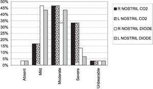
3. Pain during the procedure: When we compared both techniques we realized that pain ranges during the procedure varied from moderate to severe being that CO2 laser presents moderate to severe range of pain, whereas in diode laser predominates mild pain.
Statistically speaking, the comparison between CO2 and diode laser resulted in statistically significant differences favorable to diode laser (p=0.004) (Table 1).
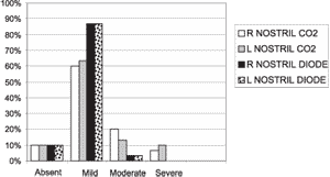
4. Bleeding: Laser diode presented lower bleeding rate, as provided by Graph 1. In both types, intraoperative bleeding was mild, but the CO2 group presented a larger number of patients with moderate and severe bleeding.
We found statistically significant difference favorable to diode laser with p=0.05 (Table 2).
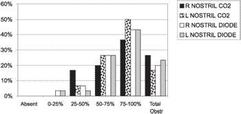
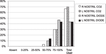
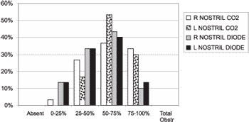
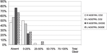
5. Nasal obstruction: In Graphs 2 and 3 we can see the worsening of nasal obstruction. Graph 4 shows the comparisons between the 10th day with predominant improvement of obstruction of 75% of total obstruction to a predominant range between 25 and 100%. No patients presented complete obstruction.
The comparison of preoperative and 5th day postoperative nasal obstruction between the methods did not show statistically significant differences with p = 0.96 (Table 3) and p = 0.07 (Table 4), respectively.
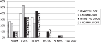
Between day 10 and 30, there was a deviation of the obstruction range to absent to 50%. We detected a greater evolution in reduction of the obstruction in the group of laser CO2 (Graphs 5 and 6).
As to statistical analysis, we detected that on day 10 there was p = 0.02 and CO2 group had nasal obstruction levels greater than the diode laser group (Table 5).
On day 30, we also found statistically significant difference with p = 0.02 but in this case the CO2 group presented obstruction means significantly lower than the diode laser (Table 6).
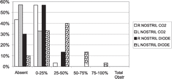
On day 60, it was possible to notice the curve of the patients in group CO2, since it was close to the results of six months whereas the diode laser presented slower evolution with greater percentage of patients between 25% and 100% of obstruction (Graph 7).
There were no statistically significant differences with p = 0.09 (Table 7).
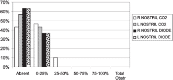
After 90 days, the results with both groups were close to absent to 25% in all patients, except for one nostril of a patient in group CO2 (Graph 8). We did not find statistically significant differences with p = 0.22 (Table 8).
At 6-mohth postoperative, some patients presented worsening of the symptoms but similarly in both groups (Graph 9). We did not find statistically significant differences with p = 0.13 (Table 9).
Graph 1. Pain scale - comparing both techniques
Graph 2. Bleeding scale - comparing both techniques
Graph 3. Preoperative nasal obstruction - comparing both techniques
Graph 4. Nasal obstruction day 5 - comparing both techniques
Graph 5. Nasal obstruction day 10 - comparing both techniques
Graph 6. Nasal obstruction day 30 - comparing both techniques
Graph 7. Nasal obstruction day 60 - comparing both techniques
Graph 8. Nasal obstruction day 90
Graph 9. Nasal obstruction six months - comparing both techniques
DISCUSSION
Laser is one more instrument that can help in surgically treating inferior nasal concha hypertrophy. There are different types of laser and the most normally used ones are Ho: YAG, Nd: YAG, CO2, KTP/532 and Diode.
Lasers can be used in the inferior nasal concha in two different ways: the one that produces ablation of soft tissues of the concha, causing damage to the surface of the nasal concha for later scarring, or interstitial damage, such as linear cauterizations, in which the optical fiber is introduced in the nasal parenchyma and the power is applied intra-parenchyma, which does not cause external lesion, since there is only damage in the introduction site of the optical fiber.
This study compared interstitial technique with laser diode and mucosa ablation technique with laser CO2.
In the sample presented here the diode laser showed the following:
1. Surgical time: it is very reduced. Derowe et al.11 using an application technique over the mucosa of the inferior concha, reported the need of 17.76 minutes to make the surgery. In our study, the mean time was 10.3 minutes including the duration of anesthesia, making the procedure as comfortable as possible for outpatient application. The variation in the surgical time was due to: 1) learning curve, being that longer times were more common in the beginning of the procedures, reducing the time with the acquired experience, and the last procedures were faster; 2) type of patient and size of the nasal concha, the more energy required, the longer the surgical time.
2. Used energy: the variation depended on volume to be reduced, since larger conchae required more energy.
3. Intraoperative pain with diode laser ranged from mild (45%) to moderate (38%), and only one child presented unbearable pain. The procedure is effective to be performed as an outpatient procedure, even in the absence of sedation.
4. Intraoperative bleeding: we showed the classification adopted as absence (10%) or mild (87%), which amounted to 97% of the patients. Only in one nostril of one patient (3%) there was moderate bleeding. It is important to point out that the patients underwent the technique without previous administration of vasoconstriction.
5. As to nasal obstruction, the patients assigned to the group had initial classification over 50% of obstruction to complete obstruction in 91.5%, taking both nostrils into account. Six months after the surgery, 100% of the patients presented classification between absent and 25% obstruction in the right nostril and only 2 patients (6%) presented obstruction between 25 and 50% in the left nostril. Such patients, later analyzed, proved to be allergic and the postoperative control was not efficient.
Unfortunately, we could not compare our results with diode laser with the literature, since there were no similar studies reported.
The results with CO2 laser technique (group 2) led us to the following inferences:
1. Surgical time: Derowe et. al.11 in a study reported the mean time of 23.14 minutes with CO2 laser. In our study the time was 22 minutes, quite acceptable by the patients, especially because it included the anesthesia and the fact that the stopwatch was not stopped after the procedure started.
2. Used energy: the procedure was performed with powers 10W and 20W, which was also reported by Kawamura et. al.12 and Mc. Combe et.al.13.
3. Intraoperative pain was classified as moderate (47%) to severe (33%). When both nostrils were considered together, most of the patients presented pain classified as severe or unbearable. We did not find any literature report that addressed intraoperative pain. Upon reviewing the surgical descriptions, we realized that such patients were submitted only to local anesthesia, with no sedation support, which makes us realize that sedation is an important factor for the performance of the surgery, allowing that the procedure be conducted in the outpatient unit.
4. Bleeding: the levels of intraoperative bleeding found with ablation of mucosa with CO2 laser were mild (61.5%) to moderate (21.5%), which avoided the use of postoperative nasal packing, providing more comfort to patients. Derowe et al.11 reported postoperative bleeding in 3 patients in a series of 14 patients. In our series, we did not observe postoperative bleeding. Eight and a half percent of the patients presented bleeding classified as severe during the procedure, which was controlled with packing soaked in oxymetazolin removed before the discharge, if the patient had no posterior bleeding.
5. Nasal obstruction: the group assessed presented preoperative obstruction over 50% in 84% of the patients, considering both nostrils. After 6 months of follow-up, there was 98.5% of the patients with 25% of obstruction. Only one patient (1.5%), considering both nostrils, presented obstruction between 25 and 50%. Upon comparing the results of the literature, there is a study by Lippert & Werner14 in 112 patients with 2-year follow-up in which they were satisfied in 87.5% of the cases after 6 months, 82.1% within 1 year and 80.4% within 2 years; Kawamura et al.12 reported that in 72 patients studied over 2 years, the results were excellent or good in 85%. Fukutake et al.15 (1986) reported in patients followed up for one year 77% of improvement in 35 patients. Elwany & Harrison16 reported improvement in 77% of the patients. Derowe et al.11 reported improvement rate in 57 patients with mean follow-up of 15 months. We concluded that our results were better than expected based on the literature, but we followed them up for a period of 6 months. We would like to follow them up for a continuous period of 5 to 10 years.
Comparing both techniques, we compiled the following data:
a) Surgical time: The shortest surgical time was obtained with laser diode when compared to CO2 laser, which is mainly resultant from the way it is applied, since CO2 creates a carbonized surface over the bed of the application, hindering its action. The carbonized area does not have water, which is the main action element of CO2. Diode laser is applied interstitially and the main action element is blood, the main element in the inferior nasal concha.
b) Used energy: It was possible only to assess with laser diode, since CO2 does not allow quantification of energy employed.
c) Intraoperative pain: The statistical analysis showed that diode laser was superior to CO2 laser in this aspect. Probably the pain reduction with diode laser is correlated with longer duration of the procedure with CO2, which increases discomfort, plus the need to remove the carbonization crusts. Another factor that could explain the pain would be the need to have simultaneous introduction of CO2 fiber with endoscope through the nasal cavity.
d) Bleeding: The statistical analysis showed us that diode laser is more hemostatic than CO2, probably owing to its greater affinity with hemoglobin.
In both cases, it was not necessary to use postoperative packing, differently from conventional techniques in which packing is required for up to 6 days 17. This is an essential factor for the comfort of patients, reducing morbidity of the surgical procedure.
e) Nasal obstruction: it is important to assess this item in addition to the evolution of the obstruction throughout the postoperative days. Initial obstruction is more important considering the formation of fibrin crusts whose removal improve obstruction.
The presence of more obstruction statistically confirmed with CO2 laser was due to greater formation of crusts originated from the lesion of the nasal mucosa surface.
The faster nasal obstruction improvement after the 30th day with CO2 laser can be a result of the edema caused by the interstitial lesion that takes longer to be absorbed than the superficial lesion, in which the deep lesion is smaller. As months go by, the curves tend to balance up to six months.
The worsening that some patients had at 6 months with both types of lasers is probably owed to allergic or vasomotor pathology and to difficulties in making patients comply with the postoperative clinical treatment.
In the literature, there are few studied that compare the turbinectomy techniques, especially with laser. In 1990, Elwany & Harrison16 in a study comparing four techniques - partial turbinectomy, turbinectomy with CO2 laser, inferior turbinoplasty and cryoturbinectomy in 80 patients divided into 4 groups reported improvement with laser in 80% of the patients, improvement of 75% with inferior turbinectomy, 50% with inferior turbinoplasty and 45% with cryoturbinectomy. In the same study, the authors reported improvement of preoperative hyposmia in patients in the group submitted to laser and inferior turbinectomy, whereas there were no improvements in the groups submitted to turbinoplasty and cryoturbinectomy.
In 1992 Mc. Combe et.al.13, in a randomized prospective study, compared turbinectomy with laser with submucous cauterization conducted in 29 patients comparing the results in 3 days and 6 weeks, with greater improvement in 3 days in the laser group and in 6 weeks there were no significant changes. They concluded that the surgery with laser was a surgical alternative superior to submucous cauterization.
In 1998 Derowe et. al.11 in a randomized study with 46 patients divided into 3 groups submitted to surgery with Nd: YAG, diode and CO2 laser techniques, with endoscopic control and one-year follow-up reported improvement in 41% with diode on the mucosa surface, 47% with Nd: YAG and 57% with CO2, data that did not produce statistically significant differences.
Salam & Wengraf18, in a study comparing conchoantropexis on one side and total inferior turbinectomy on the other side did not find differences in the efficacy of nasal obstruction between both techniques.
Meredith19 in the study with 162 patients that responded to questionnaires sent 33 months after the procedure reported 86% of improvement after partial resection and 69% of improvement with electrocauterization associated with concha luxation.
Interstitial surgery with diode laser is a recent technique based on linear cauterization and the technique described by Li et. al. in 1998 for radiofrequency, using the advantages of laser, which according to this study, have similar results of CO2 laser technique already studied and analyzed by many different studies conducted.
Turbinectomy with CO2 laser in the study proved to be more painful than diode laser, therefore, requiring sedation as adjuvant to local anesthesia.
The main disadvantages of these techniques are the cost of the device and the interstitial technique and the fact that the results are still unknown in the long run.
It is important to point out that the procedures should be conducted by trained professionals, both physicians and the support team (nurses, support staff, scrub nurses), so that maximum protection is provided to the patient minimizing the risks of complications.
More studies should be conducted with objective and subjective comparison also in conventional techniques and with more prolonged follow-up to define whether the cost-benefit ratio for patients justify the investment in laser devices.
CONCLUSIONS
The surgery of inferior nasal concha through the laser technique presents a great advantage: minimum level of bleeding, which favors the procedure to be conducted at outpatient level, which does not require the use of postoperative packing, a great comfort to patients.
As to duration of surgery, pain scale and intraoperative bleeding, the diode technique proved to be superior.
The level of satisfaction concerning the nasal obstruction presented 6 months after the surgery seemed to be similar with both techniques.
REFERENCES
1. Anon JB, Rontal M, Zinreich SJ. Pré- and Postnasal Morphogenesis of nose and Paranasal Sinuses em Anatomy of the Paranasal Sinuses. New York: Thieme; 1996.
2. Carlini D. Rinometria acústica na avaliação de pacientes entre 7 e 13 anos de idade com Nasal obstruction por rinite crônica hipertrófica. São Paulo 1999. 75p. Tese (Mestrado) - Universidade Fedral de São Paulo - Escola Paulista de Medicina.
3. Lenz H, Eichler J, Schafer G, Salk J. Parameters argon laser surgery of the lower human turbinates. In vitro experiments. Acta Otolaryngol 1977; (Stockh.)83:360.
4. Lenz H, Eichler J, Knof J, Salk J, Schafer C. Endonasal Ar+ -laser beam guide system and first clinical application in vasomotor rhinitis [German]. Laryngol Rhinol Otol 1977; (Stuttg.) 56:749.
5. Mittelman H. CO2 laser turbinectomies for chronic obstructive rhinitis. Lasers Surg Med 1982; 2:29-36.
6. ENT Applications with the Holmiun: YAG Laser Trimedyne Inc.
7. Rathfoot CJ, Duncavage J, Shapshay SM. Laser use in paranasal sinuses. Otolaryngologic Clinics of North America 1996; 29(6):943-7.
8. Jacques SL, Rastegar S, Montamedi M. Liver photocoagulation with diode laser (805 micrometers) vs. Nd:YAG laser (1064nm). In: Proceedings of SPIE 1992; p.1646.
9. Wymana. Laser tissue interactions of the diode laser at 805nm. Lasers Surg Med 1992; 4 (Suppl) Abstr: 384.
10. Manni J. Journal of Clinical Laser Medicine & Surgery 1992; 10(5):377-80.
11. Derowe A, Landsberg R, Leonov Y, Katzir A, Ophir D. Subjective comparison of ND:YAG diode and CO2 lasers for endoscopically guided inferior turbinate reduction surgery Am J Rhinol 1998; 12(3):209-12.
12. Kawamura S, Fukutake T, Kubo N, Yamashita T, Kumazawa T. Subjective results of laser surgery for allergic rhinitis. Acta Otolaryngol 1993; Suppl (Stockh.) 500:109-12.
13. Mc Combe AW, Cook J, Jones AS. A comparison of laser cautery and sub-mucosal diathermy for rhinitis. Clin Otolaryngol 1992; 17 (4):297-9.
14. Lippert BM. & Werner J. A. Reduction of hyperplastic turbinates with CO2 laser. Adv Olorhinolaryngol 1995; 49:119.
15. Fukutake T, Yamashita T, Tomoda K, Kumazawa T. Laser surgery for allergic rhinitis. Arch Otolaryngol Head Neck Surg 1986; 112:1280.
16. Elwany S. & Harrison R. Inferior turbinectomy: Comparison of four techniques. J Laryngol Otol 1990; 104:206-9.
17. Kawai M, Kim Y, Okyama T, Yoshida M. Modified method of submucosal turbinectomy mucosal flap method. Acta Oto-Laryngologica (Suppl.) Stockh 1994; 511:228-32.
18. Salam MA & Wengraf C. Concho-antropecxy or total inferior turbinectomy for hypertrophy of the inferior turbinates? A prospective randomized study. J Laryngol Otol 1993; 107:1125.
19. Meredith GM JR. Surgical reduction of hypertrophied inferior turbinates: A comparison of electro - fulguration and partial resection. Plast Reconstr Surg 1988; 81:891.
1Master in Otorhinolaryngology, Medical School, Ribeirão Preto - University of Sao Paulo
2Assistant Professor, Department of Otorhinolaryngology, Ophthalmology and Head and Neck Surgery, Medical School, Ribeirão Preto - University of Sao Paulo
Article submitted on March 24, 2003. Article accepted on August 08, 2003.


