

Year: 2003 Vol. 69 Ed. 4 - (12º)
Artigo Original
Pages: 513 to 519
 PDF PT
 Surgical treatment of otitis media with effusion: ventilation tube versus topical application of mitomycin C
Author(s):
Celso G. Becker1,
Alcino L. da Silva2,
Roberto E. S. Guimarães3,
Helena M. G. Becker3,
Iolanda M. Barra4,
Wanessa D. Oliveira4
Keywords: otitis media with effusion/ surgery, middle ear ventilation tube, mitomycin/ therapeutic use
Abstract:
The insertion of the ventilation tube (VT), which is the surgical treatment for otitis media with effusion (OME), is not free from complications and also limits social life because of the need of abandoning immersion baths. Mitomycin C is an antineoblastic that delays fibrosis and avoids stenosis in scars by its topical use. It permitted a longer ventilation of the middle ear in guinea pigs by delaying tympanostomies closure similarly to the ventilation tubes. Study Design: Prospective clinical trial. Aim: Observe and compare the efficacy of tympanostomy, effusion suction and insertion of the ventilation tube (VT group) versus results of tympanostomy, effusion suction and topic application of mitomycin C(MMC group), and also the duration of tympanotomy and incidence of complications in both groups. Results: MMC group showed a significant lower efficacy (52% versus 80%) than that of VT group (p=0,34). The occurrence of type B curve in the tympanometry and tube pharyngeal ostium free from adenoid tissue in the preoperative period represent poor prognosis factors. The topic application of mitomycin on the tympanostomy permitted a patency period of two to three weeks. On the VT group, otorrhea was observed in 13,3 % of the patients. On the MMC group, despite its smaller efficacy, there were no complications or need of missing immersion baths. Conclusion: We concluded that VT showed better efficacy than mitomycin on this trial. However more study is needed specially focusing on higher mitomycin C concentrations, longer periods of application or its serial use, for the treatment of otitis media with effusion.
![]()
Introduction
Otitis media with effusion (OME) is characterized by the chronic presence of secretion in the middle ear cavity, with intact tympanic membrane (TM) and without signs of acute inflammation, which persists for a minimum of eight weeks. It is one of the most frequent affections in children 1, representing high social-economic impact.
OME has multifactorial etiopathogenesis and the main factors are auditory tube dysfunction, infections and immune abnormalities. Auditory tube dysfunction, leading to middle ear hypoventilation, is the main etiopathogenic factor. Chronic hypoxia promotes metaplasia of the tympanic cavity epithelium, increasing the number of goblet cells and excessive production of mucus.
Clinical treatment with empirical antibiotic regimens is recommended 2. The use of antibiotics produces short-term improvement, but it does not influence the course of the disease in the long-run, whereas oral corticoids, anti-histaminic drugs and decongestants do not present evidence of efficacy 3.
Surgical treatment is indicated in failures of clinical treatment, with persistence of effusion and hearing loss for more than three months, or in the presence of complications, such as labyrinth disorders 1, 4. The options of surgical treatment are tympanotomy with aspiration of effusion, laser tympanotomy, thermal myringotomy and tympanotomy, effusion aspiration and insertion of ventilation tubes.
The removal of adenoids, hyperplasic or not, is recommended associated with ventilation tube insertion to reduce morbidity in children with OME and to modify the natural history of the disease, whereas the association of tonsillectomy does not provide any additional benefit to the progression of the case2, 5, 6.
Simple tympanotomy with aspiration of effusion tends to close in few days, and it does not allow reversion of metaplastic abnormalities, with build up of effusion 7.
Laser tympanotomy allows aeration of the middle ear of intermediate duration 7, 8, maintaining the opening for approximately three weeks and promoting improvement of OME in 78% of the patients 9. The use of laser is effective, safe and can be conducted in the medical office 9, but it represents a high initial cost for its acquisition.
Thermal myringotomy is easy to perform and allows aeration for over eight weeks, but it has high recurrence rates (40%) of effusion, being less effective than VT placement 10.
Tympanotomy, aspiration of effusion and placement of VT in the middle ear is the surgical treatment gold standard of OME 5. The insertion aims at preventing quick closure by scarring repair of the tympanum and reversion of the metaplastic abnormalities of the mucosa, being very effective in improving hearing and preventing recurrences.
VT placement, however, is not exempt from complications such as occlusion, otorrhea, early extrusion, residual perforation, TM atrophy, tympanosclerosis, formation of granulation tissue, implementation of cholesteatoma and medial migration 11, 12. It promotes social limitations that prevent children from going swimming and requires special care during the shower, avoiding wetting of the ears.
Mitomycin C (MMC) is an antineoblastic antibiotic derived from Streptomyces caespitosus. Its topical application presents anti-proliferative effect over fibroblasts, with confirmed efficacy in delaying scarring and, consequently, postoperative cicatricial stenosis, allowing greater duration of the induced fistula 13-15.
Its topical use, in various anatomical sites, has been used both in animal models and in human beings, at concentrations that vary from 0.02 mg/mL to 2.0 mg/mL according to the local application and the object of study 8, 13, 14-28. The associated surgical techniques to topical application of MMC in animals were sclerotomoty with laser in glaucoma treatment 16, maxillary antrostomy 10, dilation of induced subglottic stenosis 21, 23, and laryngotracheal reconstructions 22. In the ears, the use of mitomycin C associated with laser in tympanotomy in guinea pigs, demonstrated significant efficacy in prolonging the time of opening of tympanotomy, when compared to the exclusive use of laser 8, 14, 24, 26, 28.
In human beings, the topical use of MMC for the treatment of different diseases is well established, and it has been employed in tracheal reconstruction 19, trabeculectomy in treating glaucoma 17, external dacryocistorhinostomy 25, or primary and revision endonasal endoscopic surgery 20, decompression and shunt of endolymphatic sac in Meniere's disease 13, and laser exeresis of glottic and subglottic stenosis 15.
The clinical use of topical application of MMC in human TM has not been evaluated yet. A recent publication assessed the anti-proliferative effects and induction of apoptosis over the culture of fibroblasts in human TM, revealing dose-relation and time-dependency with significant relations between concentrations of 0.4 mg/mL and 0.1 mg/mL and ten and five minutes of application 29. Its association with tympanotomy with aspiration of effusion could cause a delay in scarring of TM with greater time of aeration of middle ear, favoring the normalization of the auditory tube and the epithelium, the example of what happens when VT are inserted. The efficacy of the procedure would avoid the large periods of social limitation in permanence of VT, as well as the risks of complications or sequelae resultant from insertions.
The purpose of the present study was to compare the efficacy of the two surgical procedures in the treatment of OME tympanotomy, effusion aspirate and insertion of ventilation tube versus tympanotomy, effusion aspirate and topical application of mitomycin C.
The specific objectives were to correlate clinical manifestations and preoperative ENT tests data with final results of surgical procedures; to determine the duration of pervious opening of tympanotomy, after topical application of mitomycin C, and to determine the frequency of complications and sequelae after both surgical procedures.
MATERIAL AND METHOD
Fifty-five children aged 6 months to 12 years with clinical and audiometric diagnosis of OME seen by the Service of Otorhinolaryngology at Hospital das Clinicas, UFMG, and Nucleo de Otorrino BH, were randomly selected from a numeric table, and after signing the informed consent term, they were assigned to one of the following groups: VT group (submitted to tympanotomy, aspiration of effusion and placement of ventilation tube; and group MMC - submitted to tympanotomy, effusion aspiration and topical application of MMC. The final distribution of the 55 patients assigned 30 patients to VT group and 25 in the MMC group.
We collected data using a clinical questionnaire comprising the following areas: identification, clinical manifestations - symptoms and duration of diagnosis, surgical technique employed and prospective follow-up data.
Patients were submitted to antiseptic cleaning of the external auditory canal with povidine-iodine detergent, bilateral tympanotomy, under microscopy on the anterior-inferior quadrant of the TM, followed by aspiration of effusion. They were submitted to placement of short-term VT, model Shepard (group VT) or to widening of tympanotomy by separating the borders up to about 2 mm wide, with bayonet microhook and insertion of a fragment of cotton embedded in MMC solution at the concentration of 0.5 mg/mL in the incision for five minutes (MMC group). We used mitomycin C (Mitocin®) produced by Bristol-Myers Squibb Brasil, in vials that have 5mg of powder for dilution.
We used simultaneously all and any indispensable procedures to improve nasal, nasopharyngeal and tube pervious capacity by previous fibronasolaryngoscopic assessment, such as adenoidectomy, adenotonsillectomy, cauterization of nasal turbinate, turbinoplasty, turbinectomy or the association of some of these surgical procedures.
In a prospective follow-up, using otoscopy or otomicroscopy, patients in group VT were assessed in the first week after insertion and then followed-up monthly up to spontaneous extrusion and closure of TM. In the MMC group, we assessed them weekly up to complete closure of tympanotomy.
Patients in both groups were followed up bimonthly for at least six months after closure of TM, through otoscopy, pneumatic otoscopy, immittanciometry and, wherever possible, pure tone audiometry to assess normalization or not of the middle ear in a prospective follow-up. Whenever necessary, owing to complications or clarifications of questions, patients were reassessed.
In the analysis of parameters and the studied population we used the following statistical tests: chi-square Pearson's test - categorical variable; Fisher's exact test - categorical variable or at least one frequency expected below five, and t Student's test - comparison of means in two quantitative or continuous variables. In the multivariate analysis, we used the logistic regression model, starting with four variables that presented significant association with the abnormal final result in the middle ear: group MMC; presence of preoperative otalgia; absence of preoperative impairment of auditory tube pharyngeal ostium, and type B tympanometric curve.
The project and Informed Consent Term (post-informed, post-clarified, consented, free and agreed) were analyzed and approved by the Research Ethics Committee of UFMG by issuing Opinion ETIC 234/02.
Results
The mean age at surgical procedures was 47.8 ± 23.2 months, minimum age f 14 months and maximum of 103 months. The mean age at the onset of the otitis episodes was 21.6 ± 15.2 months.
The presence of symptoms was reported by 76.4% of the patients and the most frequent ones were hypoacusis (74.5%), lack of attention, loud TV volume and difficulty to understand, sporadic mild otalgia (52.7%) and language disorders (18.2%). No patient or family member reported the presence of balance disorders.
The most frequent findings in the preoperative exam were opaque TM, increased radial vascularization relative to otoscopy; reduction of TM movement at pneumatic otoscopy; type B immittance curve; mixed hearing loss, absence of nasal secretion, and bilateral impairment of auditory tube pharyngeal ostium. The mean percentage of occlusion of the choanae by adenoid tissue was 79.1% ± 12.6. Among the data from preoperative ENT examination, otoscopy, pneumatic otoscopy, presence of nasal secretion, degree of tonsillary growth and nasal turbinate characteristics did not show association with middle ear final outcome after surgical treatment.
Adenoidectomy isolated or associated with other procedures was conducted in all patients of the study.
The closure of TM took place 1 to 3 weeks in all patients in group MMC. In group VT, the shortest time between extrusion of VT and closure of MT was four months.
Out of the variables analyzed in the questionnaire - ENT examination, data from audiometric tests - and the surgical technique used, only immittanciometry, impairment of auditory tube pharyngeal ostium by adenoid vegetation and group of surgical technique presented correlation with the middle ear outcomes, respectively, p= 0.041, p= 0.035 and p= 0.028.
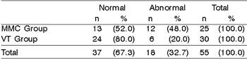
The resolution of OME in patients of groups VT and MMC was respectively 80% and 52%. The group variable presented association with final middle ear outcome, more efficiently in group VT (Table 1). Despite the fact that the final outcome observed in the middle ear was associated with the group, the individual analysis of each group did produce association between final outcome and duration of TM opening in group MMC nor in group VT (respectively, 0.404 and p= 0.166).
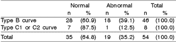
The presence of type B tympanometric curve was associated with poor prognosis factor for middle ear final outcome (Table 2).
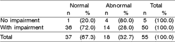
The absence of auditory tube impairment by adenoid tissue was associated with poor prognosis for middle ear final outcome (Table 3).
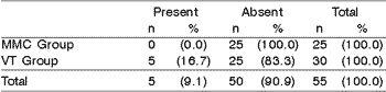
The incidence of otorrhea (13.3%) was statistically greater in the group submitted to VT insertion, compared to the MMC group that did not present any episodes (Table 4). Tympanosclerosis was observed only in patients in group VT (16.7%), being non-existent in group MMC. No patient presented VT occlusion, residual perforation of TM, cholesteatoma or medial migration of VT.
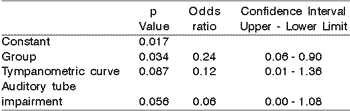
Multivariate analysis involving variables that presented statistical association (p<0.05) with the final outcome of normalization or not observed in the middle ear revealed the possibility of the middle ear being normal: 4.26 times greater in the VT group than in the MMC group; 8.36 times greater in patients with preoperative type C1 and C2 curve than in patients with type B curve by Jerger; 16.7 times greater in patients with impairment than in those without preoperative auditory tube impairment (Table 5).Table 1. Comparison between the final result of the middle ear, six months after the closure of the tympanic membrane, in the group of tympanotomy with ventilation tube insertion (VT) and in those with application of mitomycin C.
p= 0.028; OR= 0.271; IC= (0.082 - 0.890).
Table 2. Correlation between middle ear final outcome six months after closure of tympanic membrane and preoperative tympanometric curve in patients with otitis media with effusion (n=54).
p=0.041; OR= 0.778; IC= (0.653 - 0.926).
Table 3. Correlation between middle ear final outcome six months after tympanic closure and preoperative impairment of pharyngeal ostium of the auditory tube in patients with otitis media with effusion (n=55).
p= 0.035; OR= 0.097; IC= (0.010 - 0.947).
Table 4. Comparison of the incidence of otorrhea observed in the group treated with ventilation tube (n=30) and the one submitted to topical application of mitomycin C (n=25).
p= 0.041
Table 5. Analysis of logistic regression between the significant variables of group, preoperative impairment of pharyngeal ostium of the auditory tube and tympanometric curve obtained with immittanciometry.
Discussion
The difference in mean age at onset of otitis (21.6 ± 15.2 months) and age at the time of the operation (47.8 ± 23.2 months) was 26.2 months, revealing an average delay of more than two years in the performance of the surgical procedures. We did not observe, however, association between surgical act delay and middle ear final outcome in the studied patients (p = 0.374).
The possibility of OME manifest as an asymptomatic or little symptomatic affection, with late diagnosis in one fourth of the children, reinforces the importance of the pediatrician in screening and detecting the disease. The presence or absence of symptoms, however, did not influence the middle ear final outcome (p = 0.311).
The presence of otalgia suggested a tendency of predictive factor of poor prognosis in the middle ear final outcome p= 0.043; OR= 3.413; IC= (1.008 - 11.551), which, however, lost its statistical significance after adjustment of the multivariate analysis for the exclusion of misleading factors.
The size of palatine tonsils and the their potential removal did not present association with the final result of the surgical procedure over the middle ear, confirming the previous studies 1, 2, 5, 6, which demonstrated that only adenoidectomy reduces morbidity and modifies the natural history of children with OME and that the association with tonsillectomy does not provide additional benefits.
The presence of tympanometric curves C1 and C2 was associated with good prognosis factor for middle ear final outcome (p = 0.041), differently from type B curve that represented a factor of poor prognosis (Table 2).
Preoperative impairment of pharyngeal ostium of ipsilateral auditory tube by adenoid tissue presented an association with middle ear final outcome (Table 3). Children with impairment of auditory tube pharyngeal ostium had greater change of normalizing the middle ear in six months of follow-up, when compared to children with no previous tube impairment (p = 0.035). The literature presents a well-defined relationship in many studies 1, 3, 4, 6, between adenoidectomy and elimination of the tube impairment and progressive evolution of OME. The preoperative impairment of tube ostium by adenoid tissue is known to be a poor prognosis factor for the installation and maintenance of OME, and its removal is recommended. The importance of our study is that in an inverted viewpoint, without conceptual changes, the patients with OME whose preoperative fibronasolaryngoscopy did not reveal auditory tube impairment, presented poor prognosis factor regardless of the surgical procedures conducted, including adenoidectomy. In such cases, the adenoid factor was not responsible for the persistence of effusion. Studies that correlate patients with OME without auditory tube impairment by adenoid tissue should be conducted to assess the progressions of the disease, analyze other possible factors that interfere in the auditory tube dysfunction and the influence they have over the progressive evolution of the disease. In our study, it was not possible to assess the association between adenoidectomy and final outcome of OME seen in all patients submitted to surgery.
The duration of TM opening corresponded to the duration of middle ear aeration, providing reversion of metaplastic abnormalities or the tympanic cavity epithelium. In the 25 patients of the MMC group, closure happened two (40%) or three weeks (100%) after tympanotomy. Similarly to what happened in guinea pigs, in which the association of the topical application of MMC to tympanotomy induced by laser prolonged significantly the opening of TM 8, 14, 24, 26, in our study, the topical application of MMC prolonged the opening time of tympanotomy for a period of up to 21 days. The TC closure in the MCC group, however, was much faster than in the VT group, which probably influenced negatively the final outcome of the group compared to the other (p= 0.028, Table 1).
The action mechanism of MMC resulted from the cellular division inhibition, protein synthesis of DNA, inhibiting the replication of epithelial cells and specially fibroblasts 15, 24, 29. The closure time of the tense portion of the TM depended on the cicatricial repair of the three layers: external or epithelial, medial or connective and internal or mucosa. Since only the medial layer of the TM presents fibroblasts, the action of proliferation delay by topical application of MMC and consequent maintenance of perviousness of tympanotomy, seem to be less effective when compared to other anatomical sites, in which the formation of cicatricial collagen is important, and it can be extremely reduced by this form of application.
In the 30 patients of group VT, closure was detected in 0.0%, 36.7%, 66.7%, 90.0% and 100.0% of the right TM within 3, 6, 9, 12 and 15 months, respectively, after tympanotomy. The shortest time since VT extrusion and closure of MT was four months, in only two patients. The mean time from extrusion of VT model Shepard was 8.3 months, similarly to what was observed by Weigel et al.30, for the same VT model.
OME resolution, six months after closure of TM, in patients submitted to tympanotomy with VT insertion or application of MMC was 80% and 52%, respectively, significantly greater efficacy in the VT group compared to the MMC (p=0.028, Table 1). Thus, after a six month follow-up, the likelihood of the middle ear to normalize with VT placement was significantly greater than with topical application of MMC.
In our study, the efficacy in 13 patients (52%) of group MMC was close to 60%, obtained with thermal myringotomy 10 and inferior to that observed by Silverstein et al.9, that employed similar opening TM period (3 weeks) induced by laser and observed efficacy of 78%.
We did not observe significant difference between TM closure time in up to two weeks or three weeks from final result of Group MMC (p= 0.404) and this finding is explained by the minimum difference between these two periods of closure.
Previous studies revealed that the concentration 8, 26, duration of exposure 8 or serial reapplication of MMC do not influence significantly the period of maintenance of the opening of tympanotomy in guinea pigs. A recently published article 29, after the conclusion of our study, however, confirmed our opinion that further studies are required, since greater concentrations, time and frequency of topical application of MMC in humans can prolong the delay in scarring of TM, providing better efficacy in resolution of OME.
The patient in group VT were regrouped according to duration of tube placement, spontaneous extrusion and closure of TM in three subgroups: 0 to 6 months, 7 to 9 months and over 9 months, based on mean time of VT permanence of 8 months 11, 30 and in the mean of 8.3 months obtained in our study, showing time of early elimination, habitual and late time of VT. There was no association between middle ear final outcome and closure time of TM in group VT (p=0.166).
Among acute complications resultant from surgical procedures in the middle ear, only otorrhea was observed in 13.3% of the patients in group VT, being that in MMC group no patients presented any episodes (p= 0.041, Table 5). The highest incidence of otorrhea in group VT can be explained by discrepancy between maintenance of opening compared to the MMC group, providing greater risk of middle ear contamination. All episodes of otorrhea were late, between two and eight months from placement of VT and related to the accumulation of water in the external auditory canal or upper airway infections. The incidence of otorrhea was below the one described in the literature which varied from 21% to 50%, with a study in Brazil that reported 26.3% 12.
There was no occlusion in 30 patients in group VT contrary to the occlusion findings in 11% of the patients 30, with the same VT model.
There was no residual perforation of TM in patients in both groups, agreeing with Weigel et al.30 study. The presence of calcification of the middle layer of TM was observed in 16.7% of the TM only in group VT, suggesting that it was resultant from the local trauma caused by the presence of drainage. No patient presented installation of cholesteatoma, nor medial migration of VT.
The previous choice of VT model Shepard was based on literature reports that identified this model as effective in the resolution of OME, presenting low incidence of complications, such as spontaneous extrusion in most patients, and its use is indicated in the habitual pediatric population 11, 30, such as the one in our study.
The lowest efficacy in group MMC does not exclude its future use since there are advantages such as low incidence of complications or sequelae, low period of restriction of baths, which is socially important in a tropical country, possibility of high efficacy in patients that require short term tympanotomy, such as barotraumas or candidates to hyperbaric therapy with auditory tube dysfunction.
Multivariate analysis started with four variables that presented significant association (p<0.05), with abnormal middle ear final outcome: MMC group, presence of preoperative otalgia, absence of preoperative impairment of auditory tube pharyngeal ostium, and preoperative type V tympanometric curve. The analysis used the logistic regression model when the presence of preoperative otalgia lost its significance. The other variables - the group, preoperative impairment of auditory tube pharyngeal ostium and tympanometric curve - were important to explain the final outcomes, allowing us to determine that six months after closure of tympanotomy: 1) the likelihood of the middle ear being normal is 4.26 times greater in patients in group VT than in MMC group patients; 2) the likelihood of the middle ear being normal is 8.3 times higher in patients with preoperative tympanometric curve type C1 or C2 than in patients with type B of Jerger; 3) the likelihood of the middle ear being normal is 16.7 times higher in patients with impairment than in those without preoperative auditory tube impairment (Table 5).
Conclusion
Treatment of otitis media with effusion through tympanotomy, aspiration of effusion and topical application of mitomycin C presented less efficacy than what was observed for the group with ventilation tube placement.
Type B tympanometric curve and absence of preoperative auditory tube pharyngeal ostium impairment by adenoid tissue represented poor prognosis factors concerning the final outcome observed for the middle ear after six months of follow-up.
The topical application of mitomycin C provided maintenance of the opening of the tympanic membrane for a period that varied from two to three weeks.
Recurrence of effusion, six months after closure of the tympanic membrane, occurred in 20% to 48% , respectively, in the ventilation tube and in the mitomycin C groups. No one in the mitomycin C group presented complications, whereas in the ventilation tube group, otorrhea and tympanosclerosis were observed in 13.3% and 16.7% of the cases, respectively.
References
1. Handler SD. Current indications for tympanostomy tubes. Am J Otolaryngol 1994; 15:103-108.
2. Bluestone CD, Klein JO. Clinical practice guidelines on otitis media with effusion in young children: strengs and weakness. Otolaringol Head Neck Surg 1995; 112:507-11.
3. Mills R. The management of childhood otitis media with effusion. J R Soc Med 1996; 89:132-134.
4. Gates GA. Adenoidectomy for otitis media with effusion. Ann Otol Rhinol Laryngol Suppl 1994; 103:54-58.
5. Gates GA, Avery CA, Cooper JC, Prihoda TJ. Chronic secretory otitis media: effect of surgical management. Ann Otol Rhinol Laringol 1992; 101:866-69.
6. Maw AR, Bawden R. The long term outcome of secretory otitis media in children and the effects of surgical treatment: a ten year study. Acta Otorhinolaryngol Belg 1994; 48(4):317-24.
7. Goode RL. CO2 laser myringotomy. Laryngoscope 1982; 92:420-23.
8. Estrem AS, Batra TJ. Preventing myringotomy closurte with topical mitomycin C in rats. Otolaryngol Head Neck Surg 1999; 120(6):794-98.
9. Silverstein H, Kuhn J, Choo D, Krespi YP, Rosenberg SI, Rowan PT. Laser-assisted tympanostomy. Laryngoscope 1996; 106(9):1067-74.
10. Kent SE. Thermal myringotomy versus grommets in the management of secretory otitis media. Int J Pediatr Otorhinolaryngol 1989; 17:31-5.
11. Isaacson G, Rosenfeld RM. Care of the child with tympanostomy tubes. Pediatr Clin North Am 1996; 43(6):1183-193.
12. Souza NJA, Mangabeira Albernaz PL, Fukuda Y. Otite média secretora: análise de 232 casos operados. Acta AWHO 2000; 19(3):116-24.
13. Yazawa Y, Suzuki M, Kitano H, Kitajima, K. Intraoperative mitomycin C in endolymphatic sac surgery for Meniere's disease: a pilot study. J Otorhinolaryngol Relat Spec 1999; 61(4):188-94.
14. Estrem SA, Baker TJ. Preapplication of mitomycin C for enhanced patency of myringotomy. Otolaryngol Head Neck Surg 2000; 122(3):346-48.
15. Rahbar R, Valdez TA, Shapshay SM. Preliminary results of intraoperative mitomycin-C in the treatment and prevention of glottic and subglottic stenosis. J Voice 2000; 14(2):282-86.
16. Iliev ME, Van Der Zypen E, Frankhauser F, England C. Transconjunctival application of mitomycin C in combination with laser sclerostomy ab interno: a long-term morphological study of the postoperative healing process. Exp Eye Res 1997; 64(6):1013-26.
17. Ayyala RS, Pieroth L, Vinals AF, Goldstein MH, Schuman JS, Netland PA, Dreyer EB, Cooper ML, Mattox C, Frangie JP, Wu HK, Zurakoxski D. Comparison of mitomycin C trabeculectomy, glaucoma drainage device implantation, and laser neodymium: YAG cyclophotocoagulation in the management of intractable glaucoma after penetrating keratoplasty. Ophthalmology 1998; 105(8):1550-56.
18. Ingrams DR, Volk MS, Biesman BS, Pankratov MM, Shapshay SM. Sinus surgery: does Mitomycin C reduce stenosis? Laryngoscope 1998; 108:883-86.
19. Ward RF, April MM. Mitomycin-C in the treatment of tracheal cicatrix after tracheal reconstruction. Int J Pediatr Otorhinolaryngol 1998; 44(3):221-26.
20. Zilelioglu G, Ugurbas SH, Anadolu Y, Akiner M, Akturk T. Adjunctive use of mitomycin C on endoscopic lacrimal surgery. Br J Ophthalmol 1998; 82(1):63-66.
21. Spector JE, Werkhaven JA, Spector NC, Huang S, Page RN, Baranowski B, Luther M, McGehee B, Reinisch, L. Preservation of function and histologic appearance in the injured glottis with topical mitomycin-C. Laryngoscope 1999; 109(7):1125-129.
22. Coppit G, Perkins J, Munaretto J, Nielsen R, McKinney L, Ulnick K. The effects of mitomycin-C and stenting on airway wound healing after laryngotracheal reconstruction in a pig model. Int J Pediatr Otorhinolaryngol 2000; 53(2):125-35.
23. Eliashar R, Eliachar I, Esclamado R, Gramlich T, Strome M. Can topical mitomycin prevent laryngotracheal stenosis? Laryngoscope 1999; 109 (10)1594-600.
24. Estrem SA, Vanleeuxen RN. Use of mitomycin C for maintaining myringotomy patency. Otolaryngol Head Neck Surg 2000; 122(1):8-10.
25. Liao SL, Kao SC, Tseng JH, Chen MS, Hou PK. Results of intraoperative mitomycin C application in dacryocystorhinostomy. Br J Ophthalmol 2000; 84(8):903-6.
26. Yucel OT. Topical use of mitomycin C in laser myringotomy: an experimental study in rats. Int J Pediatr Otorhinolaryngol 2000; 54(2/3):93-6.
27. O'Reilly RC, Goldman SA, Widner SA, Cass SP. Creating a stable tympanic membrane perforation using mitomycin C. Otolaryngol. Head Neck Surg 2001; 124(1):40-45.
28. Jassir D, Buchman CA, Gomez-Marin O. Safety and efficacy of topical mitomycin in myringotomy patency. Otolaryngol. Head Neck Surg 2001; 124(4):368-73.
29. Jang, HJ, Song CS, Pak, SC. Effect of exposure to mitomycin C on cultured tympanic membrane fibroblasts. Int J Pediatr Otorhinolaryngol 2003; 67:173-76.
30. Weigel MT, Parker, MY, Goldsmith MM, Postma DS, Pillsbury, HC. A prospective randomized study of four commonly used tympanostomy tubes. Laryngoscope 1989; 99:252-56.
1 Assistant Professor, Department of Ophthalmology, Otorhinolaryngology and Speech and Hearing Pathology, Medical School, Federal University of Minas Gerais.
2 Faculty Professor, Department of Surgery, Medical School, Federal University of Minas Gerais.
3 Joint Professor, Department of Ophthalmology, Otorhinolaryngology and Speech and Hearing Pathology, Medical School, Federal University of Minas Gerais.
4 Resident Physician, Núcleo de Otorrino-BH.
Address correspondence to: Celso Becker - Rua Otoni 909, sala 502 Santa Efigênia Belo Horizonte MG 30150-270
Tel (55 31) 3273-4635 Fax (55 31) 3281-4604 - E-mail: cbecker@medicina.ufmg.br
Study conducted and partially financed by the Division of Otorhinolaryngology, Hospital das Clínicas, UFMG and Núcleo de Otorrino BH.
Article submitted on March 16, 2003. Article accepted on July 01, 2003.
Print: ![]()