

Year: 2003 Vol. 69 Ed. 4 - (8º)
Artigo Original
Pages: 485 to 489
 PDF PT
 Parotid pleomorphic adenoma: clinical, diagnostic and therapeutical aspects
Author(s):
Romualdo Suzano Louzeiro Tiago1,
Gilson Araújo Castro2,
Luiz Artur da Costa Ricardo3,
Rogério Borghi Bühler4,
Antônio Sérgio Fava5
Keywords: parotid, pleomorphic adenoma, parotidectomy
Abstract:
The neoplasias of the parotid gland are a heterogeneous group with more than 30 histological kinds already defined, and the pleomorphic adenoma is the most common benign tumor. Aim: In this study we present our case of parotid gland pleomorphic adenoma, with the purpose of discussing the clinical presentation, the diagnosis and the more suitable surgical techniques to treat this disorder. Material and Method: A retrospective clinical study was carried out on a group of 68 patients with histological diagnosis of parotid gland pleomorphic adenoma who underwent surgery at the Department of Otolaryngology of the HSPE-FMO, from January 1982 to June 2002. The data regarding their age, sex, more frequent symptoms, supplementary tests performed, location, size, surgical technique used, treatment complications and evolution were gathered. Results: In our case we observed a higher incidence in female patients, being more frequent in their fifth decade of life. The most common clinical presentation was of a nodule at the parotid region, this sign being present in 100% of the cases. Superficial parotidectomy by identifying and preserving the facial nerve was the more common surgery (91.2 % of the cases). The more frequent post-surgical complications were paresis (14.7%) and paralysis (7.3%) of the facial nerve, followed by the Frey's syndrome (4.4%). Conclusion: The pleomorphic adenoma is the most common benign neoplasia of the parotid gland, being more frequent in female patients from their fifth decade of life. It is a clinically diagnosable neoplasia, to be confirmed with a histopathological examination. The superficial parotidectomy is the basic procedure to diagnose and treat it, being low the recurrence index with this surgery.
![]()
Introduction
Neoplasia of the parotid gland consists of a heterogeneous group with more than 30 histological types already defined according to the classification of WHO (World Health Organization), published in 19911. The incidence of this disorder in US is 1(one) case in 100,000 (one hundred thousand) individuals annually, with approximately 3% of the tumors located in the head and neck and 0.6% in the human body 2-4. The pleomorphic adenoma is the most common benign neoplasia of parotid gland accounting for approximately 60 to 70%, and with increased incidence from the 4th to the 6th decade of life on. 5,6
Clinically, the pleomorphic adenoma of the parotid gland has the aspect of a single nodular lesion, with clearly demarcated, lobulated surface, solid consistency, mobile and painless upon palpation. Most of the cases (90%) involve the surface lobe of the gland, with 80% located in the lower portion 6. Bilateral tumors are extremely rare7. Microscopically, the variety of types of cells is the major characteristic of pleomorphic adenoma, not only among different tumors, but also in different parts of the same tumor. It is formed by epithelial, myoepithelial and mesenchymal elements, involved by a condromyxoidstroma or even condrosteoidstroma 4.
The diagnosis of neoplasia of parotid gland is confirmed with detailed clinical history and physical examination. Imaging tests, particularly ultrasound and CT scan are not essential and could be performed in selected cases for treatment planning purposes3. Fine needle aspiration biopsy is another diagnostic method that can be used to determine if the tumor is benign or malignant, however it is not used to define therapeutic management8. Definitive diagnosis of pleomorphic adenoma is obtained with histopathological analysis in wax sections collected from parotidectomy with identification and preservation of facial nerve6.
This study presents the analysis of 68 patients with pleomorphic adenoma of parotid gland with the purpose of discussing clinical presentation, diagnosis and the most adequate surgical techniques to treat such disease.
MATERIAL AND METHOD
The retrospective clinical trial was conducted in a group of 68 patients with histopathological analysis of pleomorphic adenoma of parotid gland, which have undergone surgery at the Otorhinolaryngology Service of Hospital do Servidor Público Estadual de Sao Paulo - "Francisco Morato de Oliveira", from January 1982 to June 2002. Data included information related to age, gender and most frequent symptoms, additional tests (parotid gland ultrasound and fine needle aspiration biopsy), location, size, surgical technique applied, complications of the treatment and outcomes.
RESULTS
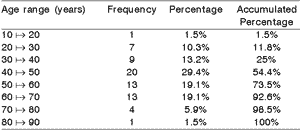
In this study we found higher incidence in women, ratio of 3.5:1. The age of the patient at the moment of diagnosis ranged from 18 years to 84 years (mean age: 48.7 years). Tumors were more frequent in the 5th decade followed by the 6th and 7th decades (Table 1). The elapsed time between onset of symptoms and the appointment with the doctor ranged from 1 month to 18 years, with average time of 46 month (3 years and 10 months).
The most common clinical symptom was nodule in the parotid gland region, and this was present in 100% of the cases followed by otalgia (2.9%) and facial paralysis (1.4%). Increased frequency of pleomorphic adenoma was found in the left parotid gland (61.5%), with 1.6:1 ratio compared to the right side.
Ultrasound of parotid gland was performed in 28 patients. The ultrasound most common finding was solid nodule (78.6%), followed by cystic (17.8%) and mixed (3.6%) aspect.
Cytological analysis with fine needle aspiration biopsy was conducted in 24 patients, and 15 of them were suggestive cases of pleomorphic adenoma, with concordance rate of 62.5% of cases.
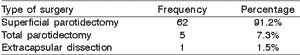
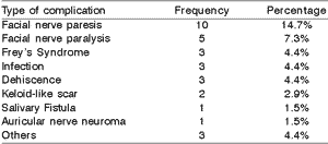
Surgical treatment was applied in all cases. Surface parotidectomy with identification and spare of facial nerve was the most common surgery performed (Table 2). We found out that 92.7% of pleomorphic adenomas were located in the surface lobe of the parotid gland and 7.3 were located in the deep lobe. Surgical indication of radical parotidectomy was reserved to neoplasias located in the deep lobe of the parotid gland (5 cases). The average size of the nodules in relation to their higher diameter was 3.8 cm. Most common post-operative complications were facial nerve paresis and paralysis followed by Frey's syndrome, infection of the surgical site and dehiscence (Table 3). Recurrence of pleomorphic adenoma occurred in two cases (2.9%). Malignant transformation was not found.Table 1. Frequency of pleomorphic adenoma of the parotid gland, according to age range.
Table 2. Surgical approach of pleomorphic adenoma of the parotid gland.
Table 3. Complication of the surgical treatment of pleomorphic adenoma of the parotid gland.
DISCUSSION
Pleomorphic adenoma is the most common benign neoplasia of parotid gland, occurring predominantly in women (1.5:1 ratio), and average age varying from 36 to 45.7 years6,8. Occurrence in women was three times higher when compared to male patients and predominantly in the 5th decade of life or approximately at 48.7 years of age.
It is a slow-growth tumor and the progress of the nodule in the parotid gland region ranged from 1 to 18 years with average of 3 to 10 months. The average size of the lesion was 3.8 cm. Malignant tumors tend to present a faster growth rate in shorter time, and generally are associated with other signs and symptoms such as: facial paralysis, fixation to skin, trism, lymph node metastasis and local pain 3. Another important piece of data is that malignant tumors of salivary gland tend to occur in the 6th decade of life ranging from 53 to 65 years of age. 9. We showed one (1) case of facial paralysis in a patient that had a nodule of approximately 2 cm in its larger diameter in the first medical visit; during surface parotidectomy, we noticed close proximity of the lesion to the facial nerve branch.
Data such as symptom duration, growth rate, age and gender are important as guidelines to clinical rationale and likely diagnosis of parotid nodule.
The use of imaging tests is not essential, but in certain cases they might help to determine the location and extension of the lesion, therefore contributing to surgical planning.
Sialography has not been used since it has a risk of disseminating tumor cells 10. Ultrasound is a low cost non-invasive technique, painless and easy to perform that can help to define if the lesion is solid or cystic 3. In our case study, 78.6% of the pleomorphic adenomas were presented as solid nodules. In our sample we did not order CT scan of the parotid region as a routine practice, and the lesions were apparently restricted to the surface pole of the gland upon clinical examination. Nonetheless, CT scan is very useful to assess lesions that infiltrate to parapharyngeal space which present facial paralysis, cases of suspected malignancy, fixed or semi-fixed nodule to deep plans or tumor recurrence 3. MRI is more useful to evaluate parapharyngeal space than CT Scan since it allows better identification of the internal architecture of the parotid gland and better definition of the limits of the lesion without using iodated contrast or exposure to radiation 11.
The use of fine needle aspiration in the evaluation of parotid masses is quite controversial and depends on great expertise from the pathologist to prevent misdiagnosis. False positive and false negative results are not very common in cases of pleomorphic adenoma and it may help the surgeon in determining if the lesion is benign or malignant within a sensitivity rate ranging from 93.3% to 95.7%, and with specificity level ranging from 98% to 100%12-14. Batsakis et al. (1992) reported a concordance rate of 74% between fine needle aspiration and final histopathological analysis of parotid neoplasias 15. In twenty-four of the patients that underwent the procedure in our case study, the concordance rate with histopathological analysis was 62.5% (15 cases out of 24).
If any question about the nature of the lesion remains after preliminary investigation, the next minimal diagnostic procedure should be surface parotidectomy with identification and facial nerve preservation followed by frosting. Incision biopsy should be avoided because in addition to generating a scar that will have to be removed in a definitive procedure, it has increased risk of tumor dissemination and causing lesion of facial nerve3,16.
Surface parotidectomy consists of the resection of a portion of the parotid gland laterally located to the facial nerve after careful identification and preservation of this nerve. Nodular lesion is removed with exposure of its capsule, recovered by normal glandular tissue, with a margin of at least 2 cm (except if the tumor is located near the facial nerve)6. This procedure is required due to the characteristic of pleomorphic adenoma to have small digitations (pseudopodia) that transpose the capsule and are responsible for recurrence of the tumor if a surgery smaller that surface parotidectomy is performed. Patients with lesions smaller than 4 cm that are mobile and located in the surface lobe of the parotid gland are candidates for surface parotidectomy. This procedure was performed in 91.2% of our cases.
Total parotidectomy removes all glandular tissue laterally and medially located to the facial nerve and is primarily indicated for cases involving deep lobe of the parotid gland 6. The procedure was performed in 5 cases with involvement of the deep lobe corresponding to 7.3% of the surgeries.
Surgeries like enucleation or extracapsular dissection of the tumor should be avoided since these procedures do not identify the facial nerve. According to Witt (2002), since resection of the adenoma is performed very close to the capsule, the likelihood of capsular rupture is very high resulting in a statistically significant percentage of recurrence against surface parotidectomy or total parotidectomy 6,17. Enucleation results in a recurrence rate of 25% or 9 times higher than surface parotidectomy (3%). In this study recurrence rate of pleomorphic adenoma affected 2 cases or 2.9%.
The most common complication resulting from the treatment of parotid gland tumors is facial nerve dysfunction, either transient or permanent. Facial nerve paresis is 2.3 times more frequent in total parotidectomy (60%) than in surface parotidectomy (26%)6. It happens due to the increased handling of the ramus of the nerve in total parotidectomy. The incidence of facial nerve paresis after surface parotidectomy ranges from 16 to 47%18-20. This study reported 14.7% of paresis of facial nerve, thus, within the range reported in the literature. The occurrence of permanent paresis in the literature ranged from 0% to 9%18-20. In our study it was approximately 7.3% and paralysis was more common in the marginal ramus of the mandible. Surely the age of the patient and expertise of the surgeon are determinant factors for the occurrence of facial nerve dysfunction 20.
Another complication that occurs after parotidectomy is Frey's Syndrome. This syndrome was described by neurologist Lucie Frey after observing the occurrence of facial sweating and hyperemia in soldiers that were victims of wound injury in the ipsilateral region of the parotid gland21. The syndrome was also referred as auriculotemporal syndrome resulting in discontinuation of parasympathetic post-ganglionar fibers of the IX cranial pair (glossopharyngeal nerve) that travel by the auriculotemporal nerve and by the V cranial pair (trigeminal nerve), that regenerate and infiltrate into sweating glands located along the skin immediately above the surgical bed after parotidectomy. The stimuli that should result in salivary secretion cause sweating of the parotid gland region 18. It occurs in 35% to 60% of the patients and is closely related to the amount of glandular tissue removed6,22. This syndrome is very common after total parotidectomy (47%), occurring in 17% of surface parotidectomy procedures and in 10% of partial surface parotidectomy surgeries (when the tumor is located in the inferior pole and superficially to the gland)6. The incidence observed in this study was 4.4%, considerably lower than the rate found in the literature, probably due to the lack of direct questioning to the patients.
CONCLUSIONS
Pleomorphic adenoma is the most common benign neoplasia of the parotid gland and it is more frequent in women from the 5th decade of life on. It is a neoplasia of clinical diagnosis that is confirmed with histopathological analysis. Surface parotidectomy is the minimal procedure required for diagnosis and management plan, leading to low rate of recurrence.
REFERENCES
1. Seifert G, Sobin L. Histological typing of salivary gland tumors. World Health Organization. International histological classification of tumors, 2nd ed., Berlin: Springer-Verlag; 1991.
2. Hugo NE, MCKinney P, Grifish BH. Management of tumors of the parotid gland. North American Surgical Clinic 1973; 53:105-11.
3. Kamal SA, Othman EO. Diagnosis and treatment of parotid tumours. J Laringol Otol 1997; 111:316-21.
4. Van der Wal JE, Leverstein H, Snow GB, Kraaijenhagen HA, Van der Waal I. Parotid gland tumors: histologic reevaluation and reclassification of 478 cases. Head Neck 1998; 20:204-7.
5. Spiro RH. Salivary neoplasms: overview of a 35-year experience with 2807 patients. Head Neck Surg 1986; 8:177-84.
6. Witt RL. The significance of the margin in parotid surgery for pleomorphic adenoma. Laryngoscope 2002; 112:2141-54.
7. Turnbull AD, Frazell EL. Multiple tumors of the major salivary glands. Am J Surg 1969; 118:787-9.
8. Tsai SC, Hsu H. Parotid neoplasms: diagnosis, treatment, and intraparotid facial nerve anatomy. J Laringol Otol 2002; 116:359-62.
9. Skolnik EM, Friedman M, Becker S, Sisson GA, Keyes GA. Tumors of the major salivary glands. Laryngoscope 1977; 87:843-61.
10. Touquet R, Mackenzie IJ, Carruth JAS. Management of parotid pleomorphic adenoma, the problem of exposing tumor tissue at operation. The logical pursuit of treatment policies. Br J Oral and Maxillofac Surg 1990; 28:404-8.
11. Miller FR, Wanamaker JR, Lavertu P, Wood BG. Magnetic resonance imaging and the management of the parapharyngeal space tumors. Head Neck 1996; 18:67-77.
12. Shaha AR, Webber C, DiMaio T, Jaffe BM. Needle aspiration biopsy in salivary gland lesions. Am J Surg 1990; 160:373-6.
13. Frable MAS, Frable WJ. Fine-needle aspiration biopsy of salivary glands. Laryngoscope 1991; 101:245-9.
14. Candel A, Gatuso P, Reddy V, Matz G, Castelli M. Is fine needle aspiration biopsy of salivary gland masses really necessary? Ear Nose Throat J 1993; 72:485-9.
15. Batsakis JG, Sneide N, El-Naggar AK. Fine-needle aspiration of salivary glands: its utility and tissue effects. Ann Otol Rhinol Laryngol 1992; 1001:185-8.
16. Jonhson J. Salivary glands. In Gluckmann J, Gullane P, Jonhson J. Practical approach to head and neck tumors, New York: Raven Press; 1994. Cap. 2.
17. Henriksson G, Westrin KM, Carlsoo B, Silversward C. Recurrent primary pleomorhic adenomas of salivary gland origin: intrasurgical rupture, histopathologic features, and psudopodia. Cancer 1998; 82:617-20.
18. Langdon JD. Complications of parotid gland surgery. J Maxillofac Surg 1984; 12:225-9.
19. Owen ER, Banerjee AK, Kissin M, Kark AE. Complications of parotid surgery: the need for selectivity. Br J Surg 1989; 76:1034-5.
20. Mra Z, Komisar A, Blaugrund SM. Functional facial nerve weakness after surgery for benign parotid tumors: a multivariate statistical analysis. Head Neck 1993; 15:147-52.
21. Harper KE, Spielvolgel RL. Frey's syndrome. Int J Dermatol 1986; 25:224-6.
22. Gordon AB, Fiddian RV. Frey's syndrome after parotid surgery. Am J Surg 1976; 132:54-8.
1 Ph.D. studies under course, Post-Graduation Program in Otorhinolaryngology and Head and Neck Surgery,
Federal University of Sao Paulo - Escola Paulista de Medicina. Assistant Physician, Service of Otorhinolaryngology, HSPE-FMO-IAMSPE.
2 Resident Physician, Service of Otorhinolaryngology, HSPE-FMO-IAMSPE.
3 Master studies under course, Post-Graduation Program in Otorhinolaryngology, HSPE-FMO-IAMSPE.
4 Master studied under course, Post-Graduation Program in Otorhinolaryngology, Medical School, Santa Casa de Sao Paulo.
Assistant Physician, Service of Otorhinolaryngology, HSPE-FMO-IAMSPE.
5 Ph.D. in Medicine, Medical School, University of Sao Paulo. Head of the Division of Head and Neck, Service of Otorhinolaryngology, HSPE-FMO-IAMSPE.
Affiliation: Service of Otorhinolaryngology, Hospital do Servidor Público Estadual - "Francisco Morato de Oliveira" - Instituto de Assistência Médica ao Servidor Público Estadual - Sao Paulo SP.
Address correspondence to: Romualdo Suzano L. Tiago - Rua Estado de Israel 493, ap. 51 Vila Clementino Sao Paulo SP 04022-001
Tel (55 11) 5084-7725 - E-mail: romualdotiago@uol.com.br
Article submitted on January 27, 2003. Article accepted on July 10, 2003.
Print: ![]()