
Year: 2001 Vol. 67 Ed. 5 - (2º)
Artigos Originais
Pages: 612 to 616
Treatment of benign paroxysmal positional vertigo with repositioning manevers
Author(s):
Roberto A. Maia 1,
Flávia L. Diniz 2,
Agnaldo Carlesse 2
Keywords: vertigo, treatment repositioning maneuvers
Abstract:
Introduction: Benign Paroxysmal Positional Vertigo (BPPV) is among the most common vestibular disorders. It is characterized by recurrent episodes of vertigo induced by changes in head position. The condition is readly diagnosed by performing the Dix-Hallpike maneuver. Nystagmus is always present by this way. Study design: Prospective results clinical not randomized. Material and method: A total of seven patients diagnosed with BPPV received the repositioning maneuver of Epley. Five out of seven patients had excellent recovery, two patients had good results and one had a bad result. For this last one different treatments are discussed. Conclusion: We performed otoneurological examination in all patients; this test seems to be an intersting prognosis method for seeking the results of treatment. The repositioning maneuver of Epley is an excellent treatment for BPPV. Most of the patients of our series were successfuly treated by this method.
![]()
INTRODUCTION
Definition: Benign positional paroxysmal vertigo (BPPV) is one of the most frequent affections of the peripheral vestibular system. In 1921, Barany1 described it and later Dix and Hallpike5 detailed it, in 1952. Its typical clinical characteristic is sudden vertigo episode, sometimes severe and short, with total remission of symptoms within 45 seconds. The vertigo crisis is always triggered by a change in the position of the head. Some typical triggering movements are: lying down, standing up from bed, turning to one side when on bed, moving the head up and down. The situation tends to persist for weeks to months and sometimes for years. The diagnosis of BPPV is always confirmed by the classical maneuver of Dix-Hallpike, which triggers an evident nystagmus with short duration and latency in the patients.
Pathophysiology: Pathophysiology of BPPV is explained by the presence of calcium carbonate crystals that are the degenerated fragments of utriculus otoconia, displaced to the region of the semicircular canals, almost always in the posterior semicircular canal. Two theories are hypothesized. In the first theory, named Cupulolithiasis, fragments are adhered to the cupula of the posterior semicircular canal. This theory was written and evidenced in 1962 by Schuknecht10, who found these crystals deposited on the purface of the cupula of the posterior semicircular canal in two patients with BPPV. The second theory, named Canalithiasis or Ductolithiasis suggests that the degenerated fragments are not adhered to the cupula but rather fluctuating in the endolymph of the posterior semicircular canal. In both theories, the triggering movement of the patient's head put the fragments into action, which in turn, leads to inappropriate stimulation of the cupula of the posterior semicircular canal and the excitation of the posterior ampullar nerve, with symptoms of vertigo.
Symptomatology: Diagnostic investigation of BPPV is conducted by the maneuver of Dix-Hallpike. This maneuver consists of the movement of the had of the patient in order to promote a displacement of endolymph and consequently, of the cupula of the posterior semicircular canal. In the Dix-Hallpike maneuver the patient is initially seated, with the head laterally rotated (to the right or left, according to the tested side), at approximately 45 degrees. The examiner holds the head of the patient, making a sudden and quick movement of lying down, in horizontal supine position. When the patient is lied down, since there is horizontal fixation of the head, it lied pending backwards at approximately 30 degrees. The patient is immobilized in this position and the eyes are opened and maintain a fixed stare. In BPPV patients, there is an evident nystagmus, some seconds after the stimulation (with latency), which lasts about 45 seconds (finishable). Otoneurological study, represented by the classical caloric tests, does not show typical findings of BPPV. In such patients, caloric tests may be normal or show hyper or hyporeflexive responses.
Treatment: BPPV is self-limited and in most of the cases it regresses spontaneously, with no treatment, within a period of time that varies from case to case. Some distinct types of treatment are proposed:
1. Vestibular habituation drills. Proposed by Brandt and Daroffz,3, is consists of drills made by the patient at home many times a day for two to three weeks. Head movements are exercised as an attempt to create the habit. Many patients believe it is difficult to proceed with it, because of the frequent sensation of vertigo triggered by the drills.
2. Labyrinthic sedative medication. They attenuate the symptoms and expect a gradual regression of the episodes of BPPV.
3. Surgical ablation of the posterior semicircular canals. It consists in blocking the endolymphatic flow in this canal, preventing the triggering of the symptomatology. Some surgical techniques are proposed and there is always the risk of further aggression to the inner ear, with consequent hearing loss. In 1997, Pulec9 described a technical variation of the classical surgery in a series of 17 patients, with no cases of hearing loss.
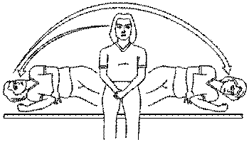
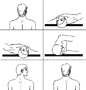
4. Repositioning maneuvers. Currently, this treatment approach has gained some space when compared to the others. It is the result of a practical approach, easy to handle and highly successful. Two classical maneuvers appeared: the liberatory maneuver by Semont11, described in 1988 (Figure 1), and Epley's canaliculi repositioning, described in 1992 (Figure 2). Different modifications of these techniques have been presented in past years4,7,12. In all maneuvers, the purpose is to make the degenerated fragments of otoconia, abnormally present in the posterior semicircular canal, to return to the utriculus by the action of gravity. Semont presented a series of 711 patients treated with a cure rate of 84% after the first session, and 93% after the second session. Many authors believe that Semont's liberatory maneuver is aggressive because it triggers severe dizziness many times, and consequently, it is not well tolerated by patients. In turn, Epley presented a series of 400 patients treated with the repositioning maneuver with a cure rate of 95%. Epley's maneuver, with some possible alterations, is the most widely adopted approach, especially in the USA, probably because it is more tolerated by patients. In general, the use of a small vibrator is recommended. It is placed close to the mastoid region on the affected side, which will help the displacement of fragments from the posterior semicircular canal. After the maneuver, the patient is instructed to try to avoid for 48 hours lying down with the head lower than the body and to rest preferably seated. The patient should also refrain from suddenly moving the head back and forth. These instructions intend to minimize the tendency of fragments to migrate back to the posterior semicircular canal. Some authors recommend the use of cervical collar for variable periods of time to meet the same needs.
MATERIAL AND METHOD
Seven patients with diagnosis of BPPV based on typical clinical history, confirmed by the presence of nystagmus in the maneuver of Dix Hallpike, were described. The classical repositioning maneuver of Epley was performed in all cases, in variable number of sessions. The mastoid oscillator was used and patients were instructed to prevent the lying down position, head down, for two days. All patients were followed up for a prolonged period of time, evidencing the results and the difficulties in some specific cases.
Case 1 - female 19-year-old patient
She had a car accident, with cranial damage, 20 days before. Since then, she experienced rotation dizziness triggered by various head movements. Normal otoneurological study. Dix-Hallpike maneuver to the right showed nystagmus. Repositioning was conducted. Repeated Hallpike's maneuver and the patient still showed nystagmus. Reevaluated 15 and 30 days later, after the second repositioning and the patient has remained asymptomatic since then.
Figure 1. Semont's maneuver. The patient is seated and the examiner is in from of him. The patient is quickly forced to lie down on the affected side. The patient should remain in position for 4 minutes. Next, he is quickly turned to the other side, always keeping the ear downwards. The patient is again maintained in the position for 4 minutes.
Case 2 - female 30-year-old patient
Rotation dizziness triggered when lying down, noticed for two days. Similar episodes six months before with spontaneous and gradual remission within approximately 15 days. Normal otoneurological study. Dix-Hallpike maneuver to the right resulted in nystagmus. Repositioning was conducted. Reevaluated three and 20 days later, the patient remained asymptomatic.
Case 3 - male 44 year-old patient
Rotation dizziness triggered by turning to the left side on the bed, for 14 days. The patient did not refer previous episodes of dizziness. Normal otoneurological study. Dix-Hallpike maneuver to the left presented nystagmus. Repositioning was conducted. Followed up for 30 days, the patient was asymptomatic.
Figure 2. Epley's maneuver. The patient is observed by the examiner, who is behind him/he.
Position 1 - Patient is seated on the examining table, lying down so as to leave the head pending from the table. The head will always be conducted and supported by the examiner.
Position 2 - Patient lies on his back, and his/her head is laterally rotated 45° to the affected labyrinth (left), and next it is hyperextended to the back, pending outside from the examining table.
Position 3 - Head turned to the other side (right) at 45°.
Position 4 - Head and body are rotated to the same side (right), so that the patient-can look to the floor.
Position 5 - Upon maintaining the lateral rotation of the head (right), the patient is brought back to the seated position.
Position 6 - Restore the points of the initial position, looking straight forward. Turn the head to the initial position and maintain a steady stare.
Case 4 - female 59-year-old patient
Rotation dizziness triggered by movement, backward elevation of the head and by turning to the left side on bed, for 7 days. She did not report previous episodes of dizziness. Normal otoneurological study. Dix-Hallpike maneuver to the left presented nystagmus. Repositioning was conducted. She was followed up for 5 months and remained asymptomatic with no relapse.
Case 5 - female 50-year-old patient
Rotation dizziness, followed by nausea and triggered by turning the head to the right, for 10 days. Otoneurological study: bilateral vestibular, hyporeflexia in caloric tests. Dix-Hallpike maneuver to the right presented nystagmus. Repositioning was conducted. She was reevaluated after 15 days and referred that she had a "50% improvement of the disease". Dix-Hallpike maneuver was repeated and produced no nystagmus. She was reevaluated 40 days after repositioning and reported "a 90% improvement of dizziness", whereas 60 days later she was asymptomatic.
Case 6 - male 57-year-old patient
Rotation dizziness triggered by movements of lowering and elevating the head and turning the body on bed, for 30 days. Otoneurological study: caloric tests with predominance of significant right labyrinth. Dix-Hallpike maneuver to the left showed nystagtnus. Repositioning was conducted. He remained asymptomatic for two days, had recurrence of dizziness on the third day, when Dix-Hallpike maneuver was repeated to the left and produced nystagmus. Repositioning was conducted once again. The patient remained asymptomatic for one year and then started to show relapse of dizziness episodes. Dix-Hallpike maneuver to the left produced nystagmus. Repositioning was conducted. On the evaluation .7 days after, he presented partial improvement of dizziness. New Dix-Hallpike maneuver presented nystagmus. Repositioning was conducted again. On the 15th and 30th follow-up day he remained asymptomatic.
Case 7 - female 45-year-old patient
Rotation dizziness followed by nausea and triggered by head movement that had started a little time before. Otoneurological study with mild bilateral vestibular hypereflexir-in the caloric tests. Normal magnetic resonance imaging. Normal clinical hematological tests. Dix-Hallpike maneuver on the right triggered a very evident nystagmus. Repositioning was conducted. The patient was followed up monthly for 4 months. Significant initial improvement, but the patient maintained the complaint of moderate positional dizziness. In each follow-up visit, repositioning was always repeated. Six months after, symptoms were still present and Dix-Hallpike maneuver was positive. The patient was referred to using labyrinthic medication and practicing vestibular habituation drills.
RESULTS
DISCUSSION
The clinical history of patients presented here was always compatible with BPPV, that is to say, there was dizziness triggered by movement - in some cases, very specific movements. Clinical examination presented the basic condition for diagnosis in all cases: presence of nystagmus with Dix-Hallpike maneuver.
The otoneurological study contributed little to diagnosis, but in our sample it served as a prognostic tool. In patients with normal otoneurological study, the response to repositioning maneuver was excellent, with no recurrence of the episodes. In patients with abnormal response to the caloric tests, either the response to the maneuver was not immediate or the condition presented relevant recurrence.
Results presented after Dix-Hallpike maneuver showed that all patients presented some improvement in the short-run. Most of the cases showed complete remission with no recurrence. In some patients, the improvement was partial or there were recurrence episodes of BPPV confirmed by the positive Dix-Hallpike maneuver. In such cases, repositioning showed good results in most of the cases. These facts show the importance of early reevaluation of repositioned patients with the repetition of Dix-Hallpike maneuver, followed by repositioning, if necessary.
Repositioning maneuvers were well accepted in all cases. Even in patients submitted to it in different visits in which nystagmus and vertigo sensation wer6 triggered, the maneuver was well tolerated and accepted.
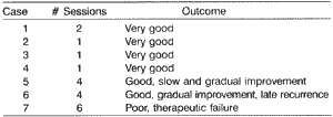
The results presented in the table clearly show that in most patients the curative effect of the repositioning maneuver is very evident. In one single visit, there was complete and immediate remission of positional dizziness episodes. In Case 5, we noticed, gradual remission of the symptomatology with slow regression, questioning the efficacy of the maneuver since the improvement could result from the natural regression of the process. Case 6 is interesting because after the second repositioning, we noticed immediate regression of symptomatology, showing efficacy of the approach. However, one year later, there was recurrence of the condition following the same standards, and there were the same positive results after new repositioning. Case 7 showed repositioning therapy failure: even after insisting on the method for 6 months, there was relevant symptomatology. In this case, Dix-Hallpike maneuver was always positive and present in all repetitions, showing the persistence of degenerated otoconia fragments in the posterior semicircular canal, that is, the method failed to remove them from there. Other forms of treatment were indicated in the case.
CONCLUSION
1. Diagnosis of BPPV is basically clinical. Patients' history and the examination using Dix-Hallpike maneuver conclude the diagnosis. Complementary tests do not contribute significantly to the diagnosis.
2. Otoneurological study with caloric tests proved to be an important prognostic method. Patients with good responses to repositioning maneuver presented normal responses in the caloric tests.
3. Repetition of the Dix-Hallpike maneuver some days after repositioning was useful to observe the results obtained and, if necessary, to repeat repositioning.
4. Repositioning maneuver proved to be an effective method for the treatment of BPPV; the majority of the patients reported here presented complete remission of symptoms after the conduction of one repositioning session.
REFERENCES
1. BARANY, R., cited by Dix, R., Hallpike, C.S. - Diagnose von Krankheitserscheinungen im Bereiche des Otolithenapparates. Acta Otolaryngol 2:434-7, 1921.
2. BRANDT, T.; DAROFF, R.G.B. - Physical therapy for benign paroxysmal positional vertigo. Arch Otolaryngol 106: 484-5, 1980.
3. BRANDT, T.; STEDDEN, S.; DAROFF, R.B. - Therapy for benign paroxysmal positional vertigo, revisited. Neurology 44:796-800, 1994.
4. COHEN, H. S.; JERABEK, J. - Efficacy of treatments for posterior canal benign paroxysmal positional vertigo. Laryngoscope 109:900 3, 1999.
5. DIX, M.R.; HALLPIKE, C.S. - The pathology, symptomatology and diagnosis of certain common disorders of the vestibular system. Proc Soc Med 45:431-354, 1952.
6. EPLEY, J. M. - The canalith repositioning procedure: for treatment of benign paroxysmal positional vertigo. Otolaryngol Head Neck Surg 107: 399-404, 1992.
7. LEMPERT, T.; WILCK, K.T. - A positional maneuver for treatment of horizontal canal benign positional vertigo. Laryngoscope 106: 476 8, 1996.
8. PARNES, L.S.; MC CLURE, J.A. - Posterior semicircular canal occlusion for intractable benign paroxysmal positional vertigo. Ann. Otol. Rhinol. Laryngol., 99:330-4, 1990.
9. PULEC, J.L. - Ablation of posterior semicircular canal for benign paroxysmal positional vertigo. ENT J 76:17-24, 1997.
10. SCHUKNECHT, H.F. - Cupulolithiasis. Arch. Otolaryngol., 90:765-78, 1969.
11. SEMONT, A.; FREYESS, G.; VITTE, E. - Curing the BPPV with a liberatory maneuver. Adv. Otol. Rhinol. Laryngol., 42.290-3, 1988.
12. WOLF, J.S.; MATTOX, D.E. - Success of the modified Epley maneuver in treating benign paroxysmal positional vertigo. Laryngoscope; 109:584-90, 1999.
1 Assistant Physician of Clinical Otorhinolaryngology, Medical School, Universidade de Santo Amato.
2 Resident Physician, Clinical Otorhinolaryngology, Medical School, Universidade de Santo Amaro.
Study carried out at the Clinical Otorhinolaryngology Department, Medical School, Universidade de Santo Amaro.
Address correspondence to: Dr. Roberto Alcantara Maia - Rua Jerônimo da Veiga 164, cjt°. 9 A - 04536-000 São Paulo/ SP.
Article submitted on February 16, 2001. Article accepted on May 30, 2001.


