

Year: 2002 Vol. 68 Ed. 5 - (19º)
Artigo Original
Pages: 722 to 728
 PDF PT
 Strange animated bodies in othorinolaringology
Author(s):
Ricardo R. Figueiredo 1,
Sandro Dorf 2,
Márcia S. Couri 3,
Andréia A. Azevedo 4,
Fernanda Mossumez 5
Keywords: strange animated bodies, insects, otolaryngology
Abstract:
Study design: Clinical retrospective. Material and method: Fufty-six cases of animated foreign bodies collected inside human ears (55 insects and 1 arachnid) and one case collected in nasal fossae (insect) were reported. The material was collected in the Emergency sector of Souza Aguiar Hospital, in Rio de Janeiro, between 1998 and 2000, and was identified by zoologists of Museu Nacional, Rio de Janeiro. Most of the cases had occurred in Nova Iguaçu and Campo Grande, suburbs of Rio de Janeiro. Clinical features and complications were analyzed. Results: The recorded insects are: 30,35% Blattaria (cockroaches); 25% Diptera (flies and mosquitos); 12,5% Lepidoptera (butterflies and moths); 10,7% Coleoptera (beetles); 7,15% Hemiptera (bugs, cicads, aphids, etc.), 5,35% Hymenoptera (wasps, bees, ants, and sawflies) and 5,31 % others.
![]()
INTRODUCTION
Different types of organisms may invade the human body in many different manners. They may be inhaled, ingested with contaminated food or water, or through sexual contact, thus penetrating the natural cavities of the human body. Consequences may range from a slight discomfort to severe clinical conditions, if they are pathogenic to humans. There are reports in the literature of injuries and ulcerations in cavities, such as infestations of the oral mucosa, among others.
Some species of arthropod animals may invade the human body through its natural openings, especially the mouth, nostrils and external acoustic duct. These invaders include insects, both in their adult and young forms. One example of human invasion by insects is myiasis, that is, the infestation of live or dead tissues by the larvae of flies. Larvae may cause many types of injuries, depending on the species and circumstances of the infestation, such as secondary bacterial infections and the patient's immune status.
In human beings, the ears, nose and throat are the most commonly infested areas, and deadly cases have been reported in the literature (Guimarães & Papavero, 1999).
The objective of this study was to identify the main invaders in the State of Rio de Janeiro, classify them, describe the clinical changes they cause, and their treatment, so as to prevent future invasions.
MATERIAL AND METHODS
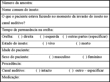
The specimens studied were collected in the Emergency Room of the Otorhinolaryngology Clinic of Municipal Hospital Souza Aguiar, in Central Rio de Janeiro, between 1998 and 2000. It comprises 56 samples of invading organisms preserved in test tubes with formaldehyde. For each case, a form was filled out recording information such as common name of the insect (given by the patient or staff), what the patient was doing when the invasion occurred (lying, walking, etc.), time elapsed since the invasion, and others (Figure 1).
Specimens were collected by means of an otologic lavage or with the help of Hartmann forceps. Some of the specimens were examined with the help of a microscope, after checking whether the insect was alive or dead. When they were alive, they were first killed with the instillation of ether, except the larvae. Given the technical difficulties to remove them in some cases, certain samples were extremely damaged, thus making impossible the identification to the level of species.
The information on the samples has been summarized in Table 1, which also includes the identification of the organisms, separated by Order. Researchers of the Departments of Entomology and Invertebrates of the National Museum of Rio de Janeiro identified the organisms. All the specimens are stored in the collection of the National Museum.
Table 1. Animated foreign bodies in the ear of patients at the ER of Hospital Souza Aguiar - identification and information about the patient and the insect.
Note: MT: tympanic membrane; M/F: male and female; side R/L: right and left.
RESULT AND DISCUSSION
Biological Aspects
All of the material examined included insects, except for samples 7 and 45, which were arachnids. Most of them were identified to the level of species or genus, some to the level of family, either due to the lack of experts in that certain group of insects, or due to the state of the sample. Table 1 shows the identifications.
The data show that most (about 55.35%) of the invaders were either Blattidae (cockroaches) or Diptera (flies and mosquitoes), which accounted, respectively, for 30.35% and 25% of the cases. Next, were the Lepidoptera (butterflies and moths), 12.50%; Coleoptera (beetles), 10.70%; Hemiptera (bedbugs, cicadas, etc.), 7.15%; Hymenoptera (wasps, bees, ants), 5.35%; Isoptera (termites) and Araneae (spiders), 3.60% and only 1.71% was Thysanura.
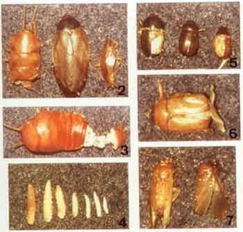
The Blattidae (Figures 2-3) were represented chiefly by Periplaneta spp. (47.5%) and Blattella spp. (29.5%), which are household species. These cockroaches look for food remains inside households and may account for the transmission of bacterial and protozoal diseases and food contamination. They may also invade the labial mucosa of people during sleep, looking for food remains, causing a form of herpes known as "Herpes blattae" (Costa Lima, 1938). All of the Blattella specimens were B. germanica (Linnaeus). Other species found were Pycnoscelus surinamensis (17%) and Ischnoptera spp. (6%).
Almost all the dipteran found belong to the genus Cochliomyia Townsend (25%), which is endemic in the New World. Most of them were (54%) C. hominivorax (Coquerel) (Figure 4) and only 7.7% were C. macellaria (Fabricius). Both species were collected in their larvae stage; they belong to the family Calliphoridae, and are two of the most abundant and widespread species of this genus. The larvae are obligatory parasites that cause myiasis in men and in other animals; they usually develop between the dermis and subcutaneous layer. C. hominivorax is found from southern USA to northern Chile, Argentina, and Uruguay. It is an obligatory parasite of wounds in mammals, feed on living tissues, and infest almost all forms of domestic animals, wild animals, and human beings (Guimarães and Papavero, 1999). C. macellaria is common in the tropical and subtropical regions of the Western Hemisphere. They may act as secondary invaders of wounds. One of the cases of C. hominivorax occurred in both nasal cavities of a beggar.
Other dipterans are four mosquitoes of the family Culicidae and one fly of the family Sarcophagidae, Oxysarcodexia amorosa (Townsend). The members of the family Sarcophagidae are, without exception, either viviparous or oviparous. Their larvae develop in carcasses, excrement or decaying matter. Males are frequent visitors of flowers, but the female of some species are parasites of other invertebrates, sometimes destroying the host's eggs or larvae (Shewell, 1987). There are reports of accidental cases of myiasis by larvae of Sarcophagidae, including a case of auricular myiasis caused by Bercaea haemorrhoidalis (Fallen) in Argentina (Guimarães & Papavero, 1999).
The coleopterans (Figures 5-6) collected belong to the family Scarabaeidae, which comprises phytophagous species, easily attracted by mercury-vapor lamps. Here, they have been represented by the sub-families Rutelinae (60 %) and Dynastinae (40%). 40% of the invader beetles were Leucothereus spp.. Lepidoptera is one of the largest orders of predominantly phytophagous insects, and here were represented by 7 families (Nielsen & Common, 1991).
Figura 1. Formulário com os dados do paciente e do inseto
Figure 2. (left to the right): Periplaneta sp (sample 58); Pycnoscelus surinamensis (sample 56); Blatella germanica (sample 17). Figure 3. Ischnoptera sp (sample 50). Figure 4. Larvae of Cocchliomyia hominivorax (various samples). Figure 5. (left to the right): Eutheola humilis (sample 53); Leucothyreus sp (sample 26); Leucothyreus sp (sample 08). Figure 6. Geniates sp (sample 4). Figure 7. (left to the right): Ardiosterus sp (sample 51); Noctuidae (sample 11).
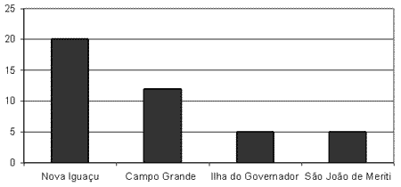
Figure 8. Area where most of the cases were recorded (%).
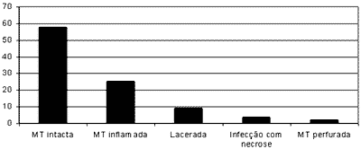
Figure 9. Status of the auditory canal (%) (MT = tympanic membrane).
Of the hemipteran collected, 25% were Macyconalia cavifrons (Stal), 25% Cicadellidae, 25% Cydnidae, and 25% Cimex hemipterus (Fabricius). The latter, together with other species of the same genus, is known as the common bedbug. They are tropical parasites of birds and mammals, but they often attack humans to suck their blood. Cimex hemipterus live in households, next to the beds of humans, and come out at night to feed, which they do rapidly, becoming engorged in a few minutes. They typically produce several bites, a short distance away from each other, in a linear configuration. Normally, humans do not feel the bites, and they do not prefer a specific part (although the face is the most commonly affected area, as it is more often uncovered during sleep). Some people are asymptomatic, but others may present edema and pruritus (Waterhouse, 1991).
The hymenopteran were the least represented group of insects (5.35%). Most of the specimens collected belonged to the families Formicidae (ants 66.60%) and Apidae (bees 33.40%). Both families are sinanthropic (species that adjust well to the conditions created by humans, keeping their independence, and adapting very well to urban environments), and very common in urban areas.
Isopterans (termites) were represented by two specimens of Coptotermes hevilandi (Horlmargren). It is a relatively small Order, closely related to the Blattidae, which occurs predominantly in tropical and subtropical regions. These insects feed on wood-whether healthy or not-dry grass, fungi, and other materials of vegetal origin. The main material used is pulp, but some species are capable of degrading lignin (Watson & Gay, 1991).
The small and cosmopolitan Order of the Thysanura was represented by a sample of Btenolepisma longicaudata (Escherich). Most of the Thysanura live freely and are extremely agile. They are hominivorous and very small, measuring between 3 and 7 mm in length (Smith & Watson, 1991).
The Order Araneae was represented by the families Gnaphosidae, which lives only in the soil, and hardly ever gets in contact with humans, and Salticidae, which is sinanthropic. Plexippus paykulli (Audoin) is known as eat-fly, because they move a lot to eat, as they do not make a web. They are domestic and very common.
Embioptera and Psocoptera (1.8% each) are insects without medical relevance. Embioptera is one of the smallest and least studied insect Orders; it is essentially tropical and feeds exclusively on vegetables. The Psocoptera are found in all zoogeographic regions, on leaves, branches, barks, under stones, in human households and in stored products. (Smithers, 1991).
Clinical Aspects
There were no significant differences related to gender, 51.79% being female and 48.21% male.
As to age, 25% were aged between 0 and 12, 12.5% were between 13 and 21 years of age, 58.92% between 22 and 59, and only 1.78% was above 60 years of age. The youngest occurrence was a case of auricular myiasis in a one-year-old child.
The average time elapsed between the entrance of the invader and its removal was about nine hours. The quickest case was a cockroach in a person who lived in the Center (close to the hospital), which took about half an hour; and the longest was another cockroach in a 59-year-old patient from Campo Grande (western outskirts of Rio), who took about 7 days to seek care. There were 4 cases in which the person did not know when the invasion had occurred, two cases of myiasis, one of a mosquito (Culex sp.), and one bee (Apidae). Normally, patients had been to two or more hospitals before getting to Souza Aguiar, due to the serious lack of ENT care in the State of Rio de Janeiro, and to the fact that Hospital Souza Aguiar is a reference for strange bodies in ENT.
As to the site of invasion, 51.79% were in the right ear, 46.42% in the left ear, and there was one case of nasal myiasis (1.79%).
Concerning the state of the invader, 73.22% were dead when removed and 26.78% were alive. Among the alive, the most frequent were Diptera (53.33%, of which 75% were myiasis), followed by the Blattidae (26.66%), Hemiptera, Lepidoptera and Thysanura (6.66% each). As to the activity people were engaged in at the moment of invasion, it is worth stressing that 23.21% were lying in bed, 16.07% were lying on the floor, and 7.14% were next to areas of abundant vegetation or trees. Therefore, being immobile and lying down, in addition to being close to green areas, were factors that proved relevant for invasion.
The regions of the Baixada Fluminense (Nova Iguaçu) and the West Zone (Campo Grande) accounted for most of the cases (Figure 8).
About 58% of the cases did not present complications; however, when they occurred, the most frequent were hyperemia of the external acoustic duct, laceration of the external acoustic duct, secondary infection and necrosis of the external ear, and tympanic perforation (Figure 9). Most of the complications were not severe and were treated with topic (otologic drops) and symptomatic medication. The cases of tympanic perforation were prescribed antibiotics and followed up at outpatient level. Most of the severe cases were myiasis, caused chiefly by the larvae of C.
hominivorax. In the case of auricular myiasis, the greatest possible number of larvae was removed, preferably under microscopic vision. A dressing was placed so as to close the lumen of the external acoustic duct (so as to reduce the air supply to the larvae). These dressings used calomel, a mercury byproduct, which, in spite of the many reports as to its possible neuro and hepato-toxicity, has shown to be totally safe when used in ENT, as our experience has shown us. In our opinion, auricular myiasis can be perfectly treated at outpatient level, except when social factors make it impossible. Dressings were changed every other day, and antibiotics (cephalexin) were prescribed, until the complete remission of the symptoms. None of the cases left sequelae, except those that already had a chronic inflammatory pathology, such as otitis media and conditions affecting the sinuses. Children with chronic otitis media are frequent targets, due to their low socio-economic status.
The most serious case was certainly that of a beggar with nasal myiasis, who presented suppuration, necrosis of the septal cartilage and lower turbinate. Nasal myiasis is common among elderly, psychiatric, and alcoholic patients, and bearers of nasal granulomatosis, particularly those who live in rural regions. Additionally, frequent falls, associated to little mobility, cause bleeding that attract some species of flies. In these cases, patients have to be hospitalized for parenteral antibiotic therapy and surgical debridement of the lesions, which very frequently leaves sequelae, with large septal perforations. We do not have experience with the use of ivermectin, anti-parasitic drug mentioned by Ramalho et al. (2001).
CONCLUSION
Cases of invasions of the ear, nose and throat by insects and other organisms are not rare. Most of them can be easily solved and do not leave sequelae, but some may evolve into severe complications. Most of the insects collected in this study were attracted either by mercury-vapor light or food leftovers. Therefore, one way to minimize the invasions of human cavities by insects is to reduce the use of mercury-vapor light and to adopt cleaning and hygiene measures, such as the elimination of food remains, for instance. In order to avoid complications, it is also important not to try to have them removed by non-qualified professional staff or in the absence of the appropriate material.
ACKNOWLEDGEMENTS
We thank researchers of the Departments of Entomology and Invertebrates of the National Museum, UFRJ - Sonia Maria Lopes Fraga, Paulo Magno, Gabriel Mejdalani, Renner Luiz Cerqueira Baptista, Denise Pamplona, and Maria Antonieta Pereira de Azevedo, for their help in identifying the specimens, and all otorhinolaryngologists of Municipal Hospital Souza Aguiar.
REFERENCES
1. Costa Lima AM. Insetos do Brasil. 1º Tomo - Série didática, nº 2 da Escola Nacional de Agronomia do Rio de Janeiro; 1938. p. 1-470.
2. Guimarães JH & Papavero N. Myiasis in man and animals in the neotropical region; Bibliographic database. São Paulo: Editora Plêiade/Fapesp; 1999. p.1-308.
3. Nielsen ES & Common IFB. Lepidoptera: 817-915. In: The Insects of Australia. A textbook for students and research workers. Volume II, Second Edition, Division of Entomology CSIRO Australia; 1991. p.543-1132.
4. Ramalho JRO et al. Miíase nasal: relato de um caso. Revista Brasileira de Otorrinolaringologia 2001;67(4):581-84.
5. Ross ES. Embioptera (Embiidina): 405-400. In: The Insects of Australia - A textbook for students and research workers. Volume I, Second Edition, Division of Entomology CSIRO Australia; 1991. p.1-542.
6. Shewell GE. Sarcophagidae: 1159-1186. In: Manual of the Nearctic Diptera Vol. II. Otawa, Agriculture Canada, Research Branch Monograph, 28, 1987. vi + 675-1332 p.
7. Smith GB & Watson JA. Thysanura (Zygentoma): 275-278. In: The Insects of Australia. A textbook for students and research workers. Volume I, Second Edition, Division of Entomology CSIRO Australia; 1991. p.1-542.
8. Smithers CN. Psocoptera: 412-420. In: The Insects of Australia. A textbook for students and research workers. Volume I, Second Edition, Division of Entomology CSIRO Australia; 1991. p.1-542.
9. Waterhouse DF. Insects and humans in Australia: 221-235. In: The Insects of Australia. A textbook for students and research workers. Volume I, Second Edition, Division of Entomology CSIRO Australia; 1991. p.1-542.
10. Watson JAL & Gay FJ. Isoptera: 330-347. In: The Insects of Australia A textbook for students and research workers. Volume I, Second Edition, Division of Entomology CSIRO Australia; 1991 p.1-542.
1 Physician, Service of Otorhinolaryngology, Municipal Hospital Souza Aguiar, Rio de Janeiro.
2 Intern of Scientific Studies, Museu Nacional, UFRJ; undergraduate of Medical School, Souza Marques.
3 Joint Professor, Museu Nacional, UFRJ; Researcher with CNPq; Ph.D. in Veterinarian Parasitology; Rural Federal University of Rio de Janeiro.
4 Physician, intern of the Service of Otorhinolaryngology, Municipal Hospital Souza Aguiar, Rio de Janeiro.
5 Resident physician, Service of Otorhinolaryngology, Municipal Hospital Souza Aguiar, Rio de Janeiro.
Study conducted at Municipal Hospital Souza Aguiar, Rio de Janeiro and Museu Nacional, Rio de Janeiro.
Address correspondence to: Ricardo R. Figueiredo - Rua 60, nº 1680, apto 202
Bairro Sessenta - Volta Redonda - RJ - CEP 27255-650 - Tel. (55 24) 3342-9073 - E-mail: otosul@ig.com.br
Article submitted on November 03, 2002. Article accepted on June 20, 2002
Print: ![]()