

Year: 2014 Vol. 80 Ed. 2 - (17º)
Relato de Caso
Pages: 180 to 181
 PDF PT
  PDF EN
Neuroendocrine adenoma of the middle ear confused with congenital cholesteatoma
Author(s): Yee Hyuk Kim1; Sang Heun Lee2; Da Jung Jung3
DOI: 10.5935/1808-8694.20140036
![]()
INTRODUCTION
Neuroendocrine adenomas of the middle ear (NEAME) are rare tumors.1-4 Whereas neuroendocrine cells are normally found in the lung and gastrointestinal tract, they are normally not found in the middle ear (ME) mucosa.1-6 Although neuroendocrine tumors were initially believed to originate from the neuroendocrine cell system, it is now known that they may originate from tissue that lacks neuroendocrine cells, such as the ME mucosa, because these tumors are derived from pluripotent stem cells that are capable of neuroendocrine differentiation.1,5
CASE PRESENTATION
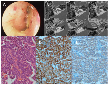
A 27-year-old female presented with a three-month history of fullness in the right ear. The patient visited a local hospital and was diagnosed as congenital cholesteatoma. The patient was referred to this hospital. Upon otoscopic examination, the right tympanic membrane (TM) was found to be bulging, with a whitish ME mass (fig. 1A). Pure tone audiometry revealed air and bone conduction thresholds of 20 and 12.5dB HL, respectively, on the right side. However, the thresholds at 4 kHz and 8 kHz were elevated. The computed tomography (CT) scan showed soft tissue density in the ME cavity, and there was no evidence of bony destruction (fig. 1B).
Figure 1 A, Preoperative otoscopic finding shows whitish mass behind the tympanic membrane. B, Preoperative axial view of temporal bone CT shows soft tissue density in the middle ear cavity. C, Tumor is composed of trabeculars lined by multiple layers of small and monotonous cells with an intraluminal eosinophilic secretion (H&E stain ×200). D, Immunohistochemical stain for chromogranin shows strong positivity within tumor cells (chromogranin × 400). E, Immunohistochemical stain for synaptophysin shows strong positivity within tumor cells (synaptophysin × 400).
A canal wall up mastoidectomy and tympanoplasty were performed. During the operation, a soft, pale, well-encapsulated mass was found filling the tympanum and encasing the stapes. The incus and stapes were extracted for complete removal of tumor. The footplate of the stapes was repositioned; the incus was remodeled and then positioned on the stapes footplate. The specimen was soft and relatively tougher than cholesteatoma, and there was no keratin material.
The histopathologic results revealed a well-differentiated neuroendocrine neoplasm. Routine light microscopy revealed a proliferation of small monotonous cells arranged in trabecular patterns. There were no findings of necrosis, nuclear atypia, and mitosis (fig. 1C). Immunohistochemical stainings were positive for chromogranin, synaptophysin, neuron specific enolase, vimentin, CD56, and cytokeratin (fig. 1D-E).
The final diagnosis was revised from congenital cholesteatoma to NEAME. The patient did not present any symptoms related to carcinoid syndrome. Chest and abdominal CT were performed, as well as an assessment of the urinary catecholamines and their metabolites for investigating distant metastases and simultaneous carcinoid tumor in other organs. The catecholamines and their metabolites were found within the normal limits and chest and abdominal CT did not reveal any mass lesions. The patient has been followed for 30 months without evidence of recurrent disease.
DISCUSSION
NEAME can be confused with glomus tympanicum and congenital cholesteatoma, as was the case with the present patient. It is difficult to diagnose NEAME before the final histological examination, since the clinical manifestations and radiographic findings are nonspecific.
The pathologic diagnosis of NEAME is primarily based on light microscopy with hematoxylin-eosin staining; the diagnosis is confirmed by immunohistochemical investigation.1,2 On immunohistochemical staining, epithelial markers include cytokeratins, and neuroendocrine markers include chromogranin, synaptophysin, neuron specific enolase, vimentin, and CD56.5 Angouridakis et al. suggested that NEAME were positive for epithelial and neuroendocrine markers, while carcinoid tumors of the ME were negative for epithelial markers and positive for neuroendocrine markers.6 Saliba et al. proposed that NEAME were positive for immunohistochemistry and there was no evidence of metastasis, whereas carcinoid tumors of the ME were positive for immunohistochemistry and there was evidence of metastasis or carcinoid syndrome.5 In the present case, the tumor cells were positive for epithelial marker and neuroendocrine markers. There was no evidence of metastasis and carcinoid syndrome. Therefore, the tumor of present case is best described by the term NEAME.5,6
FINAL REMARKS
NEAME are uncommon. The diagnosis of NEAME is made by the presence or absence of immunohistochemical markers, metastases, and carcinoid syndrome.
CONFLICTS OF INTEREST
The authors declare no conflicts of interest.
REFERENCES
1. Sahan M, Yildirim N, Arslanoglu A, Karslioglu Y, Kazikdass KC. Carcinoid tumor of the middle ear: report of a case. Am J Otolaryngol. 2008;29:352-6.
2. Ferlito A, Devaney KO, Rinaldo A. Primary carcinoid tumor of the middle ear: A potentially metastasizing tumor. Acta Otolaryngol. 2006;126:228-31.
3. Ramsey MJ, Nadol JB, Jr., Pilch BZ, McKenna MJ. Carcinoid tumor of the middle ear: Clinical features, recurrences, and metastases. Laryngoscope. 2005;115:1660-6.
4. Chan KC, Wu CM, Huang SF. Carcinoid tumor of the middle ear: a case report. Am J Otolaryngol. 2005;26:57-9.
5. Saliba I, Evrard AS. Middle ear glandular neoplasms adenoma, carcinoma or adenoma with neuroendocrine differentiation: a case series. Cases J. 2009;2:6508.
6. Angouridakis N, Hytiroglou P, Markou K, Bouzakis A, Vital V. Middle ear adenoma/carcinoid tumor: a case report and review of the literature. Rev Laryngol Otol Rhinol (Bord). 2009;130:199-202.
1. Department of Otorhinolaryngology-Head and Neck Surgery, School of Medicine, Catholic University of Daegu, Daegu, Korea
2. Department of Otorhinolaryngology, Daegu Veterans Hospital, Daegu, Korea
3. Department of Otorhinolaryngology-Head and Neck Surgery, School of Medicine, Kyungpook National University, Daegu, Korea
Corresponding author.
S.H. Lee
E-mail corin9525@hanmail.net
Received 29 October 2012.
Accepted 23 March 2013.
Print: ![]()
All rights reserved - 1933 /
2026
© - Associação Brasileira de Otorrinolaringologia e Cirurgia Cérvico Facial