
Year: 2014 Vol. 80 Ed. 1 - (5º)
Artigo Original
Pages: 11 to 17
Olfaction disorders: retrospective study
Author(s): Luciano Lobato Gregorio; Fábio Caparroz; Leonardo Mendes Acatauassú Nunes; Luciano Rodrigues Neves; Eduardo Kosugi Macoto
DOI: 10.5935/1808-8694.20140005
Keywords: Smell; Olfaction disorders; Olfactory perception
Abstract:
INTRODUCTION: The smell, subjective phenomenon of great importance, is poorly understood and studied in humans. Physicians with more knowledge about smell disorders tend to consider the phenomenon important and to better manage the diagnosis and its treatment.
AIMS: First to describe a sample of patients presenting with main complaint of disturbances of smell. And second, to show our experience on management and treatment of this disease.
DESIGN: Retrospective cross-sectional cohort study.
MATERIALS AND METHODS: Sample description and assessment of treatment response in patients with main complaint of hyposmia or anosmia from January 2005 to October 2011.
RESULTS: From 38 patients presented with main complaint of an olfactory disorder, 68.4% of the patients were presented with hyposmia and 31,5% with anosmia, with a mean duration of 30.8 months. The main etiologic diagnoses were idiopathic (31.5%), rhinitis (28.9%) and CRS with polyps (10.5%). Responses to treatment with topical steroids and alpha-lipoic acid were variable, as well as in the literature.
CONCLUSION: Greater importance should be given to disorders of smell in practice of otolaryngologists, since its large differential diagnosis and the fact that could increase morbidity to patients, impacting on their quality of life.
![]()
INTRODUCTION
Olfaction disorders have attained great importance in recent years. In humans, the sense of smell is probably the least understood, being mostly a subjective phenomenon.1 Anosmia and hyposmia are terms that refer to the complete and partial loss of smell, respectively. The term dysosmia, in turn, describes an altered sense of smell, both in response to environmental stimuli (an alteration termed parosmia) and spontaneous events (termed phantosmia).2
The loss or impairment of olfaction is a relatively common condition, especially in the elderly. Doty et al. estimated that approximately 75% of individuals older than 80 years and 50% of those between 65 and 80 years suffer from considerable decrease in olfactory function.3 However, the prevalence of olfaction disorders in the general population was and has been underestimated, as indicated by some recent studies. In a study with a representative sample of the population (n = 1,387) performed in Sweden, a prevalence of 19% was calculated for olfaction disorders in general.4
Moreover, it is well established that the sense of smell strongly contributes to taste perception, so that patients with hyposmia or anosmia have great difficulty in perceiving the taste of food, thus losing the appetite for and pleasure from food.1 The olfactory impulses propagate to the limbic system, as well as to the higher cortical areas. Certain olfactory stimuli may trigger diverse emotional responses and create different cortical associations with other senses; this process is connected to the individual's memory and the perception of emotional stimulus quality, i.e., the feeling of being pleasant or unpleasant.1
Another factor to consider is that hyposmia may cause a significant impact on the quality of life of patients, bringing difficulties in activities of daily living, mood disorders, decreased appetite, and even work problems.5
Standardized olfactory tests were created in an attempt to objectify a subjective symptom since olfaction disorders are sources of several medical complaints and result in thousands of annual physician visits.6 In this sense, olfactory dysfunction has been investigated as one of the earliest pre-clinical signs in both Alzheimer's disease (AD) and in sporadic Parkinson's disease (PD).7
In a recent study performed with questionnaires applied to otolaryngologists (n = 231), it was observed that only 7.3% of participants reported using olfaction assessment tests in daily clinical practice. It was demonstrated that most physicians observed tests being applied and received training on testing for olfaction disorders during the residency period. In addition, physicians with greater knowledge about these disorders tend to consider the disease to be more important, and therefore achieve better diagnosis and treatment.8
This study aimed to describe the sample of patients with a complaint of olfaction disorders, report this service's experience in the management of these patients, and review the literature on olfaction disorders with emphasis on olfaction assessment methods, etiological diagnosis, and conduct.
METHOD
This was a retrospective historical cohort study of patients treated at a rhinology outpatient clinic of a tertiary hospital with a chief complaint of olfaction disorder (anosmia and hyposmia) during the period of January of 2005 to October of 2011. The following were evaluated: age, gender, duration of complaint, symptoms at diagnosis (nasal obstruction, anterior or posterior rhinorrhea, epistaxis, headache, among others), comorbidities, as well as findings during the otorhinolaryngology examination and findings at the complementary imaging assessment. The percentage of improvement with different treatments according to each diagnostic hypothesis was also evaluated.
On physical examination, the following were considered to be positive findings: presence of nasal polyps at any grade and location; septal deviation grades II and III; inferior turbinate hypertrophy with pale mucosa; and presence of secretion in the middle meatus. Regarding the computed tomography (CT) findings of the paranasal sinuses, the following were considered as positive: obstruction of the ostium-meatal complex; partial or complete opacification of one or more paranasal sinuses; and presence of concha bullosa. No standardized routine olfactory tests were applied during patient assessment.
RESULTS
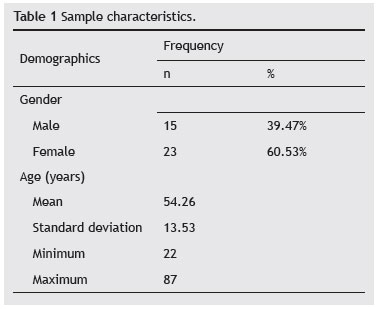
The study was submitted to and approved by the Ethics Committee of Plataforma Brasil under CAAE No. 07390412.9.0000.5505. All outpatients complaining of olfaction disorders treated at the rhinology outpatient clinic of a tertiary hospital from January of 2005 to October of 2011 (n = 38) were included. Patients with minor complaints of anosmia or hyposmia and those who had incomplete data in medical records were excluded. Sample characteristics are shown in Table 1.
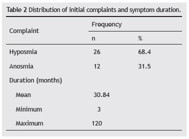
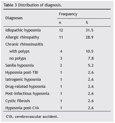
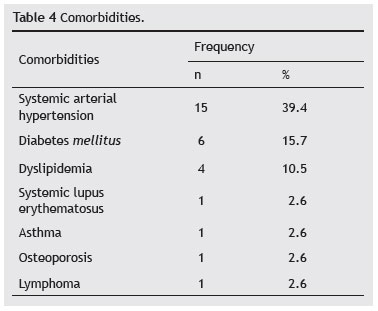
The initial complaints and their duration are described in Table 2. The distribution of diagnoses found in the evaluation of patients is shown in Table 3. Patients' comorbidities are shown in Table 4.
Regarding the physical examination findings, 18 (47.3%) of 38 patients had one or more criteria at physical examination described as positive. Regarding the CT examination, it was requested for 20 patients (52.6%), of whom seven were positive, and 13 showed no alterations. It is important to mention that no other alterations or anatomical variations that could influence ostium-meatal complex drainage (e.g., paradoxical middle turbinate, Haller cells) were observed in any of the cases.
Of all the patients in the sample who sought the outpatient clinic with complaints of olfaction disorders, 8 (21%) had undergone previous treatment at another service.
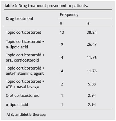
Of these, only four (50%) had partial improvement. Thirty-four patients (89.4%) received drug treatment in this service. The therapeutic options are detailed in Table 5.
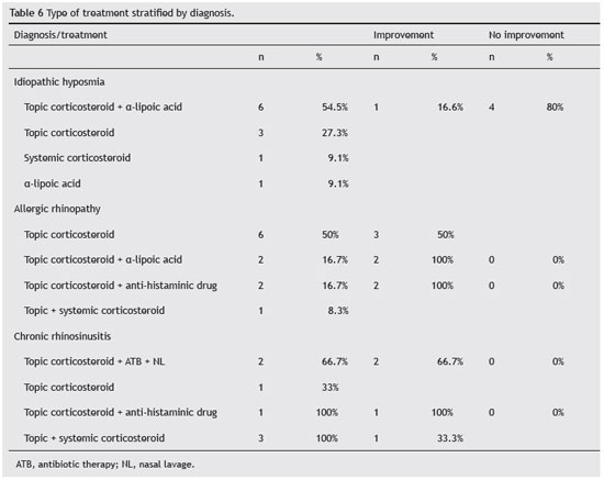
Table 6 shows the type of treatment in the main etiological diagnoses of the patients in this sample, with the respective improvement percentages.
A minimum follow-up of 2 months was attained in 57.8% of patients to assess treatment response. Of these, 16 (72.7%) patients showed partial or full response to treatment.
DISCUSSION
The sense of smell is quite undervalued in humans, despite its usefulness not only in monitoring the entry of harmful agents in the upper airways, but also in largely determining taste and palatability of foods or drinks.7 A normal perception of smell is important for the safety, nutritional status, and quality of life of an individual.9 Hyposmia or anosmia are associated with several conditions, such as PD, AD, Down's syndrome, multiple sclerosis, and schizophrenia, among others.10 Recently, neuroimaging studies determined that patients with anosmia show loss of cortical gray matter volume in certain areas (such as the medial prefrontal cortex), thus postulating an association between the development of neurodegenerative diseases and patients with anosmia.11,12 Nevertheless, few otolaryngologists have enough knowledge on olfaction disorders.8 A recent study in Turkey indicated that 83.5% of otolaryngologists did not observe the implementation of standardized olfaction assessment tests during the residency period.8 The lack of knowledge and training of physicians in their daily practice can lead to misdiagnosis and mismanagement of olfaction disorders.
When assessing the patient, the clinical history is the most important tool to attain the diagnosis. Important questions are: time of loss, associated concomitant factors (e.g., history of head trauma or a history of upper respiratory tract infections [URTIs]), characterization of total (anosmia) or partial loss (hyposmia), uni/bilaterality, presence of olfactory sense fluctuation, other associated nasal symptoms, characterization of the onset as sudden or insidious, and the presence of concomitant neurological signs and symptoms.
The supplementary examination can indicate underlying liver, kidney, or even neurological diseases. The history of past or current use of medications is also important.2 In the present sample, a case of hyposmia of probable drug-related etiology was observed in a patient with underlying diagnosis of rheumatoid arthritis using methotrexate.
The physical examination involves complete otorhinolaryngological examination, including flexible endoscope, focusing on narrowing alterations; changes in the epithelium; or presence of abnormalities in the olfactory area. The neurological examination should help in cases directed by the clinical history.2 The CT scan of the paranasal sinuses can help in the diagnosis of chronic rhinosinusitis and disclose areas of hypodensity in the olfactory region. Magnetic resonance imaging (MRI), in turn, can be useful in cases of nasal tumors invading the lamina cribrosa and central nervous system, or in cases of olfactory bulb underdevelopment, or even when neurodegenerative processes are suspected.13
Studies have shown that MRI presents good cost-effectiveness in the etiological diagnosis of olfactory disorders, as it can disclose information relevant to the diagnosis in up to 25% of cases of idiopathic dysosmia.14 In the present study, CT was requested for 52.6% of patients. MRI was less often requested (for only one patient) due to the high cost and difficulty of access to the examination.
Regarding the olfaction assessment methods, a large variety of tests have been developed in recent decades to clinically evaluate the presence or absence of hyposmia or anosmia. The originally developed clinical measure consisted of presenting odoriferous substances to patients and asking them to name these substances sequentially. This method was shown to be inadequate, as even normal patients have difficulties in naming certain types of odors, whereas certain substances may produce irritation of the nasal mucosa and stimulate the perception of patients with hyposmia. Other methods were shown to be inefficient, as they are too time-consuming for clinical use, involving complex equipment and showing lack of accuracy in the diagnosis of hyposmia - such as the measure of the olfactory evoked potentials.10
Therefore, standardized olfaction assessment tests were developed by large specialized centers, which were disseminated for use in clinical assessment due to their practical applicability.10 The most commonly used test is the University of Pennsylvania Smell Identification Test (UPSIT), which tests for the most common odoriferous particles, encapsulated, including 40 items.2 After being presented each odor from a list of four items, patients choose the one that best describes the odor they perceived. The total number of odors identified is the UPSIT score. This score allows for the classification of olfactory function as normosmia, hyposmia (mild, moderate, and severe) and anosmia.6 This test can be self-applied and takes approximately 15 to 20 minutes. It has high reliability, as described in the literature. This test helps to solve the problem of concomitant stimulation of the trigeminal nerve when presenting odors.6 Its limitations refer primarily to patients with cognitive deficits and language problems.10
Other problems were also observed in these standardized tests, such as the presence of false negatives. Deems et al. demonstrated that up to 29% of patients complaining of loss of smell had no alterations on standardized tests.2 Recently, the UPSIT-Br2 questionnaire was created and adapted to the Brazilian sociocultural context;9 it was shown to be a sensitive tool in the assessment of olfaction disorders in Brazil. In a recent study, the findings of a study that applied this questionnaire were consistent with those obtained in the literature, showing a better performance in females compared to males in identifying odors.9
Regarding the etiology, over 200 causes of olfactory disorders have been reported. Among these, four main causes stand out, excluding age-related hyposmia: traumatic brain injury (TBI), URTIs, chronic rhinosinusitis (CRS) with or without nasal polyps, and the idiopathic form. These four causes account for 70% to 85% of all causes of hyposmia.2 These causes can be divided into two large groups according to their physiopathology: sensorineural or conduction diseases. The former includes olfactory nerve or central nervous system injury, damage from upper airway infections, traumatic brain injury, neurotoxins, and lastly, CRS without polyps.
The causes of conduction, in turn, include nasal tumors, traumatic septal deviations affecting the cribriform plate, and CRS with polyps.13 This classification is often confusing, as some causes can be identified as both conduction and sensorineural, such as CRS. For this reason, the authors chose not to apply this classification in the present sample.
Some studies have identified traumatic brain injury (TBI) as one of the most frequent causes. A review of large series of patients after TBI demonstrated that 5% to 7% of all trauma cases had anosmia, and occipital or frontal trauma was the most common type related to olfaction disorder.2 Although the trauma intensity is usually directly proportional to the degree of olfactory loss, loss of consciousness may not necessarily occur; in addition, mild trauma can cause anosmia or hyposmia. Young adult males are most commonly affected, as they constitute a risk group to cranial trauma. Anosmia is found in up to three-quarters of these patients.2
The post-viral cause is diagnosed when there is loss of olfaction after an upper airway infection, without other apparent cause. These patients typically refer to the URTI as "the worst I have ever had." They tend to be older, with a predominance of females at a ratio of 2:1, for still unknown reasons. In this case, hyposmia is more common and dysosmia can be observed in up to two-thirds of patients.2 Some studies suggest that post-viral hyposmia or anosmia should always be investigated through imaging exams, treating these as diagnosis of exclusion cases, as some underlying disease may be present, including intracranial tumors.15
The sinonasal diseases, in turn, are mentioned as causes of hyposmia in approximately 15% of cases.2 Of the patients with CRS, approximately 65% to 80% have olfactory dysfunction.16 Nasal obstruction preventing airflow in the olfactory fossa is often identified as the etiological factor in these cases, in addition to damage to the neuroepithelium due to chronic inflammation.
For instance, the presence of large polyps in the olfactory area prevents the molecules that stimulate the receptors from binding to them. In patients with CRS, a study demonstrated that the presence of nasal polyps, septal deviation, inferior turbinate hypertrophy, and (to a lesser extent) smoking and allergic rhinitis were predictors of olfactory dysfunction.17 These patients may not have nasal complaints in addition to hyposmia, and the disease may not be evident in the nasal endoscopy. This makes diagnosis more difficult; thus, otolaryngologists should be aware of causes that are amenable to surgical treatment - chronic rhinosinusitis with or without polyps - before assuming the idiopathic cause of hyposmia.2 In the present sample, 18.4% of the cases were diagnosed with CRS with or without nasal polyps.
In CRS without nasal polyps, studies have demonstrated that the presence of eosinophils in the mucosa of the olfactory epithelium and basal membrane thickening are independent predictive factors of olfactory dysfunction.18 Some studies also demonstrate that improvement after functional endoscopic sinus surgery (FESS) in these patients is variable and difficult to predict. In spite of conflicting evidence, it is suggested that patients with anosmia and CRS with polyps have higher chances of recovering olfactory function after surgery than patients with hyposmia without nasal polyps.16
The tumor causes are often relatively easier to diagnose. Meningiomas of the olfactory region and suprasellar tumors have also been associated with loss of olfaction. Esthesioneuroblastoma, despite being rare, is one of the main tumor causes of hyposmia.2
Congenital anosmia (such as Kallman's syndrome), in turn, is found in 1% to 2% of cases, but it is rarely symptomatic. Other causes of hyposmia or anosmia include aging, acquired immunodeficiency syndrome (AIDS), neurodegenerative diseases, liver disease, post-operative trauma (skull base surgery), toxins, vitamin and trace element deficiencies,2 calcification of the olfactory bulb,19 and sarcoidosis,20 among others.
Regarding the treatment, there are still controversies in the literature. Randomized studies are still necessary when comparing responses to treatment between different etiological groups of olfactory disorders. In some studies, it was observed that, in idiopathic hyposmia related to sinonasal diseases and in post-infectious diseases, topical corticosteroid (mometasone) did not improve olfactory function in a statistically significant manner after one to three months of use in all three groups, while oral corticosteroids (prednisolone at initial dose of 40 mg, decreasing for 21 days) had a positive effect.21
Other studies, however, showed improved olfactory function after the use of topical corticosteroid (mometasone) in post-infectious hyposmia.22 In the present sample, topical corticosteroid alone was used in 13 patients (38.4%), with varying responses. The percentage of improvement was higher in CRS cases with or without polyps and allergic rhinitis, and it was lower in cases of idiopathic hyposmia. In a recent prospective study of 32 patients, it was observed that the use of topical corticosteroids with a nozzle system is more effective in promoting olfaction improvement when compared to the traditional spray application system.23
α-lipoic acid, in turn, has been studied for its potential antioxidant effects and release of neural factors in olfactory function improvement. A beneficial effect was demonstrated in the recovery of olfactory function after upper airway infection with a dose of 600 mg/day for a mean period of 4.5 months.24 However, there are few studies on its use in olfactory disorders. In the present study, the responses to α-lipoic acid were variable and difficult to analyze, as in most cases it was used as combined therapy.
Other substances have also been tested in the treatment of hyposmia. A recent animal study demonstrated that zinc gluconate, a substance that has been frequently used in the United States in topical preparations for the treatment of the common cold, can have a negative effect on olfactory function and cause anosmia.25
Recently, it was discovered that the olfactory cells have a remarkable capacity to regenerate and originate new neurons, a fact that has generated a great momentum in the research on olfactory epithelium cell transplantation. Grafts of tissue with olfactory cells not only survive their placement in different regions of the brain, but also conserve the characteristics of normal epithelium after transplantation.
In this sense, it was observed that the axons of olfactory sensory cells in olfactory epithelium grafts are able to grow toward the cerebral cortex. Furthermore, olfactory epithelium tissue transplanted directly into the olfactory bulb directly has high survival rates, and ultimately expresses the protein found in mature olfactory neurons. Nevertheless, several technical questions regarding the placement of the grafts are being resolved before this line of research in humans can be considered.26
CONCLUSION
Human olfaction disorders have attained great importance in recent years , being investigated as early markers of neurodegenerative diseases such as AD and PD. Nevertheless, few otolaryngologists are aware of and consider this issue, which may lead to errors in diagnosis and management. To describe the sample profile of patients presenting with a chief complaint of hyposmia or anosmia in this service, that greater emphasis should be given to the initial assessment of olfactory disorders, with more accurate diagnoses and more detailed etiological research methods, as the differential diagnosis is broad and can result in great morbidity to the patient, with an impact on their quality of life.
CONFLICTS OF INTEREST
The authors declare no conflicts of interest.
REFERENCES
1. Neto FP, Targino MN, Peixoto VS, Alcântara FB, Jesus CC, Araújo DC, et al. Anormalidades sensoriais: olfato e paladar. Int Arch Otorhinolaryngol. 2011;15:350-8.
2. Jafek BW, Murrow B, Linschoten M. Evaluation and treatment of anosmia. Curr Opin Otolaryngol Head Neck Surg. 2000;8:63-7.
3. Doty RL, Shaman P, Dann M. Development of the University of Pennsylvania Smell Identification Test: a standardized microencapsulated test of olfactory function. Physiol Behav. 1984;32:489-502.
4. Brämerson A, Johansson L, Ek L, Nordin S, Bende M. Prevalence of olfactory dysfunction: the skövde population-based study. Laryngoscope. 2004;114:733-7.
5. Temmel AP, Quint C, Schickinger-Fischer B, Klimek L, Stoller E, Hummel T. Characteristics of olfactory disorders in relation to major causes of olfactory loss. Arch Otolaryngol Head Neck Surg. 2002;128:635-41.
6. Deems DA, Doty RL, Settle RG, Mooregillon V, Shaman P, Mester AF, et al. Smell and taste disorders: A study of 750 patients from the University of Pennsylvania Smell and Taste Center. Arch Otolaryngol Head Neck Surg. 1991;117:519-28.
7. Doty RL. The olfactory system and its disorders. Semin Neurol. 2009;29:74-81.
8. Miman MC, Karakaş M, Altuntaş A, Cingi C. How smell tests experience and education affect ENT specialists' attitudes towards smell disorders? A survey study. Eur Arch Otorhinolaryngol. 2011;268:691-4.
9. Silveira-Moriyama L, Azevedo AM, Ranvaud R, Barbosa ER, Doty RL, Lees AJ. Applying a new version of the Brazilian-Portuguese UPSIT smell test in Brazil. Arq Neuropsiquiatr. 2010;68:700-5.
10. Frank RA, Dulay MF, Gesteland RC. Assessment of the Sniff Magnitude Test as a clinical test of olfactory function. Physiol Behav. 2003;78:195-204.
11. Bitter T, Gudziol H, Burmeister HP, Mentzel HJ, Guntinas-Lichius O, Gaser C. Anosmia leads to a loss of gray matter in cortical brain areas. Chem Senses. 2010;35:407-15.
12. Sun GH, Raji CA, Maceachern MP, Burke JF. Olfactory identification testing as a predictor of the development of Alzheimer's dementia: a systematic review. Laryngoscope. 2012;122:1455-62.
13. Holbrooka EH, Leopold DA. An updated review of clinical olfaction. Curr Opin Otolaryngol Head Neck Surg. 2006;14:23-8.
14. Decker JR, Meen EK, Kern RC, Chandra RK. Cost effectiveness of magnetic resonance imaging in the workup of the dysosmia patient. Int Forum Allergy Rhinol. 2013;3:56-61.
15. Obando A, Alobid I, Gastón F, Berenguer J, Marin C, Mullol J. Should postviral anosmia be further investigated?. Allergy. 2009;64:1556-7.
16. Rudmik L, Smith TL. Olfactory improvement after endoscopic sinus surgery. Curr Opin Otolaryngol Head Neck Surg. 2012;20:29-32.
17. Sánchez-Vallecillo MV, Fraire ME, Baena-Cagnani C, Zernotti ME. Olfactory dysfunction in patients with chronic rhinosinusitis. Int J Otolaryngol. 2012;2012:327206.
18. Soler ZM, Sauer DA, Mace JC, Smith TL. Ethmoid histopathology does not predict olfactory outcomes after endoscopic sinus surgery. Am J Rhinol Allergy. 2010;24:281-5.
19. Ishman SL, Loehrl TA, Smith MM. Calcification of the olfactory bulbs in three patients with hyposmia. Am J Neuroradiol. 2003;24:2097-101.
20. Reed J, deShazo RD, Houle TT, Stringer S, Wright L, Moak JS. Clinical features of sarcoid rhinosinusitis. Am J Med. 2010;123:856-62.
21. Heilmann S, Huettenbrink KB, Hummel T. Local and systemic administration of corticosteroids in the treatment of olfactory loss. Am J Rhinol. 2004;18:29-33.
22. Seo BS, Lee HJ, Mo JH, Lee CH, Rhee CS, Kim JW. Treatment of postviral olfactory loss with glucocorticoids, ginkgo biloba, and mometasone nasal spray. Arch Otolaryngol Head Neck Surg. 2009;135:1000-4.
23. Shu CH, Lee PL, Shiao AS, Chen KT, Lan MY. Topical corticosteroids applied with a squirt system are more effective than a nasal spray for steroid-dependent olfactory impairment. Laryngoscope. 2012;122:747-50.
24. Hummel T, Heilmann S, Hüttenbriuk KB. Lipoic acid in the treatment of smell dysfunction following viral infection of the upper respiratory tract. Laryngoscope. 2002;112:2076-80.
25. Duncan-Lewis CA, Lukman RL, Banks RK. Effects of zinc gluconate and 2 other divalent cationic compounds on olfactory function in mice. Comp Med. 2011;61:361-5.
26. Costanzo RM, Yagi S. Olfactory epithelial transplantation: possible mechanism for restoration of smell. Curr Opin Otolaryngol Head Neck Surg. 2011;19:54-7.
Department of Otorhinolaryngology and Head and Neck Surgery, Universidade Federal de Săo Paulo (UNIFESP-EPM), Săo Paulo, SP, Brazil
Corresponding author.
L.L. Gregorio
E-mail: gregorioluciano@me.com
Received 10 December 2012.
Accepted 12 October 2013.
