
Year: 2013 Vol. 79 Ed. 6 - (15º)
Artigo Original
Pages: 738 to 744
Variants of papillary thyroid carcinoma: association with histopathological prognostic factors
Author(s): Fábio Muradás Girardi1; Marinez Bizarro Barra2; Cláudio Galleano Zettler3
DOI: 10.5935/1808-8694.20130135
Keywords: papillary carcinoma; prognosis; thyroid neoplasms.
Abstract:
Papillary carcinoma is the most common thyroid malignancy. Many variants of this tumor have been described, with different morphological and molecular characteristics. Although most cases have excellent prognosis, the relationship between tumor architecture and its biological behavior remains controversial.
OBJECTIVE: To present the experience of a single center on the prevalence of thyroid papillary carcinoma variants and their relationship with other histopathological prognostic factors.
METHOD: Retrospective study of all the cases submitted to thyroidectomy for papillary carcinoma in the same institution over 11 years.
RESULTS: We included 517 patients, 81.9% of them were women. The average age was 47.2 years. The variants recognized to have higher aggressiveness potential corresponded to 5.6% of the sample. We found an association of tumor subtypes with greater lesion diameter, T staging, lymphovascular and gland capsule invasion.
CONCLUSION: A small percentage of papillary carcinoma cases is represented by variants recognized by their greater potential for aggression. There are associations between these variants and several other histopathological factors already recognized for their prognostic value, which may, by themselves, influence the outcome of these cases.
![]()
INTRODUCTION
Papillary carcinoma (PC) is the most common thyroid malignancy. Generally, it is an indolent disease and it has a good prognosis if completely resected. However, some cases usually related to some specific clinicopathological parameters may yield worse outcomes. The TNM classification is the most used in the risk classification of malignant tumors1. Lymph node metastasis in the lateral compartment (N1b), massive extrathyroidal disease (T4) and distant metastasis (M1) are independent factors that have been correlated with poor prognosis in these patients2.
Although some studies on the prognostic value of some PC variants have already been developed, our knowledge about the prevalence and biological behavior of these variants is still poor, especially due to sample size. In our study, we investigated the prevalence of these histological variants by means of a large institutional series, comparing clinicopathological characteristics according to histological subtype and between groups with variants recognized by their greater or lesser aggressiveness potential.
METHOD
Patients
The histopathological records of all patients who underwent thyroidectomy with final histopathological diagnosis of thyroid PC from June 2000 to December 2010 were reviewed at our institution. All patients underwent clinical and ultrasound evaluation preoperatively. Relevant cases underwent cytologic evaluation of thyroid nodules by Fine Needle Aspiration (FNA). The central or lateral compartment neck dissection is not performed electively in our institution, being reserved for patients with clinical or ultrasound suspicion of lymph node metastases. Many cases of PC, cases of poorly differentiated carcinoma, tumors with undifferentiated zones, cases without description of the size of the largest nodule, PC cases with focal insular component, as well as medullary neoplasia associated with thyroid PC, were taken off the study.
The following parameters were entered into a specific database (Microsoft® Excel 2003 version, Microsoft Corporation, Redmond, WA, USA): age, gender, concomitant Hashimoto's thyroiditis, associated lymph node dissection, detailed histopathological description with information on the tumor variant or tumor differentiation pattern, predominant nodule diameter, multifocality, extrathyroidal extension, lymph node group compromised and T and N staging. We did not evaluate distant metastasis, since only the histopathological reports alone were reviewed.
Definitions and Pathology
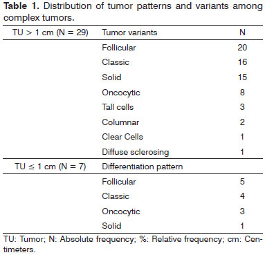
Tumors were classified into each variant or differentiation pattern, similar to that of the World Health Organization (WHO) classification3 as: Follicular, Oncocytic, Clear Cells, Diffuse Sclerosing, Tall Cells, Solid, Cribriform, Fasciitis-like, Macrofollicular, and Microcarcinoma. There were no cases belonging to the macrofollicular and cribriform variants in our sample. Besides these, we added the Columnar and the Warthin-like differentiation pattern, present in six cases. The conventional, usual or typical thyroid PC cases were classified as belonging to the Classic pattern or variant. When evaluating the microcarcinomas, we ran descriptive analysis of tumor differentiation patterns similarly to the previously described variants. For statistical purposes, we grouped differentiation patterns and tumor variants. When more than one variant or differentiation pattern were present in the same case, these were considered complex variants or tumor patterns. These were described as shown on Table 1.
Tumors were considered multifocal when two or more foci were found in one or both lobes. Hashimoto's thyroiditis has been suggested based on histopathological findings. Papillary microcarcinomas were defined as tumors with no more than 1.0 cm in diameter at the final histological examination. The Tall, Columnar, Solid or Diffuse Sclerosing Cells were defined as variants or differentiation patterns with greater aggressiveness potential; and those with the less malignancy potential were the Classical, Follicular and Oncocytic types. Other variants or patterns of tumor differentiation were excluded from the statistical calculation and are only presented descriptively.
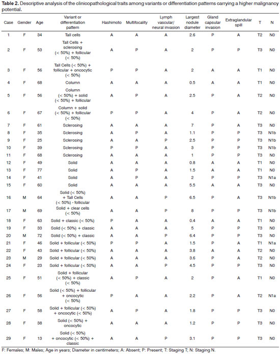
Complex tumor cases with tumor nodules or zones classified in the higher malignancy category were grouped with other similar cases for the purpose of statistical calculation and are listed on Table 2. The same was true among cases with low malignancy potential. Pathological staging was performed according to the seventh edition of the American Joint Committee on Cancer pTNM staging system4. Lymph node status was defined by pathological evidence of metastases in the lymph nodes which were removed. Extraglandular involvement was defined based on evidence of tumor infiltration beyond the capsule gland upon microscopic examination. We performed a comparative analysis of clinical and histopathological variables among the Classic, Follicular and Oncocytic variants, and among the groups with variants recognized by the highest and lowest potential for aggression, as previously agreed. All the data was collected by the same researcher (Girardi FM) and the entire pathology review was carried out by the same pathologist (Barra MB).
Statistics and Ethical Aspects
Descriptive analysis was used to summarize the data. We performed the Kolmogorov-Smirnov test to assess normality of continuous variables. Continuous variables with normal distribution were expressed as mean and standard deviation. Those with abnormal distribution were also expressed as median, minimum and maximum values. Categorical variables were expressed as absolute and relative frequency. We used Student's t test and ANOVA for comparing mean ages; the Mann-Whitney U test and Kruskal-Wallis tests were used for comparing tumor diameter and the nonparametric chi-square test was used to compare categorical variables. Statistical analysis was performed using the EpiInfo software, version 3.4.3, 2007. All tests considered significance level of 5%.
The authors guarantee data keeping and the confidentiality of the material obtained. As there were no interventions, we did not have to use the Informed Consent Form. The project was approved by the Ethics Committee of our institution (Project No 3483/11).
RESULTS
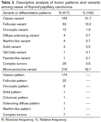
Between June 2000 and December 2010, there were 623 thyroidectomy for thyroid cancer performed in our institution. Eight cases were excluded due to lack of tumor diameter information, all corresponding to Classic PC. Altogether, 517 (82.9%) patients met the inclusion criteria. Of the total, 81.9% were women. The male:female ratio was 1:4.5. The average age was 47.20 years, range of 13-87 years. We observed that the Microcarcinoma variant was the most prevalent, with 42.1% of cases, followed by the Classical and Follicular variants, respectively (Table 3).
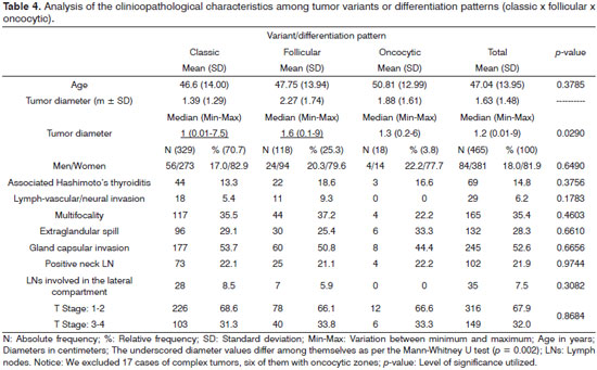
In 36 cases there were more than one tumor variant or differentiation pattern in the same gland, and the Classic variation or differentiation pattern was the most prevalent in those cases. Except for nodule size, we did not detect statistically significant differences between the Classical, Follicular and Oncocytic variants or differentiation pattern (Table 4). However, this difference was not maintained when we compared the diameter between the Classical and Oncocytic variants (p = 0.2842), as well as when we compared the Follicular and Oncocytic variants (p = 0.2129), only the Classical and Follicular variants, as per the Mann-Whitney U test (p = 0.002).
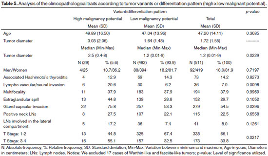
As noted on Tables 2 and 5, those variants recognized by having the highest malignancy potential accounted for only 5.6% of the sample, and the solid variant predominated. We did not observe statistically significant differences between age, gender, multifocality, Hashimoto's thyroiditis and positive neck lymph nodes between groups with lower or higher malignancy potential variants or differentiation pattern (Table 5). However, we observed their association with larger tumor size, lymphovascular and gland capsule invasion and T staging.
DISCUSSION
The most prevalent variant or differentiation pattern in our study, grouped microcarcinomas, was the Classic, followed by the Follicular variant or pattern. Together accounted for 86.2 % of the total. Similar percentages have been observed in other series5,6; and the study by Lam et al. 5 was the one that most closely approximated to the figures in our sample. Studies have shown that, despite some histological differences observed between the Classic and Follicular variants of thyroid PC, both neoplasms have favorable prognosis and similar cancer-specific survival at 10 and 15 years7. Lang et al. 7 observed fewer metastatic lymph nodes and lower extraglandular overflow rate among the Follicular variant cases when compared to the usual forms of PC. Ozdemir et al. 8 showed greater tumor diameter, although lower prevalence of capsular invasion and extraglandular extravasation among Follicular variant cases when compared to the Classic one.
Likewise, although there are differences in the literature9, in most cases involving the Oncocytic variant of thyroid PC, the tumor is confined to the gland without evidence of association with poor prognosis histological features10. There are molecular differences between Oncocytic Carcinomas and the Oncocytic variant of the thyroid PC, suggesting that both diseases have different are genetic behavior11. Moreover, under the same staging classification, there is no evidence that the thyroid PC Oncocytic variant differs from the usual forms and the PC Follicular variant in biological behavior and malignancy potential6,10.
In our study, except for tumor size, neither difference was observed between the clinical and pathological parameters involving the variants: Classic, Follicular and Oncocytic. In the analysis of subgroups, even diameter differences do not sustain, this being a different characteristic among the cases of Follicular and Classic variants in the sample, similar to the study by Ozdemir et al. 8. It is known that the follicular variant diagnosis of CP in the preoperative period is a challenge, both from the cytology point-of-view12 as well as the clinical-echographical point of view8, which may have delayed the diagnosis of this tumor variant and justify the larger diameter this histological subtype in relation to the classical form of PCin our sample.
Many different histologic variants have been described for the thyroid PC. Some variants have been associated with worse prognosis13 . Michels et al. 14 developed a study analyzing survival between patients with regular thyroid PC and those with the Tall Cells variant. The 10-year survival was 90% and 79%, respectively. In an univariate analysis, the Tall Cells variant was associated with worse outcomes, which was not confirmed in the multivariate analysis. Other authors have developed similar studies involving other histological subtypes recognized by their worst prognosis, and in all of them, the evidence that the histological variant is an independent predictor with respect to outcome is weak13. The Columnar variant, similarly to the Tall Cells variant, shows an association with advanced locoregional disease and distant metastases6. Falvo et al. 15 developed a comparative study of 83 cases of Diffuse Sclerosing variant with 183 cases of the usual forms. They concluded that the Diffuse Sclerosing variant is characterized by intrathyroidal spread and a high rate of lymph node and lung metastases. Sywak et al. 16 reported that the Solid variant has a high propensity to extrathyroidal extension and lymph node metastasis. Like the previously described studies, we observed association between variants or cases with differentiation pattern having greater potential for malignancy and various histological features historically known for their association with poor prognosis.
Despite the representative sample volume of our study, cases recognized by worst potential prognosis are rare and account for a small part of the total number of thyroid PC cases, which makes it difficult to compare the clinical and histopathological data between each variant separately. Jung et al. 17 in an institutional series of 14 years, gathered 23 cases of variants with the greatest malignancy potential (ten cases of the Tall Cell variant, five Diffuse Sclerosing, four Columnar, three of the Solid variant and one mixed case between the Columnar and Tall Cell variants) for comparative analysis of prognostic factors and outcomes in a group of cases of poorly differentiated thyroid carcinoma. Similarly, we chose to unify all cases of variants historically known for their association with poor prognosis in a single comparison group. Little is known about the role played by worse prognosis variants' foci or zones in the context of Classic, Follicular or Oncocytic variants or patterns. We chose to include them with the other cases with the greatest potential for malignancy for statistical calculation purposes. All cases that contained variants or patterns of differentiation recognized by worse prognosis are individually listed on Table 2, with discrimination of the predominant subtype, which we believe can collaborate in number for subsequent studies from other research centers, given the low incidence of these tumor variants even in large series.
There is a hypothesis that the thyroid PC has its beginnings in the Follicular Classic pattern, and with time it transforms into more malignant forms, depending on some molecular events, all the way to poorly differentiated and anaplastic tumors. Thus, it is postulated that patients with more malignant forms should be older, acquiring, in the course of the disease, all the histopathological characteristics already recognized of poor prognosis18. This model has been well studied among Tall Cell variants, represented in our study by only four cases with ages ranging between 34-64 years. In our study, we found no statistical correlation of age with tumor variant, which we believe may have been influenced by the low representation of this variant in our sample, besides excluding cases with areas of poorly differentiated carcinoma and tumors with areas of anaplasia.
CONCLUSION
The Columnar, Tall Cells, Diffuse Sclerosing, Solid variants are usually more malignant than the Classical, Follicular and Oncocytic variants of the thyroid PC. Although prognostic studies using multivariate analysis, have publicized that the histologic subtype has lost strength as an independent predictor of worse outcomes, the forms for higher malignancy potential tend to be associated with several other factors historically reported in cases of unfavorable outcomes, alerting the physician that he/she is facing a potentially malignant tumor. We believe that our study came to add to the current knowledge, especially as a substrate for further studies involving more than one research center, given the low occurrence of some of these variants.
REFERENCES
1. Sobin LH, Wittekind CH. TNM classification of malignant tumors (UICC). New York: Wiley; 2002.
2. Ito Y, Miyauchi A, Jikuzono T, Higashiyama T, Takamura Y, Miya A, et al. Risk factors contributing to a poor prognosis of papillary thyroid carcinoma: validity of UICC/AJCC TNM classification and stage grouping. World J Surg. 2007;31(4):838-48. DOI: http://dx.doi.org/10.1007/s00268-006-0455-0
3. LiVolsi VA, Albores-Saavedra J, Asa SL, Baloch ZW, Sobrinho-Simões M, Wenig B, et al. Papillary carcinoma. In: DeLellis RA, Lloyd RV, Heitz PU, Eng C, eds. Pathology and Genetics of Tumous of Endocrine Organs. Lyon: IARC Press, 2004, p.57-66.
4. Edge SE, Byrd DR, Carducci MA, Compton CA, eds. AJCC Cancer Staging Manual. New York: Springer; 2010.
5. Lam AK, Lo CY, Lam KS. Papillary carcinoma of thyroid: A 30-yr clinicopathological review of the histological variants. Endocr Pathol. 2005;16(4):323-30. DOI: http://dx.doi.org/10.1385/EP:16:4:323
6. Ito Y, Hirokawa M, Uruno T, Kihara M, Higashiyama T, Takamura Y, et al. Prevalence and biological behaviour of variants of papillary thyroid carcinoma: experience at a single institute. Pathology. 2008;40(6):617-22. DOI: http://dx.doi.org/10.1080/00313020802320630
7. Lang BH, Lo CY, Chan WF, Lam AK, Wan KY. Classical and follicular variant of papillary thyroid carcinoma: a comparative study on clinicopathologic features and long-term outcome. World J Surg. 2006;30(5):752-8. DOI: http://dx.doi.org/10.1007/s00268-005-0356-7
8. Ozdemir D, Ersoy R, Cuhaci N, Arpaci D, Ersoy EP, Korukluoglu B, et al. Classical and follicular variant papillary thyroid carcinoma: comparison of clinical, ultrasonographical, cytological, and histopathological features in 444 patients. Endocr Pathol. 2011;22(2):58-65. DOI: http://dx.doi.org/10.1007/s12022-011-9160-0
9. Gross M, Eliashar R, Ben-Yaakov A, Weinberger JM, Maly B. Clinicopathologic features and outcome of the oncocytic variant of papillary thyroid carcinoma. Ann Otol Rhinol Laryngol. 2009;118(5):374-81. PMID: 19548388
10. Montone KT, Baloch ZW, LiVolsi VA. The thyroid Hürthle (oncocytic) cell and its associated pathologic conditions: a surgical pathology and cytopathology review. Arch Pathol Lab Med. 2008;132(8):1241-50. PMID: 18684023
11. Trovisco V, Vieira de Castro I, Soares P, Máximo V, Silva P, Magalhães J, et al. BRAF mutations are associated with some histological types of papillary thyroid carcinoma. J Pathol. 2004;202(2):247-51. PMID: 14743508 DOI: http://dx.doi.org/10.1002/path.1511
12. Faquin WC. Diagnosis and reporting of follicular-patterned thyroid lesions by fine needle aspiration. Head Neck Pathol. 2009;3(1):82-5. DOI: http://dx.doi.org/10.1007/s12105-009-0104-7
13. Silver CE, Owen RP, Rodrigo JP, Rinaldo A, Devaney KO, Ferlito A. Aggressive variants of papillary thyroid carcinoma. Head Neck. 2011;33(7):1052-9. DOI: http://dx.doi.org/10.1002/hed.21494
14. Michels JJ, Jacques M, Henry-Amar M, Bardet S. Prevalence and prognostic significance of tall cell variant of papillary thyroid carcinoma. Hum Pathol. 2007;38(2):212-9. DOI: http://dx.doi.org/10.1016/j.humpath.2006.08.001
15. Falvo L, Giacomelli L, D'Andrea V, Marzullo A, Guerriero G, de Antoni E. Prognostic importance of sclerosing variant in papillary thyroid carcinoma. Am Surg. 2006;72(5):438-44. PMID: 16719201
16. Sywak M, Pasieka JL, Ogilvie T. A review of thyroid cancer with intermediate differentiation. J Surg Oncol. 2004;86(1):44-54. DOI: http://dx.doi.org/10.1002/jso.20044
17. Jung TS, Kim TY, Kim KW, Oh YL, Park do J, Cho BY, et al. Clinical features and prognostic factors for survival in patients with poorly differentiated thyroid carcinoma and comparison to the patients with the aggressive variants of papillary thyroid carcinoma. Endocr J. 2007;54(2):265-74. PMID: 17379963 DOI: http://dx.doi.org/10.1507/endocrj.K06-166
18. Das DK. Age of patients with papillary thyroid carcinoma: is it a key factor in the development of variants? Gerontology. 2005;51(3):149-54. DOI: http://dx.doi.org/10.1159/000083985
1. Head and Neck Surgeon, member of the head and neck surgery team at Santa Rita Hospital, Irmandade Santa Casa de Misericórdia de Porto Alegre
2. Pathologist; Department of Pathology, Santa Casa de Misericordia de Porto Alegre
3. PhD in Pathology, Department of Pathology, Santa Casa de Misericordia de Porto Alegre
Irmandade Santa Casa de Misericórdia de Porto Alegre, Santa Rita Hospital.
Send correspondence to:
Fábio Muradás Girardi
Avenida Independência. nº 354/901
Porto Alegre - RS. Brazil. CEP: 90035-070
Telefone: (51) 3214-8436
E-mail: fabiomgirardi@gmail.com
Paper submitted to the BJORL-SGP (Publishing Management System - Brazilian Journal of Otorhinolaryngology) on June 17, 2013.
Accepted on August 27, 2013. cod. 10968.
