

Year: 2002 Vol. 68 Ed. 4 - (15º)
Artigo Original
Pages: 546 to 549
 PDF PT
 New tracheotomy speaking valve: a Brazilian proposal
Author(s):
Carlos T. Chone 1,
Antonio Bortoleto 2,
Flávio M. Gripp 3,
Agricio N. Crespo 4
Keywords: tracheotomy, airway obstruction, speaking valve
Abstract:
Introduction: Tracheotomy is performed in conditions of upper airway obstruction or chronic pulmonary disorders. The use of tracheotomy speaking valves (TSV) has been described elsewhere but always at high cost for the patient, since all of them are imported. Objective: To demonstrate the TSV manufactured in stainless steel, developed at the State University of Campinas, and the possibility of its use in speech rehabilitation of tracheotomized patients. Material and Methods: The TSV developed was used in ten consecutive patients. The valve works with a diaphragm inside a body made of stainless steel with plastic attachments. It directs the air to the larynx during phonation and allows introduction of air through the valve during inspiration, with low pressure. Results: All ten patients are using the valve, speaking and breathing through it without distressing situations even during sleep. Discussion: The TSV improves communication, intelligibility, hygiene and humidification of the airway. The psychological condition and amount of secretions in the airway are improved too. This TSV has a low cost and can benefit many patients in Brazil. Children who have had tracheotomy could have a delay in acquisition of language. The use of TSV might be helpful for the communication and social interaction of these children. Conclusion: The TSV demonstrated is safe and allows effortless phonation without digital occlusion of the tube.
![]()
INTRODUCTION
Tracheotomy is a surgery performed by the Otorhinolaryngologist in a great variety of clinical conditions that result in upper respiratory obstruction or chronic pulmonary disease to provide lung hygiene and reduction of dead space. Among them we can mention: bronchopulmonary dysplasia, neuromuscular and congenital diseases, head injury, mechanical ventilation-dependent patients, chronic obstructive pulmonary disease, laryngotracheal stenosis, laryngotracheomalacia, vocal fold paralysis and sleep obstructive apnea.
One of the main deficits caused by tracheotomy is loss of verbal communication or its appropriate development in children1, 2, 3, 4, 5, 6. In this case, verbal communication is essential for global care, psychological condition and social interaction of the patients4. When there is communication barrier between patients and healthcare professionals, there is reduced capacity to participate in treatment planning4. Especially in children, we can witness deficit in language and articulation acquisition1, 2, 3, 5. They have difficulties to digitally occlude the tracheotome canulla. There can be 5 to 9 month delays in communication skills2, in both receptive and expressive language in the presence of tracheotomy5. The longer the time with the tracheotomy, the greater the deficit5.
Other functions developed by the upper airways can also be compromised in patients with tracheotomy, such as heating, humidifying, filtering air, coughing, sneeze, taste, olfaction, swallowing3. In the latter, there was higher aspiration of tracheotomized patients6, 7, 8, 9, 10, 11, 12, 13. Its pathophysiology is not well determined. It is attributed to multiple factors, such as reduction of laryngeal elevation, reduction of cough reflex by decrease of subglottic pressure and atrophy of swallowing muscles because of lack of use9. The most important mechanism seems to be incoordination of glottic closure during swallowing9. There is also difficulty in elevating the larynx because the canulla holds the larynx to the skin of the neck region14. The balloon of the canulla, when exaggeratedly insufflate, can also take to swallowing disorders by esophagus compression14.
Tracheotomy speaking valves (TSV) minimize these associated defects. There are the valves by Passy-Muir, Montgomery, Olympic and Kistner, but the Passy-Muir one has better vocal quality noticed by listeners as well as by patients, according to the literature4. In addition, it represents the lowest rate of mechanical problems4. TSV are unidirectional and enable air entrance in inspiration with small inspiration pressure. During phonation, there is closure and the air is directed to the larynx. They are all imported, which implies high cost to our patients, a cost not covered by the public health system or by medical healthcare plans. Therefore, we developed a national TSV at Universidade Estadual de Campinas, to compensate this lack, with improvement in quality of life of tracheotomized patients and with lower cost. The purpose of the present study was to present the new TSV, made in Brazil and already used by 10 patients.
MATERIAL AND METHOD
We conducted a retrospective study at the Division of Head and Neck, Discipline of Otorhinolaryngology, Universidade Estadual de Campinas.
Ten tracheotomized patients with metallic canullae were selected to use the speaking valve. The age of patients ranges from 5 to 58 years. They were all observed for about 30 days after placement of the TSV. Patients with severe laryngotracheal stenosis over the tracheotomy, unconscious and that needed permanently insufflated balloon were excluded from the sample. They would not benefit from phonation and they are contraindications to the use of TSV, according to the protocol by Passy-Muir6, owing to the risk of respiratory obstruction.
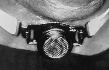
The developed TSV has a diaphragm, filter and plastic connections designed inside a body of stainless steel (Figure 1). It is adaptable to all national metallic canullae (Figure 2) by anterograde route, through coupling to TSV adapter to the head of the internal canulla of the tracheotomy. It presents one adapter for each number of tracheotomy canulla.
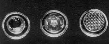
Figure 1. Stainless steel body of the speaking valve formed of three parts joined together.
Figure 2. Speaking valve adapted to the metallic tracheotomy canulla.
RESULTS
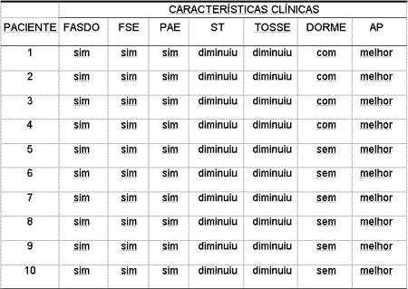
The ten patients adapted to the TSV developed by us, with appropriate and effortless phonation. They could practice sport activities, intensive walking and talk without difficulties and without the need to digitally occlude the canulla. There was reduction of the quantity of tracheal secretion produced and they reported better conditions of local hygiene, especially when coughing and when in public places. Four patients used the valve during sleep (Table 1).Table 1. Assessment of clinical characteristics by the patients after the use of speaking valve, compared to the situation without speaking valve.
Key: FASDO - appropriate phonation without digital occlusion; FSE - effortless phonation; PAE - practice of sport activities; ST - tracheal secretion; Cough - production of cough; Sleep - patient sleeps with the speaking valve; AP - activities in public.
DISCUSSION
Since 1975, techniques that enable phonation of tracheotomized patients have been tried12, 15, 16. TSV can be used in the neonatal period as of the minimal age of 13 days1. Valves allow spontaneous phonation without digital occlusion of the tracheotomy canulla. There is improvement of the psychological condition of the patients, especially concerning spontaneous phonation and reduction of tracheal secretion, with decrease of productive cough through the tracheotome, which is especially important in public spaces. The use of a filter inside the valve that can be changed weekly, such as in the TSV studied here, enables entry of less polluted air in the lower airways. Reduction in the degree of tracheotomy-related aspiration has been observed after the use of TSV 9, 13. There are studies, however, that demonstrated absence of influence of the occlusion of the external orifice of the canulla of the tracheotomy with improvement of aspiration10. It produces benefits in the cough reflex and it improves the pulmonary hygiene of patients, providing absence of formation of secretion in the tracheotome. The valves existent in the market are imported and difficult to acquire by patients in our society. The new developed TSV is of low cost and many patients can benefit from it. All patients noticed changes concerning quality of life after the use of the speaking valve. Some of them - four patients - even used it during sleep. The other patients preferred to remove it during sleep.
CONCLUSION
The speaking valve developed in the Discipline of Otorhinolaryngology and Head and Neck of Universidade Estadual de Campinas was well adapted in all patients with phonation, without digital occlusion of tracheotomy canulla.
ACKNOWLEDGEMENT
To Mr. Eduardo Luiz de Carvalho, the technical person responsible for IndusmedÒ, for the cooperation in developing and manufacturing the speaking valves used in the present study.
REFERENCES
1. Engleman SG, Carrier CT. Tolerance of the Passy-Muir speaking valve in infants and children less than 2 years of age. Pediatr Nurs 1997;23:571-573.
2. Gereau SA, Navarro GC, Cluterio B, Mullan E, Bassila M, Ruben RJ. Selection of pediatric patients for use of the Passy-Muir valve for speech production. Int J Pediatr Otorhinolaryngol 1996;35:11-17.
3. Jackson D, Albamonte S. Enhancing communication with the Passy-Muir valve. Pediatr Nurs 1994;20:149-153.
4. Leder SB. Perceptual rankings of speech quality produced with one-way tracheostomy speaking valves. J Speech Hear Res 1994;37:1308-1312.
5. Lieu JEC, Muntz HR, Prater D, Stahl MB. Passy-Muir valve in children with tracheotomy. Int J Pediatr Otorhinolaryngol 1999;50:197-203.
6. Passy V. Passy-Muir tracheostomy speaking valve. Otolaryngol Head Neck Surg 1986;95:247-8.
7. Bonnano PC. Swallowing dysfunction after tracheostomy. Ann Surg 1971;174:29-33.
8. Cameron JL, Reynolds J, Zuidema GD. Aspiration on patients with tracheostomies. Surg Gynecol Obstet 1973;136:68-70.
9. Dettelbach MA, Gross RD, Mahlmann J, Eibling DE. Effect of the Passy-Muir valve on aspiration in patients with tracheostomy. Head Neck 1995;17:297-302.
10. Leder SB. Effect of one-way valve tracheotomy speaking valve on the incidence of aspiration in previously aspirating patients with tracheotomy. Dysphagia 1999;14:73-77.
11. Muz JM, Mathog RH, Nelson R, Jones LA. Aspiration in patients with head and neck cancer and tracheostomy. Am J Otolaryngol 1989;10:282-286.
12. Nash M. Swallowing problems in tracheotomized patient. Otolaryngol Clin North Am 1988;21:701-709.
13. Stachler RJ, Hamlet SL, Choi J, Fleming S. Scintigraphic quantification of aspiration reduction with the Passy-Muir valve. Laryngoscope 1996;106:231-234.
14. Gross RD. Swallowing rehabilitation. In Myers EN, Suen JY. Cancer of the head and neck. 3rd ed. Philadelphia: W.B. Saunders; 1996. p.868-882.
15. Andersson G. The Swedish modification of the tracheostomy tube to permit speech. Paraplegia 1993;31:203-206.
16. Cowan DL. Laryngeal and tracheal stenosis: an adapted speaking aid tracheotomy tube. J Laryngol Otol 1975;89:531-534.
[1] Assistant Physician, Ph.D., Discipline of Otorhinolaryngology and Head and Neck, Medical School Universidade Estadual de Campinas (UNICAMP).
[2] Resident Physician, Discipline of Otorhinolaryngology, and Head and Neck, Medical School Universidade Estadual de Campinas (UNICAMP).
[3] Assistant Physician, Discipline of Otorhinolaryngology, and Head and Neck, Medical School, Universidade Estadual de Campinas (UNICAMP).
[4] Adjunct Professor, Ph.D. and Head of the Discipline of Otorhinolaryngology and Head and Neck, Medical School, Universidade Estadual de Campinas (UNICAMP).
Study conducted in the Discipline of Otorhinolaryngology, and Head and Neck, Medical School, Universidade Estadual de Campinas (UNICAMP).
Address correspondence to: Carlos T. Chone - Rua Major Sólon, 685 - 13024-091 Campinas - São Paulo - Tel: (55 19)3255-1966 - E-mail: carloschone@uol.com.br
Study presented at the II Congresso Triológico de Otorrinolaringologia, August 26, 2001, Goiania and at XVIII Congresso Brasileiro de Cirurgia de Cabeça e Pescoço, September 06, 2001, Recife.
Print: ![]()