

Year: 2002 Vol. 68 Ed. 4 - (11º)
Artigo Original
Pages: 522 to 525
 PDF PT
 Endoscopic anatomy of the sphenoid sinus
Author(s):
Alexandre A. Murta 1,
Christiano G. Carneiro 2,
Alexandre Felippu 3
Keywords: sphenoid sinus, endoscopic sinus surgery, internal carotid artery, maxillary nerve, optic nerve, anatomy
Abstract:
The internal anatomy of the sphenoid sinus plays a role of great importance due to its peculiar placement in the center of the head, surrounded by important adjacent structures, which make themselves transparent in its internal walls. In this study, 52 sphenoid sinus were endoscopically dissected, and the elevations and depressions presented on its internal walls, produced by the internal carotid artery and the optic, maxillary and vidian nerves, were analyzed. In 88,5% of the cases, the internal carotid artery were projected into the sinus, while the optic nerve were in 55,8%. The vidian and maxillary nerves were salient in 25% and 30% respectively. These data point us to the rich end delicate internal anatomy of the sphenoid sinus, which is progressively closer and more vulnerable to surgical maneuvers, due to the advance of surgical skills, techniques and knowledge. Thus, the study and comprehension of the sphenoid sinus internal anatomy became essential, concerning the endoscopic sinus surgery and its huge field of technical resources.
![]()
Introduction
The peculiarity of the sphenoid sinus is that it reflects the anatomical relations of its walls1. The adjacent structures, already present before its development, print a relief on its internal walls when the sinus is pneumatized and expanded, increase its contacts with these structures, or sometimes even surrounds them2, 3. Thus, at endoscopic view, we can notice a cavity whose internal walls are marked by a variable series of elevations and depressions corresponding to the impressions of adjacent anatomical structures4, and the thickness of the wall lamina can vary from compact to very thin3, or even nonexistent5. This close relation contributes to the unquestionable clinical and surgical importance of the sphenoid sinus6.
The structures that are usually part of this relation are vessels and nerves that run along the sinus, into brain afferent and efferent paths and the most important and frequent ones are the internal carotid artery and optic, maxillary and vidian (nerve of pterygoid canal) nerves7.
The present study intended to quantify the intensity and frequency of these structures projected towards the sphenoid sinus and to highlight the importance of having precise anatomical knowledge for clinical and surgical practice.
Material and Method
Fifty-two adult fresh cadavers, not formolized, were submitted to videoendoscopic surgical dissection, in the Program of Cadaver Dissection, Instituto Felippu de Rinologia (São Paulo), randomly selected and studied according to a specific protocol.
In each cadaver, after the appropriate ethmoidectomy, we proceeded to wide opening of the anterior wall of the sphenoid sinus, enabling total visualization of the sinus cavity and the relief on the internal walls. The elevations and recesses were identified and the adjacent elements were confirmed by delicate removal of bone walls.
All dissections were performed with conventional endoscopic surgical instruments, with 30 degree or 70 degree telescopes, and recorded in video for documentation and later analysis.
Results
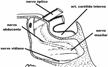
We dissected 52 sphenoid sinuses and analyzed the elevations and depressions produced by the internal carotid artery and optic, maxillary and vidian nerves on the relief of the internal walls.
Internal carotid artery presented an identifiable elevation in 46 cases (88.5%), and in 32 (61.5%), the position was considered marked, and in 8 cases (15.4%) the irregular tract - the carotid siphon - was characterized. We did not find cases of dehiscence.
The optic nerve was identified in 29 cadavers (55.8%). It is worth mentioning that in all cases in which the nerve presented a saliency on the sinus wall, the internal carotid artery had the same.
Vidian nerve (pterygoid canal nerve) proved to be visible on the sinus floor in 13 cases (25%), ranging from slight elevation to bone trabecula.
Maxillary nerve projected on the lateral wall of the sphenoid sinus in 16 cases (30.8%), many of them with the concomitant presence of the vidian nerve below it.
Figure 1.
Discussion
Thanks to the advance in imaging diagnosis techniques and endonasal surgery, paranasal sinuses and skull base anatomy have gained growing interest. In such context, sphenoid sinus has an important position owing to its singular location, practically in the center of the brain, in close contact with key structures, such as the first six cranial nerves, internal carotid artery, hypophysis, the cavernous sinus, pterygoid fossa and the brain itself3. The progressive advance of endonasal surgery reinforced the importance of knowing this anatomy, which is now seen from a different perspective - the endoscope 8. As a result of the use of endoscope, the surgeon can visualize inside the sphenoid sinus with great precision and closeness, counting on resources to changes the sight angle and letting it "look" to the sides, upward or forward using telescopes of different angles8 In addition, surgery can go beyond the bone limits, transforming the sphenoid sinus into access route for neurosurgical procedures, such as for the hypophysis and some intracranial tumors9, 10. However, its great anatomical variations are constant, providing extra value to its study and knowledge17. We analyzed the behavior of the main structures related to the sphenoid sinus - internal carotid artery and optic, maxillary and vidian nerves in 52 fresh cadavers submitted to endoscopic dissection.
Carotid internal artery leads the group, not only by frequency in which it is elevated (88.5%), but also by the degree of projection inside the sinus, since 61.5% of the cases of relief are considered marked, and in 15.4% of them, the whole path was salient. These are higher incidences than those reported by van Alyea6, who noticed the presence of the artery in 65% of the cases, being that in 53% the condition was considered marked and in 14% the path was complete. Fujii et al.5 also studied the path of the sphenoid sinus artery with similar results, in addition to mentioning the case of vessel dehiscence. The importance of these statements is enormous from a surgical perspective. The knowledge of the anatomical variations of the internal carotid artery is essential to avoid inappropriate manipulation that can lead to rupture, a very severe complication, difficult to control and with obscure prognosis. Many authors reported carotid artery lesion during the procedures of endonasal endoscopic surgery11. After its identification, hemorrhage can be immediately controlled with compressive packing, followed by emergency angiography for permanent occlusion with balloon. Occasionally, the internal carotid artery has to be sacrificed by arterial ligation of the neck, with high risk of irreversible neurological damage8. Felippu reported two cases of transoperative lesions of sphenoid sinus intra-cavernous carotid artery successfully controlled by fixation of free muco-periosteal graft of lower turbinate and compressive dressing12. Maniglia et al.13 mentioned three cases of arterial lesions that resulted in death.
We have also observed a higher number of optic nerve elevations (55.8%) than other authors, such as in the study by van Alyea6, who found 40% of the cases, or by Lang, with 42.85%14. The correlation between optic nerve and posterior sinuses attracted the attention of a number of authors. Onodi15 classified this relation in 12 groups, with 38 anatomical variations. Mellinger16 pointed out the importance of posterior sinus asymmetry concerning vulnerability of the optic nerve. Both, among others, believe that projection of the optic nerve into the sphenoid sinus could be considered as an etiological factor of optic neuritis17 and other orbital-ocular diseases18. This close relation between optic nerve and sphenoid sinus and ethmoid sinus provide great vulnerability during surgeries of the paranasal sinuses. Maniglia et al. reported a series of 20 cases of blindness as the major postoperative complication13.
In our study, vidian nerve (nerve of the pterygoid canal) proved to be salient in 25% of the cases, less frequently than the studies published by other authors that reported 48%28 and 52%19, or even the 10% rate of dehiscence of the pterygoid canal inside the sphenoid sinus19. Zuckerkandl20, in 1893, was one of the first authors to identify the saliency of the vidian nerve on the sinus floor, and Sluder21,22 and Vail23 emphasized its clinical importance. The latter concluded that irritation of vidian nerve produce symptoms of Sluder's syndrome (sphenopalatine ganglion), which consists of rhinorrhea and pain on the nasal floor, eye and behind it, and maxilla and teeth.
Maxillary sinus, second division of the 5th cranial nerve, frequently produces an elevation of the lateral wall of the sphenoid sinus in the path towards the cavernous sinus, before it reaches the round foramen24, 15, a fact observed in 40% of the cases, according to van Alyea6, when compared to 30.8% identified by us, and 28.6% by Lang19. Houser26 concluded that this anatomical proximity could affect the maxillary nerve in cases of empyema of sphenoid sinus, with neuritis, even when the nerve is immersed into the blood of the cavernous sinus. Along the same lines, Garber27 pointed out that the maxillary sinus is as close to the sphenoid sinus as to vidian nerve, which also implicated it in the sphenopalatine ganglion syndrome. Sluder28 reemphasized this argument by demonstrating permeability of sphenoid sinus walls through injection of cocaine inside it, causing maxillary sinus paralysis.
Even though it was not the objective of the present study, we emphasized that sphenoid sinus has a very rich set of anatomical variations compared to posterior ethmoid sinus, sella turcica, pterygopalatine fossa, cavernous sinus, as well as different morphology for recesses, pneumatization and symmetry29, 30.
Conclusion
Thanks to the present study, we demonstrated complexity of the sphenoid sinus anatomy and its relations with noble structures, as well as the wide range of variations. We highlight the importance of understanding this anatomy for its appropriate clinical interpretation and especially for the precise and safe surgical practice. Such understanding is particularly important when its is an endoscopic surgery, considering its vast range of technical resources.
References
1. Cope VZ. The internal structure of the sphenoid sinus. J Anat (Lond) 1917;51:127-136.
2. Salinger S. The paranasal sinuses. Arch Otolaryng 1939;30:44.
3. Van Alyea OE. Nasal Sinuses: An Anatomic and Clinic Consideration. Baltimore: The Williams and Wilkins Company; 1951. p.155.
4. Draf W. Endoscopy of Paranasal Sinuses. Berlin: Springer; 1983.
5. Fujii K, Chambers SM, Rhoton AL Jr. Neurovascular relationships of the sphenoid sinus: a microsurgical study. J Neurosurg 1979;50:31-39.
6. Van Alyea OE. Sphenoid sinus anatomic study with consideration of the clinical significance of the structural characteristics of the sphenoid sinus. Arch Otolaryng 1941;34:225-253.
7. Peele JC. Unusual anatomical variations of the sphenoid sinuses. Laryngoscope 1957;67:208-237.
8. Stammberger H. Results, problems, and complications. In: Stammberger H, Hawke M. Functional Endoscopic Sinus Surgery. The Messerklinger Technique. Philadelphia: BC Decker; 1991. p. 459-477.
9. Fahlbusch R, Buchfelder M. Present status of neurosurgery in the treatment of prolactinomas. Neurosurg Rev 1985;8:195-205.
10. Hammer G, Radberg C. Sphenoidal sinus: an anatomical and morfogenological study with reference to transsphenoid hypophysectomy. Acta Radiol (Stockh.) 1961;56:401-422.
11. Stankiewicz JA. Complications of endoscopic sinus surgery. Otolaryngol Clin North Am 1989;22:749-758.
12. Felippu A. Severe Epistaxis: The Transnasal Vessels Dissection. Instruction Course, Annual Meeting of the American Academy of Otolaryngology - Head and Neck Surgery: New Orleans; 1999.
13. Maniglia AJ. Fatal and Major Complications Secondary to Nasal and Sinus Surgery. Laryngoscope 1989;99(3):276-283.
14. Lang J. In: Clinical Anatomy of the Nose Nasal Cavity and Paranasal Sinuses. New York: Thieme Medical Publishers; 1989. p.91.
15. Onodi A. The optic nerve and the accessory sinuses of the nose. New York: William Wood & Co.; 1910.
16. Mellinger WJ. Optic nerves and their relations in the sphenoidal region. Tr. Pacific Coast Oto-Ophth Soc 1937;22:85.
17. Tunis JP. Sphenoidal sinusitis in relation to optic neuritis. Laryngoscope 1912;22:1157-1164.
18. Blum ME, Larson A. Mucocele of the sphenoidal sinus with sudden blindness. Laryngoscope 1973;83:2024-2049.
19. Lang J, Keller H. Über die hintere Pfortenregion der Fossa pterygopalatina und das Ganglion pterigopalatinum. Gegenbaurs Morph Jb. 1978;124:207-214.
20. Zuckerkandl E. Normale und pathologische Anatomie der Nasenhöhle und ihrer pneumatischen Anhänge. 2nd ed. Wien: W. Braumüller; 1893;1:368-400.
21. Sluder G. The role of the sphenopalatine (or Meckel's) ganglion in nasal headaches. Int Rec Med 1908;23:989-990.
22. Sluder G. In: Headaches and Eye Disorders of Nasal Origin. St. Louis: C. V. Mosby Co.; 1919.
23. Vail HH. Vidian Neuralgia. Ann Otol Rhin & Laryng 1932;14:837.
24. Dixon FW. A comparative study of the sphenoid sinus (a study of 1600 skulls). Ann Otol 1937;46:687-698.
25. Mosher HP. The anatomy of the sphenoid sinus and the method of approaching it from the antrum. Laryngoscope 1903;13:177-215.
26. Houser KM. Anatomic relations of the sphenoid sinus to Dorello's canal: abducens paralysis. Arch Otolaryng 1932;16:488.
27. Garber H. Sinusitis and neuralgia. Arch Otolaryng 1933;18:339.
28. Sluder G. Cited by Van Alyea.
29. Cope VZ. Internal structure of the sphenoid sinus. J Anat & Physiol 1917;51:127.
30. Congdon ED. The distribution and mode of origin of septa and walls of the sphenoid sinus. Ant Rec 1920;18:97.
[1] Responsible for the Division of Facial Plastic Surgery, Instituto Felippu de Rinologia.
[2] Responsible for the Division of Laryngology and Voice, Instituto Felippu de Rinologia.
[3] General Director of Instituto Felippu de Rinologia.
Affiliation: Instituto Felippu de Rinologia
Address correspondence to: Alexandre A. Murta - Rua Stela Marina, 46 - Brooklin Novo - 04601-090 - São Paulo - SP - Tel: (55 11)5536.5353 Fax: (55 11) 5535.7525
E-mail: alexandremurta@hotmail.com
Print: ![]()