

Year: 2002 Vol. 68 Ed. 4 - (9º)
Artigo Original
Pages: 511 to 514
 PDF PT
 Endoscopic endonasal ligation of the esphenopalatine artery for severe epistaxis
Author(s):
Rodrigo P. Santos 1,
Fernando D. Leonhardt 2,
Ricardo G. Ferri 3,
Luiz C. Gregório 4
Keywords: epistaxis, sphenopalatine artery, endoscopic surgery
Abstract:
Introduction: Severe epistaxis, usually associated to hypertension and blood dyscrasia, may benefit from surgery when the conservative treatment fails. Objective: to evaluate the results of endoscopic endonasal ligation of the sphenopalatine artery for non-responsive severe epistaxis. Material and Method: chart review of twelve patients submitted to endoscopic endonasal ligation of the sphenopalatine artery regarding clinical aspects, predisposing factors and complications of the procedure. Results: the mean age was 50.9 years, and the male:female: ratio was 2:1. Hypertension was presented in 33% and hematologic disorders in 16.6% of the patients. One patient, 8.3%, presented a new bleeding episode after the surgery. Discussion: the endoscopic endonasal ligation of the sphenopalatine artery, proved to be a safe surgical method and presented 8.3% of bleeding after the procedure in our study. Conclusion: avoiding the complications of previous surgical treatments for severe epistaxis, the endoscopic endonasal ligation of the sphenopalatine artery is an effective technique affordable for otolaryngologists with practice in endoscopic endonasal surgery.
![]()
Introduction
Epistaxis, defined as any bleeding from the nasal mucosa, is the most frequent emergency in Otorhinolaryngology, presenting a prevalence of about 10 to 12%1. It is estimated that approximately 60% of the population has already presented some type of nasal bleeding. Most of the times, hemorrhage is easily controlled, even without medical support. Only 6% of the cases need specialized intervention to contain bleeding and only 1% require hospitalization, with mortality rate below 0.01%2.
Epistaxes are classified into anterior and posterior (lateral wall of the nasal fossa). To localize the bleeding site, vasoconstrictor solutions can be used to facilitate visualization and once the bleeding site is defined - anterior or posterior, various types of hemostatic treatment can be administered. Conservative treatment includes chemical or thermal cauterization, normally in anterior bleedings; anterior packing, with gauze or rayon soaked in Vaseline or antibiotic ointment, or with other materials (glove finger, merocel); and antero-posterior packing which can be made with gauze tied by silk or cotton thread, in order to appropriately pack the nasopharynx, and after that, anterior packing is performed with gauze or pneumatic catheters (such as Foley probe), which are filled with sterile water in the rhinopharynx. The placement of this type of packing can be made in the outpatient unit or under general anesthesia, if necessary.
Surgical treatment is indicated when conservative treatment, as previously described, is not effective or when there is bleeding still after the removal of the packing. The nose has abundant vascular flow and a rich network of arterial anastomoses; thus, surgeries have to be performed close to the bleeding vessel. The main interventions are endoscopic cauterization and arterial ligation, which can be made with the ethmoidal arteries, lateral wall of the posterior nose arteries (sphenopalatine), maxillary artery and even external carotid artery. Hemorrhage in the ethmoid region is less common and rarely as severe as to require arterial ligation. This indication occurs when there is active bleeding in the superior region of the nasal fossa, above the middle concha, even after clinical treatment. The access can be made by transnasal route (microscopic or endoscopic) or by external route (Lynch technique). Ligation of maxillary artery can be made using transantral or intraoral access. Transantral technique is made with a Cadwell-Luc sinusectomy and the ligation is made at the entry of the nasal cavity; intraoral technique is performed through an incision in the maxillary mucosa, at the level of the second molar, at the gingivolabial sulcus, it is possible to identify the maxillary artery and its branches close to the maxillary tuberosity. Ligation of arteries emerging from sphenopalatine foramen (sphenopalatine artery and its branches) using the endoscopic route intends to make the ligation as close as possible to the bleeding site. Ligation of external carotid is a method of exception, reserved for very specific cases, especially because of the severe risks of complication involved with the technique.
Objective
To assess the results of endoscopic endonasal ligation of sphenopalatine artery in treating severe epistaxis refractory to conservative therapeutic approach.
Material and Method
During January 2000 to April 2001, we assessed prospectively 12 patients submitted to endoscopic endonasal ligation of sphenopalatine artery to treat severe epistaxis non-responsive to conservative treatment, observing clinical history, predisposing factors, progression and complications of the procedure.
The indications for the procedures considered persistence of uncontrolled posterior epistaxis after attempt with conservative maneuvers such as chemical or thermal cauterization and anterior or antero-posterior nasal packing with gauze.
Patients were hemodynamically stabilized and submitted to the procedure under general anesthesia. We used basic endonasal surgical material, together with a zero degree rigid endoscope with 4mm diameter. The ligation of the arteries emerging from the sphenopalatine foramen, sphenopalatine artery and its branches, aimed at making the ligation as close as possible to the bleeding site. Technically, we had to collect a subperiosteal nasal mucosa flap from the lateral wall, close to the posterior portion of the middle concha. By extending the flap posteriorly, we could identify the vessels that projected from the sphenopalatine foramen. After dissection, the vessels were ligated with LIGGE CLIPS (LT 200). The flap was then replaced and the patient left without nasal packing.
Results
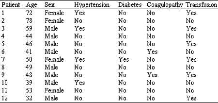
The age range varied from 32 to 78 years, mean age of 50.9 years, and gender distribution was 33% for female and 67% for male subjects.
The presence of predisposing factors, such as arterial blood hypertension, coagulopathy, previous surgery and trauma were as follows: 33% had hypertension, 16.6% had coagulopathy (hepatopathy) and no patients had trauma or previous surgery (Table 1).
All patients had been submitted to conservative methods of treatment before undergoing the endonasal endoscopic ligation of sphenopalatine artery. All of them had been submitted to anterior packing with gauze and antibiotic ointment, and except for one patient, they had also been submitted to antero-posterior packing. Only one patient, referred from a different service, had been submitted to embolization and cauterization of the sphenopalatine artery, but without success.
Only one patient (8.3%) presented new bleeding after the surgical procedure and in one patient we decided to maintain gauze with antibiotic for 24 hours after the surgery; she did not present bleeding after its removal.Table 1. Factors associated to severe epistaxis.
Discussion
Epistaxis is the most common nasal emergency that requires hospitalization3 and it has prevalence of 10-12% in the general population1. Cauterization and packing are the main therapeutic approaches to treat this affection, reserving surgical treatment for clinical presentations resistant to conservative treatment.
Epistaxis has even distribution between genders and affects all age ranges. In children, bleeding is normally originated from the anterior region of the nasal cavity, caused by local and easy to control abnormalities. In adults, most of the cases are associated with systemic abnormalities, originated from the posterior portion of the nasal cavity and normally of more difficult control.
Nasal blood irrigation is supplied by external and internal carotid systems. The external carotid artery system is the main responsible for nasal blood flow, through the maxillary and facial branches. Maxillary artery is one of the terminal arteries of the external carotid artery and close to the sphenopalatine foramen, on the lateral wall of the nose, it bifurcates into two terminal branches: septal artery and posterior lateral nasal artery (Navarro, 1996). The descending palatine artery, also the maxillary branch, penetrates the nasal cavity close to the sphenopalatine foramen, originating the major palatine artery, which penetrates into the palate, runs along the roof of the mouth and climbs up the incisor foramen, irrigating the anterior region of the septum and the nasal floor. The facial artery is divided into the superior labial branch that penetrates the nasal cavity and is distributed in the anterior nasal wall. The internal carotid artery system originates the ophthalmic artery, whose terminal branches are the anterior and posterior ethmoid arteries. Anterior ethmoid artery is thicker than the posterior one and it is responsible for irrigating the anterior third of the lateral nasal wall and part of the septum. The posterior ethmoid artery irrigates the superior concha and part of the septum. The junction of the internal and external carotid systems is at the anterior region of the septum, through the terminal branches of ethmoid arteries, superior labial artery, major palatine artery and posterior branches of the nose. This arterial junction is called Kisselbach plexus.
Etiology of epistaxis is divided into local and systemic causes. Among the local causes we can included: inflammatory-infectious (rhinitis, sinusitis, etc.), traumas (digital, fracture, nasal surgeries), anatomic (septal deviation and perforation), foreign body, chemical or weather agents, and nasal tumors (nasoangiofibroma, nasal polyposis, inverted papilloma, carcinoma). Systemic causes are drugs (AAS, anticoagulants, non-hormonal antiinflammatory, antibiotics), blood dyscrasia, systemic arterial hypertension, neoplasm and others.
Transantral ligation of maxillary artery, initially described by Seiffert4 in 1928 and made popular in 1965 by Chandler and Serrins5, presents a therapeutic failure rate of 0.5-15%6, and complications such as edema and facial anesthesia, oro-antral fistula and dental desensitization. Sphenopalatine artery ligation using middle antrostomy and surgical microscope was described by Prades7 in 1976. In 1992, Budrovich and Saetti8 described the use of nasal endoscope to make ligation of the sphenopalatine artery, a procedure that interrupts the nasal flow in a terminal position, avoiding local and retrograde bleeding, as well as anastomoses of the bilateral carotid systems and complications of the techniques of the maxillary artery ligation 9,10.
Endonasal endoscopic ligation of sphenopalatine artery represents a safe approach, because it prevents the complications of the maxillary artery ligation and ensures satisfactory control of bleeding, with re-bleeding rates within the expected levels for the maxillary artery ligation, of 0.5-15%6, 11. Among our cases, only one (8.3%) presented bleeding after the surgical procedure and he was submitted to revisional surgery with ligation of ethmoid arteries; in another case, we decided to have the packing with gauze and antibiotic ointment for 24 hours after ligation, and the patient did not present any further bleeding.
Arterial embolization is another therapeutic alternative for severe epistaxis that does not respond to conservative or surgical treatment, using angiography to identify the bleeding site. The embolization should be performed as close as possible to the bleeding site, and there is 88% efficacy rate. Complications are detected in 17-27% of the patients and most of them are transient, rarely leading to cerebral ischemia or its complications.
Severe epistaxis is normally associated to predisposing factors such as hypertension, coagulopathy, nasal trauma and previous surgeries10. Herkner et al.12 showed higher blood pressure mean in patients admitted to the emergency room because of epistaxis when compared to other causes of admission. In our study, 33% of the patients had hypertension. Two patients (16.6%) had coagulopathy, being alcoholic hepatopathy the cause of the coagulation abnormalities in these patients. No patient had history of previous trauma or surgery.
The need for blood transfusion is high among patients that have severe epistaxis 13, showing the high morbidity of the affection. In our sample, 50% of the patients needed transfusion.
Conclusion
Severe epistaxis is a challenging nosological entity, because it is normally associated to predisposing factors such as hypertension and coagulopathy and requires blood transfusion. Surgical approach is reserved to cases that do not respond to conservative treatment, cauterization and nasal packing. Endonasal endoscopic ligation of sphenopalatine artery represents the appropriate surgical option, because it does not present the complications of the previous techniques, reaches satisfactory control of bleeding and can be performed by otorhinolaryngologists used to nasal endoscopic surgery.
REFERENCES
1. Shaheen OH. Epistaxis in the middle age and elderly. [Thesis] London, 1987, University of London.
2. Dann L. Severe epistaxis. Aust Fam Physician 1994;23(2):153-155.
3. Small M, Maran AGD. Epistaxis and arterial ligation. J Laryngol Otol 1984;98:281-284.
4. Seiffert A. Unterbindung der Ateria Maxillaris interna. Zeitschrift fur Hals-, Nasen-, und Ohrenheilkunde 1928;22:323-325.
5. Chandler JR, Serrins AJ. Transantral ligation of the internal maxillary artery for epistaxis. Laryngoscope 1965;75:1151-1159.
6. Winstead W. Sphenopalatine artery ligation: an alternative to internal maxillary artery ligation for intractable posterior epistaxis. Laryngoscope 1996;106:667-669.
7. Prades J. Abord endonasal de la fosse pterygo-maxillaire. LXXIII Cong. Franc. Compt. Rendus des Seanc 1976:290-296.
8. Budrovich M, Saetti R. Microscopic and endoscopic ligature of the sphenopalatine artery. Laryngoscope 1992;102:1390-1394.
9. Sharp HR et al. Endoscopic ligation or diametry of the sphenopalatine artery in persistent epistaxis. J Laryngol Otol 1997;111:1047-1050.
10. Voegels RL et al. Endoscopic ligature of the sphenopalatine artery for severe posterior epistaxis. Otolaryngol Head Neck Surg 2001;124:464-467.
11. Snyderman CH et al. Endoscopic sphenopalatine artery ligation is an effective method of treatment for posterior epistaxis. Am J Rhinol 1999;13:137-140.
12. Herkner H et al. Hypertension in patients with epistaxis. Ann Emerg Med 2000;35(2):126-130.
13. Barlow DW, Deleyiannis FWB, Pinczower EF. Effectiveness of surgical management of epistaxis at a tertiary care center. Laryngoscope 1997;107:21-24.
14. Navarro JAC. Cavidade do nariz e seios paranasais: bases anatômicas para as microcirurgias e cirurgias endoscópicas. Bauru: All Dent; 1996.
[1] Doctorate Studies under course, Discipline of Otorhinolaryngology, Federal University of São Paulo - Escola Paulista de Medicina UNIFESP - EPM. Former-Fellow, Department of Otorhinolaryngology, University of Graz, Austria.
[2] Master Studies under course, Discipline of Otorhinolaryngology, Federal University of São Paulo - Escola Paulista de Medicina UNIFESP - EPM.
[3] Master Studies under course, Discipline of Otorhinolaryngology, Federal University of São Paulo - Escola Paulista de Medicina UNIFESP - EPM.
[4] Head of the Division of Rhinology, Discipline of Otorhinolaryngology, Federal University of São Paulo - Escola Paulista de Medicina UNIFESP - EPM.
Study presented as Free Communication at II Congresso Triológico de Otorrinolaringologia, held on August 22 - 26, 2001 in Goiania, GO.
Affiliation: Federal University of São Paulo - Escola Paulista de Medicina.
Discipline of Otorhinolaryngology - Division of Rhinology.
Address correspondence to: Fernando Danelon Leonhardt - R. Bandeira Paulista, 142 - apto. 22
04532-000 - São Paulo - SP - Tel: (55 11) 3168-0103 - E-mail: fernandodanelon@uol.com.br
Print: ![]()