
Year: 2002 Vol. 68 Ed. 3 - (12º)
Artigo Original
Pages: 369 to 375
Larynx electromyography: study of the diagnostic contribution in 30 patients carrying vocal fold immobility
Author(s):
Agrício N. Crespo(1),
Aline E. Wolf(2),
Paulo A. Kimaid(3),
Elizabeth Quagliato(4),
Maura Viana(5)
Keywords: Eletromiography, larynx, paralysis
Abstract:
Introduction: Electromyography (EMG) is a technique developed and used in neurology for diagnosis and prognostic definition of neuromuscular diseases. Study design: Clinical prospective. Material and method: Thirty (30) patients with vocal fold immobility have been grouped according to the diagnostic hypothesis clinically established. Results: EMGL diagnosed peripherical neuropathic injury, central neuropathic injury or fixation of the cricoarterytenoideous in all patients who presented vocal fold immobility with no defined cause. In those patients carrying vocal fold immobility on account of mechanical traumatic cause, clinically so defined, EMGL confirmed peripheral neuropathic injury in 70% of the cases and for the remaining 30% of the cases, it determined other causes such as peripheral neuropathic of compression, myopia and fixation of the cricoarterytenoideous. Conclusion: EMGL confirmed a chronic neuropathic injury in those patients carrying vocal fold immobility by virtue of compression caused by a possible clinically defined tumor.
![]()
INTRODUCTION
Electromyography (EMG) is a technique widely developed and used in neurology to diagnose and define the prognosis of different neuromuscular diseases. It consists of the recording of muscle action potentials through electrodes placed on the surface or inserted into the muscles. Laryngeal electromyography (LEMG) is not frequently used in the clinical practice in Brazil. According to the literature, LEMG brings relevant information to diagnosis and prognosis6, 7, 10, 12, 15 and topodiagnosis 16 of laryngeal mobility affections. It also contributes to the localization of the intrinsic muscles for botulinum toxin infections25.
The first reference to LEMG made in the literature appeared in 1944 and it defined the technique as a complementary resource to clinical assessment26. Since then, the study of clinical activity of laryngeal muscles has raised the interest of researchers.
LEMG may determine type of disease, contribute to topodiagnosis and differentiate peripheral and central disorders16. Electromyography assessment of thyroarytenoid (TA), cricothyroid (CT) and posterior cricoarytenoid (CAP) muscles provides the differentiation between neuropathic, myopathic or cartilage fixation affections28.
Normal electromyography tracings indicate cricoarytenoid ankylosis or luxation. Tracings characteristic of dennervation suggest peripheral affection. Electromyography tracing without dennervation, rarefied tracing and motor unit action potential (PAUM) with normal morphology indicates compromise of the central nervous system and, finally, myopathic patterns indicate muscle impairment21.
The assessment of neurological laryngeal alterations should not be restricted to videoendoscopy. It is important to include clinical assessment, videostroboscopy, and electromyography, which becomes a valuable tool to investigate the mechanisms responsible for laryngeal motor control, determining the nature and site of lesion and influencing therapeutic decision.
In general, vocal fold immobility with no spontaneous recovery for more than six months is considered irreversible and patients undergo surgical management22. In situations in which electromyography indicates unfavorable prognosis, this observation period may be skipped.
LEMG has been infrequently used in Brazilian clinical practices. We believe this fact is due to technical difficulties concerning the insertion technique, types of electrodes and anatomical variation, and difficulties in interpreting the electromyography tracing because the Otorhinolaryngologist is familiarized with laryngeal anatomy and physiology but not necessarily with electromyography findings. It is a test that requires a team of otorhinolaryngologist and electrophysiologist. In addition, there are still doubts concerning its true efficacy as complementary resource to diagnosis and therapy management25.
The time between beginning of immobility and performance of electromyography is important for data interpretation. After performing LEMG, we can see tracings that indicate acute or chronic damage. LEMG loses some of its value if analyzed without clinical findings and patient history.
The purpose of the present study was to assess the diagnostic contribution of electromyography to vocal fold mobility disorders.
MATERIAL AND METHOD
We assessed 30 patients with vocal fold immobility seen at the Center of Otorhinolaryngology and Head and Neck Surgery of Campinas, from May 2000 to May 2001. Ages ranged from 18 to 79 years (mean age 53.7), 15 male and 15 female subjects. The inclusion criteria considered presence of vocal fold immobility, diagnosis confirmed by videolaryngoscopy by the same Otorhinolaryngologist, and age over 18 years.
To perform a study concerning the contribution of LEMG, we divided the patients into 3 groups, according to the diagnostic hypothesis clinically defined, based on history, physical examination and supplementary tests: group A (no defined cause - 16 cases); group B (immobility caused by trauma - 10 cases, 9 from surgical trauma and one by external compression), and group C (immobility caused by likely tumor compression - 4 cases).
All patients were submitted to LEMG with standardized tests using the same equipment and performed by the specialist team comprised by the authors of the present study (Otorhinolaryngologist and Electroneuromyographist).
We studied thyroarytenoid (TA) muscle in all patients, cricothyroid (CT) and posterior cricoarytenoid (CAP) muscles if technically possible, that is, if surface landmarks enabled the precise location of the muscle, and we followed the sequence:
- TA muscle puncture on the left and on the right, electrical potentials were recorded during spontaneous breathing (rest - minimum contraction) and prolonged production of /e/ (muscle contraction), at habitual pitch;
- CT muscle puncture on the left and on the right, electrical potentials were recorded during spontaneous breathing (rest - minimum contraction) and prolonged production of /i/ (muscle contraction), at high pitch;
- digital rotation of the thyroid cartilage and CAP muscle puncture, electrical potentials were recorded during spontaneous breathing (rest - minimum contraction) and prolonged production of /e/ (muscle contraction), at habitual pitch and during deep inspiration (maximum muscle contraction).
The recordings were made with Nihon Kohden equipment, model Neuropack å with 4 channels, common mode rejection higher than 94dB, with pass filter between 20 and 5000Hz, time basis of 10ms/cm and sensitivity ranging from 100 to 500 mV. We used concentric needle electrodes, Nihon Kohden model NM 131, 30 mm. All recordings were stored in diskettes.
Electromyography analysis was performed during 'rest - the least activity' and during muscle activation. Laryngeal muscles do not have agonist and antagonist activities clearly defined such as for other muscle groups. We could observe that by the absence of electrical silence during muscle rest, and because of that we used the term muscle rest to represent the least recorded activity. Upon muscle rest analysis, we observed and classified electrophysiological events: insertion activity (AI), normal or increased; high positive wave (OAP) - absent or present; fibrillation (FIB) - absent or present; fasciculation (FAS) - absent or present; complex repetitive discharge (DRC) - absent or present. During muscle activation, we observed and classified the following electrophysiological events: motor unit recruitment (REC) - normal, increased, decreased; duration of motor unit action potentials (DUR) - normal, increased, decreased; polyphasic potential (POL) - absent or present; amplitude of motor unit action potential (AMPL) - normal or increased; interference pattern of recorded electromyography tracing - rarefied (R) or interferential (INT).
Electromyography tracings determined the classification of damage according to type (neuropathic, myopathic) and duration of process (chronic, recent):
- peripheral neuropathy (2nd motor neuron) - presence of spontaneous activity and/or motor unit action potential (PAUM) with increased duration and amplitude and/or rarified maximum effort tracing;
- central neuropathy (1st motor neuron) - absence of spontaneous activity and/or motor unit action potential (PAUM) with normal morphology and rarified maximum effort tracing;
- myopathic - PAUM with reduced duration, normal or reduced amplitude and interferential maximum effort potential;
- recent - presence of spontaneous activity and normal PAUM;
- chronic - increased duration and amplitude, which may lead to polyphasic potentials.
In cases of unilateral immobility, electromyography results in contralateral muscles were used as inter-subject parameter. In cases of bilateral immobility, they were assessed on both sides.
The present study was analyzed and approved by the Research Ethics Committee of Medical School, UNICAMP.
RESULTS
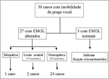
Out of the 30 studied cases, 17 had left vocal fold immobility, nine had right vocal fold immobility and four had bilateral immobility. In 27 patients electromyography showed immobility on the same side observed in the videolaryngoscopy. In three cases, tracings were normal. Of the 27 abnormal results, 24 showed neuropathic damage, two were compatible with central damage diagnosis and in one case the result indicated myopathic damage (Figure 1).
Figure 1. Flow chart of electromyographic interpretations of 30 cases of vocal fold immobility.
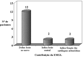
Graph 1. Contribution of LEMG to define diagnosis in Group A - With no defined cause for vocal fold immobility (n=16).
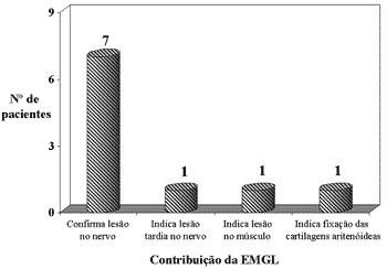
Graph 2. Contribution of LEMG to define diagnosis in Group B - Immobility caused by trauma defined by clinical history and complementary tests (n=10).
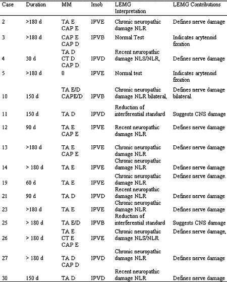
In Group A, out of the 16 patients with vocal fold immobility without defined cause, LEMG determined 12 cases of peripheral nerve damage (Cases 2, 4, 10, 12, 13, 14, 19, 21, 23, 26, 27 and 30), two cases of cricoarytenoid fixation (Cases 3 and 5), and in two cases (Cases 11 and 25) it indicated central nervous system damage (Table 1, Graph 1).
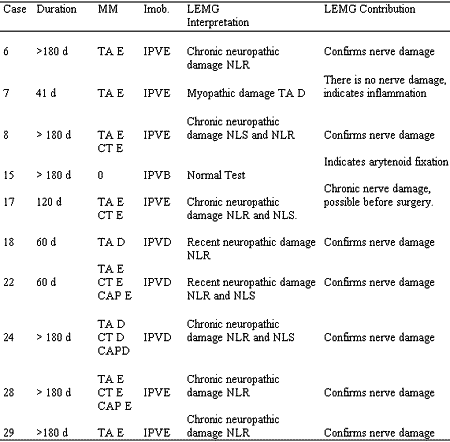
In Group B, which consisted of 10 cases with clinical history that indicated vocal fold immobility caused by trauma, we observed that LEMG confirmed the nerve damage in 7 cases (Cases 6, 8, 18, 22, 24, 28 and 29), suggested nerve lesion before a likely trauma by compression (Case 17), indicated muscle damage in one case (Case 7) and cricoarytenoid fixation in one case (Case 15) (Table 2 - Graph 2).
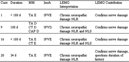
In Group C, with four cases of tumor compression as diagnostic hypothesis, LEMG revealed indications of neuropathic damage of chronic nature, suggesting compression in 100% of the cases. In Case 20, we detected a discrepancy between recent onset of symptoms and results of LEMG, which suggested an older damage (Table 3).Table 1. Group A - Vocal fold immobility with no defined cause (n=16).
Key Imob. - Immobility, IPVE - Left vocal fold immobility; IPVD - Right vocal fold immobility; IPVB - Bilateral vocal fold immobility; NLR - Recurrent laryngeal nerve; NLS - Superior laryngeal nerve; d - days; CNS - central nervous system; Duration - time from onset of complaints to electromyography; LEMG - Laryngeal Electromyography. MM. -muscles with abnormalities, TA - thyroarytenoid muscle, CT- cricoarytenoid muscle, CAP - posterior cricoarytenoid muscle, D -right, E- left, D/E - left and right, d - days.
Table 2. Group B - Vocal fold immobility caused by possible mechanical trauma (n=10).
Key Imob. - Immobility, IPVE - Left vocal fold immobility; IPVD - Right vocal fold immobility; IPVB - Bilateral vocal fold immobility; NLR - Recurrent laryngeal nerve; NLS - Superior laryngeal nerve; d - days; CNS - central nervous system; Duration - time from beginning of complaint to electromyography; LEMG - Laryngeal Electromyography. MM. -muscles with abnormalities, TA - thyroarytenoid muscle, CT- cricoarytenoid muscle, CAP - posterior cricoarytenoid muscle, D -right, E- left, D/E - left and right, d - days.
Table 3. Group C - Vocal fold immobility caused by tumor compression (n=4).
Key Imob. - Immobility, IPVE - Left vocal fold immobility; IPVD - Right vocal fold immobility; IPVB - Bilateral vocal fold immobility; NLR - Recurrent laryngeal nerve; NLS - Superior laryngeal nerve; d - days; Duration - time from beginning of complaint to electromyography; LEMG - Laryngeal Electromyography. MM. -muscles with abnormalities, TA - thyroarytenoid muscle, CT- cricoarytenoid muscle, CAP - posterior cricoarytenoid muscle, D -right, E- left.
DISCUSSION
Since the original description by Weddel and Feinstein26 in 1944, many studies have shown the importance of LEMG in the neurolaryngological assessment, both for diagnostic conclusion and for prognosis assessment3, 7, 10-23, 25-29, even though few clinicians are familiar with it in Brazil. The assessment of laryngeal neurological abnormalities is complex and requires laryngological, neurological and electrophysiological assessments that go beyond the larynx in many situations12, 20, 25.
It is a consensus among authors that one immovable vocal fold may be the result of central control, innervation, muscle affections or even poor functioning of cricoarytenoid joint. However, the expression "vocal fold paralysis" is frequently used indiscriminately. Laryngoscopy and clinical otorhinolaryngological assessment can check the presence of immobility and infer the possible cause, taking into consideration that the incidence of vocal fold immobility without clinically defined cause is high.
LEMG enables differentiation between innervation affections (paralysis), muscle impairment (myopathy) and arytenoid cartilage fixation (normal electromyography test), supporting the Otorhinolaryngologist in defining the diagnosis and clinical management.
In our study, we observed that in group A, comprising 16 patients with no clinically defined cause, LEMG determined neuropathic alterations in 12 patients, two central nervous affections and two normal tests, suggesting fixation of arytenoid cartilages.
In Group B, out of the 10 patients with vocal fold immobility by trauma, defined by clinical history and supplementary tests, 8 patients presented electromyography abnormalities suggestive of neuropathy, one had normal electromyography test (Case 15), and one presented electromyography tracing indicative of myopathy (Case 7).
Case 15 was included in Group B because he presented one immovable vocal fold and vocal abnormalities that started immediately after a trauma caused by a hanging attempt. LEMG revealed normal test, suggesting fixation of cartilages.
Case 7 presented left vocal fold immobility and vocal abnormality after thyroidectomy. Electromyography results defined myopathic cause (short-term polyphasic potentials), and the local trauma to the muscle fibers was attributed to the orotracheal intubation, as previously described in the literature28.
In Group C, comprising 4 cases of immobility possibly caused by tumor compression, LEMG confirmed clinical diagnosis in all of them.
In most cases, results were of neuropathic damage, with characteristics compatible with the period from onset of symptoms to performance of LEMG, that is, the electromyography detected chronic damage in patients whose history was older. However, in two cases - Cases 17 and 20, electromyography results were not compatible with the history reported by the patients, suggesting that the process could have been older.
In Case 20, the cause of immobility by tumor compression (thymus tumor diagnosed by CT scan), the results of LEMG were compatible with chronic damage, differently from the complaint of dysphonia reported by the patient for the past 34 days.
In Case 17, the patient was submitted to thyroidectomy and according to his report, he presented immediate vocal abnormality. LEMG revealed chronic damage to recurrent and superior laryngeal nerve, suggesting lesion before the surgery. This condition illustrated the application of the method for legal purposes.
Electromyography characteristics that were discrepant concerning duration of history in the two previous cases were compatible to the ones found in the other subjects, whose etiology was tumor compression. Although few cases have been assessed earlier, electromyography abnormalities in traumatic cases, in which the lesion is suddenly present, were consistent with the onset of clinical complaints. It made us conclude that in cases of tumor compression, the clinical manifestation may surge much longer after the electromyography abnormalities. This fact is probably due to slow and gradual impairment of axons, enabling sprouting of collateral prolongation in the myoneural junction and the clinical compensation that the other laryngeal muscles, by increased synergetic action or reduction of antagonist, may provide to the beginning of the process.
In cases in which the installation of the neuropathic process took place suddenly, the clinical repercussion was evident and it was always reported by the patient, allowing no room for the adaptation process described above.
Prognostic definition depends on the comparison with expected electromyography patterns in the different evolution stages of the paralysis, similarly to what happens with the facial nerve. Many authors experimentally studied the prognosis in animal larynges13, 14, 19. However, these studies present conflicting results. Which criteria provide clinical spontaneous recovery of immovable vocal folds? Which criteria indicate unfavorable clinical evolution? Is the restoration of vocal fold mobility always followed by normal electrophysiology? Do electromyographic signs indicative of recovery correspond to clinical improvement of the laryngeal function? These are questions still imprecisely answered.
The prognostic study of laryngeal immobility depends on the electromyography result determined chronologically at representative intervals of nervous degeneration and regeneration, already known for other nerves1, 2, 4, 5.
Out of the 30 studied patients, 24 underwent electromyography 90 days after the onset of symptoms. The distribution of our sample (second columns of Tables 1, 2, and 3) revealed that neurolaryngological assessment usually takes place late in our country. We interpreted this issue as a result of the fact that vocal manifestations found in laryngeal immobility, especially in unilateral cases, are mild and because of tolerability and social acceptance, the patients tend to postpone the visit to a specialized medical center. In addition, when the medical resources are required, some teams are not aware of the contribution that the electromyography may bring because they are not familiarized with the standardized neurolaryngological assessment and the use of LEMG as an important diagnostic tool.
CONCLUSION
Concerning the diagnostic contribution of electromyography in 30 cases of patients with vocal fold immobility, we concluded that:
- LEMG diagnosed peripheral neuropathic damage, central neuropathic damage or cricoarytenoid fixation in all cases of immovable vocal fold without defined cause;
- In cases of immovable vocal fold caused by clinically defined mechanical trauma, LEMG confirmed peripheral neuropathic damage in 70% of the cases, indicated peripheral neuropathic damage of slow progression, suggesting compression in 10% and determined other causes in 20% (myopathy and cricothyroid fixation) of the cases.
- In patients with immovable vocal fold by clinically defined tumor compression, LEMG confirmed neuropathic damage of chronic installation.
- LEMG is an important diagnostic tool and depends on clinical correlation.
- Patients with vocal fold immobility do not search specialized medical assistance upon onset of symptoms in Brazil and LEMG is not normally performed in the initial stages of the disease.
REFERENCES
1. Desmedt JE, Borenstein S. Collateral innervation of muscle fibers by motor axons of dystrophic motor units. Nature 1973;(246):500-501.
2. Dumitru D. Electrodiagnostic medicine. 1 ed. Hanley-Belfus Inc.; 1995:229-234.
3. Faaborg-Andersen K. Electromyographic investigation of intrinsic laryngeal muscle in humans. Acta Physiol Scand (suppl) 1957;41, suppl 140.
4. Gorio A, Carmignoto G, Finesso M, Polato P, Nunzi MG. Muscle reinnervation II. Sprouting, synapse formation and repression. Neuroscience 1983;(8):403-416.
5. Gorio A, Marini P, Zanoni R. Muscle reinnervation III. Motoneuron sprouting capacity, enhancement by exogenous gangliosides. Neuroscience 1983;(8):417-429.
6. Gupta SR, Bastian RW. Use of laryngeal electromyography in prediction of recovery after vocal cord paralysis. Muscle & Nerve 1993 Set;977-978.
7. Hirano M, Nozoe I, Shin T, Maeyama T. Electromyographic findings in recurrent laryngeal nerve paralysis. A study of 130 cases. Practice Otologia Kyoto 1974;(67):231-242.
8. Kimaid PAT, Resende LAL, Quagliato EMAB, Viana MA, Crespo NA, Wolf A. Eletromiografia Laríngea: Estudo normativo do potencial de unidade motora dos músculos tireoaritenoideus e cricotiroideus. Arq Neuropsiquiatr 2001;59(3B):58-59.
9. Kimura J. Electrodiagnosis in Diseases of Nerve and Muscle. 2ed. Philadelphia: F.A. Davis Company; 1989. p.249-274.
10. Kotby MN, Fadly E, Madkour O, Barakah A, Khidr A, Alloush T, Saleh M. Electromyography in neurolaryngology. Journal of Voice 1992;(2)159-187.
11. Koufman JA, Walker FO, Johardji GM. The cricothyroid muscle does not influence vocal fold position in laryngeal paralysis. Laryngoscope 1995;(105):368-372.
12. Lovelace RE, Blitzer A, Ludlow CL. Clinical Laryngeal Electromyography. In: Blitzer A, Sasaki CT, Fahn S, Brin A, Harris KS. - Neurologic Disorders of the Larynx. New York: Thieme Publishers; 1992. p.66-81.
13. Mu L, Yang S. Electromyographic study on end-to-end anastomosis of the recurrent laryngeal nerve in dogs. Laryngoscope 1990;100:1009-1017.
14. Mu L, Yang S. An experimental study on the laryngeal electromyography and visual observations in varying types of surgical injuries to the unilateral recurrent laryngeal nerve in the neck. Laryngoscope 1991;101:699-708.
15. Munin MC, Murry T, Rosen CA. Laryngeal electromyography. Diagnosis and prognostic applications. Otolaryngologic Clinics of North America 2000;4:759-771.
16. Rontal E, Rontal M, Silverman B, Kileny PR. The clinical differentiation between vocal cord paralysis and vocal cord fixation using electromyography. Laryngoscope 1993; 103:133-137.
17. Schaefer SD. Laryngeal electromyography. Otolaryngologic Clinics of North America 1991;5:1053-1057.
18. Schweizer V, Woodson GE, Bertorini TE. Single-fiber electromyography of the laryngeal muscle. Muscle & Nerve 1999;22:111-114.
19. Shindo ML, Herzon GD Hanson DG, Cain DJ, Sahgal V. Effects of d dennervation on Laryngeal muscle: A Canine Model. Laryngoscope 1992;102:663-669.
20. Simpson DM, Sternman D, Graves-Wrigth J, Sanders I. Vocal Cord paralysis: clinical and electrophysiologic features. Muscle & Nerve 1993;16:952-957.
21. Traissac L, Gioux M, Rovira HP, Henry C, Bertrand B. L'Electromyographie (EMG) du larynx dans le diagnostic des immobilités larynges spontanées ou post-thyroïdectomie. Revue de Laryngologie 1991;112(3):205-207.
22. Tucker HM. Vocal Cord Paralisis-1979: Etiology and Management. Laryngoscope 1980;89:504-507.
23. Verhulst J, Gioux M, Castro E, Quintero R, Traissac L. Intérêt et role de l'electromyographie dans l'evaluation d'um trouble de la mobilité laryngée et son pronostic. Ver Laryngol Otol Rhinol 1995;116(4):289-292.
24. Wolf AE, Crespo AN, Qualiato E, Kimaid PA, Viana M. Eletromiografia Laríngea: Aspectos Técnicos. In: 35ª Congresso Brasileiro De Otorrinolaringologia, Rio Grande do Norte, 2000. Temas Livres, Rio Grande do Norte, Sociedade Brasileira de Otorrinolaringologia, 2000. p. 95.(Resumo, TL-L-12)
25. Woodson GF. Clinical value of laryngeal EMG is dependent on experience of the clinician. Arch Otolaryngol Head Neck Surg 1998;124(4):476.
26. Weddel GB; Feinstein B. The electrical activity of the voluntary muscles in man under normal and pathological conditions. Brain 1944;67:178-242.
27. Yin SS, Qiu WW, Stucker FJ. Value of electromyography in differential diagnosis of laryngeal joint injuries after intubation. Ann Otol Rhinol Laryngol 1996;105:446-451.
28. Yin SS, Qiu WW, Stucker FJ. Major patterns of laryngeal electromyography and their clinical application. Laryngoscope 1997;107:126-136.
29. Yin S, Stucker F, Qiu WW, Batchelor BM. Clinical Evaluation of Neurolaryngological Disorders - Ann Otol Rhinol Laryngol 109:832-838, 2000.
[1] Ph.D., Professor, Coordinator of the Discipline of Otorhinolaryngology and Head and Neck Surgery, UNICAMP.
[2] Speech and voice pathologist, Master Studies in Biomedical Sciences under course, Medical School, UNICAMP.
[3] Doctorate studies in Medical Sciences - Neurology under course, UNICAMP.
[4] Ph.D., Professor, Department of Neurology, UNICAMP.
[5] Neurologist, Department of Neurology, UNICAMP.
Address correspondence to: Aline E. Wolf - Rua Jaime Sequier, 508. Parque Taquaral - Campinas - SP - 13070-140 Tel (55 19)3241-8600 / 9790-1872
Fax: (55 19) 3241-7152 - E-mail: alinewolf@aol.com
Study financially supported by CNPQ.
