

Year: 2002 Vol. 68 Ed. 3 - (7º)
Artigo Original
Pages: 332 to 335
 PDF PT
 Open rhinoplasty
Author(s):
Gilberto U. Pizarro(1),
Isabela M. DeVuono(2),
Márcia G. Moysés(3),
Reginaldo R. Fujita(4)
Keywords: rhinoplasty, open rhinoplasty, indications, surgery
Abstract:
Introduction: The open rhinoplasty began in the decade of 1930; in 1956 Secer published the technique of open rhinoplasty for the first time, even so it won credit starting from 1970.As indications of the open rhinoplasty are: nose without projection of the cartilage of the point or that requests functional progress of the nasal cartilage; twisted nose; traumatic nose; cases of secondary rhinoplasty; congenital deformities including labial rift; nasal valvuloplasty and learning of the rhinoplasty. Study design: Clinical retrospective. Material and method: In this study the authors bring the open rhinoplasty standing out its indications, advantages, disadvantages and the experience in 16 cases. The open rhinoplasty when, correct suitable, allows the global manipulation of the nasal structures and its corrections. The open rhinoplasty demonstrates to be a technique of simple manipulation and easy learning. Conclusion: In our sample complications were not observed demonstrating to be a safe surgery.
![]()
Introduction
The nose presents individual anatomical variations that require different surgical approaches according to the type of structural abnormality that is detected. Close rhinoplasty techniques are very popular but in cases of nasal deformity in which it is necessary to have three-dimensional exposure of skin and nasal substructure relation, external access enables better visualization to the surgeon. Open rhinoplasty was initially performed in the 30's by Secer (1956), the first one to publish this technique. There was gradual evolution of the technique and in 1972, it was introduced by Padovan in the North America. Anderson, Goodman, Johnson and Toriumi, among others, refined and popularized the technique in the last half of the past century. The indications for open rhinoplasty are: nose without tip cartilage projection or that requires functional advancement of nasal cartilage; twisted nose, post-trauma nose, cases of secondary rhinoplasty, congenital deformities, including cleft lip; nasal valvuloplasty and rhinoplasty learning.
The present study highlighted the indications, advantages, disadvantages and the experience in 16 cases submitted to this technique.
Material and Method
We conducted a retrospective study with 16 patients submitted to open rhinoplasty, postoperatively followed up for 5 years and 6 months, assessing the outcomes. Patients were divided in groups according to indication: isolated twisted nose, post-trauma twisted nose, secondary rhinoplasty.
Surgical technique of open rhinoplasty consisted of the following:
Initially, left and right marginal incisions that are connected with a columellar incision, enabling the elevation of the skin-perichondral envelope. Columella incision is made on the middle portion (narrowest region), with an inverted "V" on the midline, continued by bilateral marginal incisions.
After incision, we start the elevation of the skin-perichondral envelope from the bone and cartilaginous skeleton. Dissection is carefully performed removing all remaining fibrous fibers. Once the dissection reaches the caudal margin of the nasal bones, the periosteum is sectioned and elevated as part of the cover of soft tissues.
At that time, the necessary nasal deformity corrections are performed (septoplasty, nasal tip refining, reduction or enlargement of nasal dorsum, and osteotomy).
Closure starts by columella incision (nylon 6-0), followed by closure of marginal incision (catgut 4-0).
Results
In the present study, 16 patients underwent open rhinoplasty, and the indications were: isolated twisted nose (five); post-trauma twisted nose (nine), and secondary rhinoplasty (two). We observed the following: longer surgical time because the technique required careful surgical manipulation of the structures; nasal tip edema that regressed at late postoperative observation; easy surgical manipulation of cartilaginous and bone structures, and better visualization of facial and nasal structure relation. The techniques used were promptly understood by the observers because they provided better understanding of three-dimensional spatial relations of nasal skin and substructure and therefore, it was easy to correlate anatomical variations and nasal esthetics. Postoperative follow-up of these patients were conducted weekly in the first month, monthly for 5 months and annually for 5 more years. Surgical scars became imperceptible in the late postoperative period. We did not have immediate or late postoperative complications.
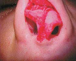
Figura 1.
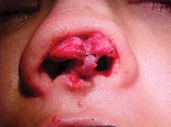
Figura 2.
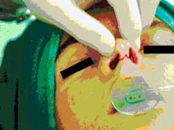
Figura 3.
Discussion
The indications for open rhinoplasty are nose without tip cartilage projection or that requires functional advancement of nasal cartilage; twisted nose; post-trauma nose; cases of secondary rhinoplasty; congenital deformities, including cleft lip; nasal valvuloplasty, and rhinoplasty learning. The open approach for rhinoplasty enables the necessary exposure to diagnose the existing deformities and to perform the maneuvers required by open rhinoplasty. After elevation of skin-perichondral envelope (Figure 1), the bone and cartilaginous framework are brought to complete view with their components and true anatomical position. Bone/cartilaginous deformities and asymmetries are apparent and the surgeon can quickly correlate the superficial topography to the anatomy of the underlying skeleton.
In open rhinoplasty, the emphasis is put on repairing the nasal contour, rather than reducing it. The open approach provides good exposure enabling precise structural modifications. Sophisticated manipulations of cartilage enable the performance of techniques such as modification of the dome through columella suture and alar cartilage grafts made with precision and symmetry, under complete view of the structures.
The projection of cartilage may be maintained or enlarged by using autologous cartilage grafts sutured at strategic positions. The surgeon has the cartilage projection under control, so he/she can determine the height of the dorsum which can be primarily modified, or the cartilage projection may be brought towards the favorable esthetical relation of the dorsum. The global result is nasal reduction maintaining the skeletal structure and making the skin adapt to the shape of the underlying structure, making the nose more harmonic, defined and elegant.
Open approach is also revolutionary in its impact on teaching of rhinoplasty. In closed rhinoplasty, experienced surgeons tend to agree that many years of experience are required to use the technique, since it relies on tactile sensations, differently from the open technique that allows direct visualization, technique demonstration and assessment of effects (Figure 2).
Open rhinoplasty has some disadvantages, such as longer surgical time caused by manipulation of nasal structure reconstruction and transcolumellar incision that requires careful dissection at the subperichondral plan. On the first postoperative days, it presents additional nasal tip edema that is rarely persistent. Esthetically speaking, the technique is normally attacked for leaving a scar on the columella (Figure 3), even though in most cases it becomes imperceptible with time.
The possible complications are skin necrosis of the columella by association of factors such as hematoma, infections and tense nasal tip, which are not observed in close rhinoplasty.
In the present study, 16 cases submitted to open rhinoplasty presented longer surgical time owing to careful manipulation required by the technique and nasal tip edema that regressed in the late postoperative period. Surgical manipulation of the cartilaginous and bone structures and observation of facial and nasal structure relations were easily achieved. The techniques used were promptly understood by the observers because they provided better understanding of three-dimensional spatial relations of nasal skin and substructure and therefore, it was easy to correlate anatomical variations and nasal esthetics. Surgical scars were imperceptible in late postoperative period. There were no early or late postoperative complications.
Conclusion
Open rhinoplasty enables global manipulation of nasal structures and the correction of its deformities, providing better visualization to the surgeon and, as a consequence, more opportunities to learn.
Our study did not show complications concerning the technique, proving that it is a safe surgery when correctly indicated and when dissection is made at the appropriate plans.
REFERENCES
1. Cachay-Velasquez H. Surgical lengthening of the short columella: Division of the depressor septi muscles. Eur Arch Otorhinolaryngol 1992;249:336.
2. Chen T-H, Chen Y-R. Extended open-tip rhinoplasty with three V-flaps for secondary correction of bilateral cleft lip nasal deformity. Ann Plast Surg 1996;37:482.
3. Cronin TD. Lengthening columella by use of skin from nasal floor and alae. Plast Reconstr Surg 1958;21:417.
4. Eskeland G, Borchgrevink H, Abyholm FE. Columella lengthening in bilateral cleft lip patients. Experience with the forked flap procedure. Scand J Plast Surg 1979;13:429.
5. Friedman GD, Gruber RP. A first look at the open rhinoplasty technique. Plast Reconstr Surg 1988;82:973.
6. Jugo S. Fundamental surgical technique. In Jugo S (ed.) Surgical atlas of external rhinoplasty: Decortication approach. Churchill Livingstone: Edinburgh; 1995. p. 32-3.
7. Maniglia AJ, Maniglia JJ, Maniglia JV. Rinoplastia estética, funcional e reconstrutora.1ª edição, Rio de Janeiro: editora Revinter; 2001. p. 182-96.
8. McComb H. Primary repair of the bilateral cleft lip nose: A 15-year review and a new treatment plan. Plast Reconstr Surg 1990;86:882.
9. Miliard DR Jr. Cleft craft II. Little, Brown: Boston; 1977. p 524, 685.
10. Millard DR. Columella lengthening by a forked flap. Plast Reconstr Surg 1958;22:454.
11. Perlman PW, Nathan MJ. Cosmetic rhinoplasty using the external approach. Ear Nose Throat J 1991,70:425.
12. Reese BR, Koltai PJ, Pames SM, Decker JW. The external rhinoplasty approach for rhinologic surgery. Ear Nose Throat J 1992;71:408.
13. Rohrich RJ, Gunter JP, Friedinan RM. Nasal tip blood supply: An anatomic study validating the safety of the transcolumellar incision in rhinoplasty. Plast Reconstr Surg 1995;95:795.
14. Shin KS, Lee CH. Columella lengthening in nasal tip plasty of orientals. Plast Reconstr Surg 1994;94:446.
15. Smith O, Goodman W. Open rhinoplasty: its past and future. J Otolaryngol 1993;22:21.
16. Teichgraeber JF, Riley WB, Russo RC. External rhinoplasties: indication for use. Br J Plast Surg 1992;45:47.
17. Toriumi DM, Mueller RA, Grosch T, Bhattacharyya TK. Larrabee WF. Vascular anatomy of the nose and the external rhinoplasty approach. Arch Otolaryngol Head Neck Surg 1996;122:24.
18. Trenite GJN, Paping RHL, Trenning AH. Rhinoplasty in the cleft lip patient. Cleft Palate Craniofac J 1997;34:63.
19. Van der Meulen JC. Columellar elongation in bilateral cleft lip repair: Early results. Plast Reconstr Surg 1992;89:1060.
[1] Master studies under course, UNIFESP - EPM.
[2] Resident Physician, Hospital Paulista de Otorrinolaringologia.
[3] Resident Physician, Hospital Paulista de Otorrinolaringologia.
[4] In charge of the Residence Service, Hospital Paulista de Otorrinolaringologia.
Affiliation: Hospital Paulista de Otorrinolaringologia
Address correspondence to: Gilberto Ulson Pizarro, rua Zacarias de Góis, 1526 - Campo Belo - São Paulo - SP - Tel.(55 11) 241.1870 Tel/fax: (55 11) 5561.4953 E-mail: gapotorrinoc@osite.com.br
Study presented at 35º Congresso Brasileiro de Otorrinolaringologia - October 2000 - Natal.
Print: ![]()