

Year: 2011 Vol. 77 Ed. 1 - (14º)
Artigo Original
Pages: 84 to 87
 PDF PT
  PDF EN
Is facial pattern a predisposing factor for otitis media with effusion in children?
Author(s): Cláudio de Góis Nery1; Fernando Stefanato Buranello2; Cícero Pereira3; Renata Cantisani Di Francesco4
Keywords: otitis media with effusion, eustachian tube, middle ear ventilation.
Abstract:
Abnormalities in craniofacial morphology are associated with Eustachian tube dysfunction and otitis media with effusion (OME). Aim: to evaluate the relationship between facial pattern and craniofacial growth direction, and OME in children with enlarged tonsils and adenoids (ETA). Methods: Clinical prospective survey in 79 children (41 male and 38 female), ranging from 4 to 10 years of age, with tonsil and adenoid enlargement (Brodsky's grades III and IV). Forty children presented with OME (study group) and 39 did not (control group). Cephalometric analysis was used to determine the facial pattern. Results: There was no correlation observed between facial pattern and OME (c 2 = 0.25 p = 0.88). Facial Axis was larger in the OME group (F(1.75) = 3.68 p = 0.05) and the Lower Anterior Facial height was smaller (F(1. 75) = 3.99 p = 0.05) in children with otitis media with effusion. Conclusions: There was no correlation between OME and facial pattern in children with ETA although a more horizontal facial growth direction, and a smaller lower anterior facial height was observed consistently among subjects in this group. This suggests that abnormal positioning of the eustachian tube influences the development of OME in children with ETA.
![]()
INTRODUCTION
Abnormalities in the craniofacial morphology are associated with Eustachian tube dysfunction and otitis media with effusion (OME)1. Pautow reported that the Eustachian Tube morphology is associated with the shape of the head2. Brachyfacial adult individuals tend to have narrower tubes, and consequently more otitis. These results corroborate the statement of other authors who reported that otitis media is common in Brachyfacial patients3,4.
One of the most frequent conditions associated with OME is enlarged pharyngeal and palatine tonsills5.
Nonetheless, not all children in school age with enlarged pharyngeal and palatine tonsils develop OME6.
Based on these statements, it is possible to propose the hypothesis that the head's shape would be a predisposing factor to OME.
The goal of the present study was to establish whether or not there are associations between the facial pattern and OME in children with enlarged pharyngeal and palatine tonsils.
Sample
This study was approved by the Ethics in Research Committee analyzing research projects - CAPPesq from the Clinical Board of the Medical School of the University of São Paulo, under protocol # 738/04.
In this clinical, prospective, case-control study we had 79 children (41 boys and 38 girls), aged between 4 and 10 years, being treated at the ENT department of the Medical School of the University of São Paulo.
All the patients had chronic upper airway obstruction because of enlarged pharyngeal and palatine tonsils. They were randomly selected from the patients' waiting list for adenotonsillectomy. 40 patients with OME and indication for ventilation tubes (study group) and 39 individuals without such indication (control group). All the individuals had at least 80% of nasopharyngeal obstruction caused by enlarged pharyngeal tonsils, established by the side x-ray taken. We included only those patients with palatine tonsil enlargement in grades III-IV, from the Brodsky classification7.
The study group was made up of 40 individuals who had had OME for more than 3 months, with air-bone gaps of at least 20 dB, no stapes reflex and flat tympanometry (type B). Thirty-nine individuals selected for the control group did not have OME diagnosed during physical exam (otoscopy) and confirmed by normal audiometry and tympanometry. The waiting list in this institution for adenotonsillectomy, with or without a ventilation tube, varied between 8 and 10 months. Usually the indications for a ventilation tube in this department follow the guidelines from the American Academy of Otorhinolaryngology concerning the insertion of ventilation tubes. The recommendation to insert a ventilation tube is a valid indicator in auditory tube dysfunction. The exclusion criteria were: personal or family history of cleft palate, or another craniofacial abnormality; chronic medical conditions; prior oral, nasal, pharyngeal or craniofacial surgery; prior or current orthodontic treatment. Syndromic patients with our without neurological disorders were taken off.
METHODS
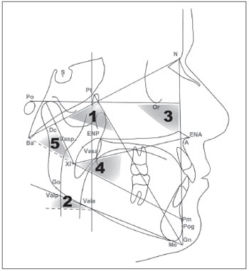
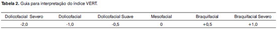
All the individuals were submitted to otolaryngological anamnesis, physical exam and side X-Ray. The radiographs were taken from each individual positioned in the cephalostate with the mandible in centric occlusion and the lips at rest. All the radiographies were digitalized and the cephalometric points were identified (Table 1). We used the Radiocef 2003, 5th edition software in order to do our analysis. The cephalometric analysis was made up exclusively from the angular measures used to establish facial typology according with Ricketts et al.8. The VERT index (vertical growth coefficient) is determined by means of an arithmetic average of the difference between the angles and their normal values8. Five factors are employed in establishing the VERT index: (1) facial axis (EF), (2) facial depth (PF), (3) mandible plane angle (PM), (4) inferior facial height (AFI), and (5) mandible arch (AM) (Figure 1). After acquisition of the cephalometric data, each factor was compared to its respective standard deviation, and to this difference we assigned a positive or negative sign to indicate direction as brachycephalic or dolicofacial. Each value was broken down by its respective standard deviation, the results from the five factors are submitted to the simple average and the result determines the VERT index, which will be compared to reference values (Table 1) in order to determine the facial pattern.
Figure 1. Cephalometric analysis.
The descriptive statistical analyses (mean ± standard deviation) were calculated from the values observed for each factor. The mean difference for independent groups was calculated using the t Student test, p values <0.05 were considered statistically significant. Correlations between the variables were made by means of the chi-square and Person's test.
The data was analyzed using the Statistical Package for Social Sciences (SPSS for Windows 12.0; SPSS Inc. Chicago, Illinois, USA).
RESULTS
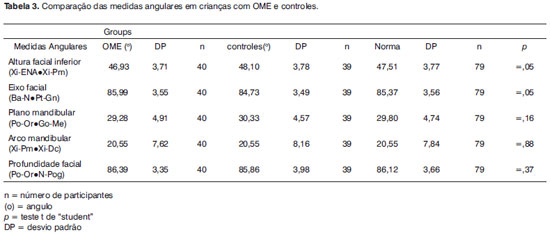
There was no relation between facial shape and otitis media with effusion (Table 2). Based on the angular measures, we noticed that the Facial Axis value, which determines mandible growth direction, was significantly larger, and the Inferior Facial Height value was significantly smaller in the OME group (Table 3), when compared to the control group.
DISCUSSION
We know that OME is more common in children because of the very characteristics of their Eustachian Tubes. During this stage of life, the tube is described as being more horizontal, in relation to the skull base, and narrower in diameter than it is in adults9.
More than half of the individuals in this study, including the control group had dolicofacial type. In other Brazilian studies, the dolicofacial type was also the predominant type4,10. The correlation between facial types and OME was not found in this study. Despite the prevalence of dolicofacial individuals in this study, there was a mild predominance of the brachyfacial pattern in the otitis group when compared to the control group. This finding is supported by other authors, who have also found a higher incidence of otitis media in the brachyfacial face pattern3,4,11. Other studies found different results12,13, with a vertical facial pattern (dolicofacial) seen in individuals with Eustachian Tube dysfunction. However, in those studies the enlargement of pharyngeal and palatine tonsils was not determined, and this may have influenced the results, as well as the sample itself. Pharyngeal obstruction and consequent oral breathing result in a more vertical facial growth in genetically predisposed individuals6,14.
The facial shape is genetically defined, because it depends on skull base shape. In this study, the sample was entirely made up of individuals with enlarged pharyngeal and palatine tonsils, with the goal of maintaining consistency among the individuals. The Facial Axis angular value was larger, while the Inferior Facial Height was lower in the OME group when compared to the control group. The facial axis describes the mandible position and, consequently, the facial development direction. It is known that the face develops in the forward and downward directions9, corroborating the findings of our previous study1. Higher Facial Axis values are associated with smaller faces. Similarly, a reduced inferior facial height may also result in a more horizontal facial growth pattern. The cartilaginous portion of the Eustachian Tube is located in the petrous-squamous portion of the temporal bone, located near the occipital and sphenoid bones15 ,16. These bones are part of the skull base and their proximity to the temporal bone explains the relationship between the head shape and the auditory tube. The skull base is the key for the growth and development of the facial structures, and its morphology can influence adjacent structures. This is known as the counterpart theory, in which the skull base determines the size of the maxilla and that of the mandible15. The auditory tube extends from the skull base (bone portion) all the way to the pharynx and its growth is associated with the development of the skull base and the nasomaxillary complex17.
The peritubal muscles (soft palate elevator and tensor muscles) go from the skull base all the way to the hard palate and cartilaginous portion of the auditory tube in the pharyngeal wall. The soft palate tensor insertion is more superior and distal to the lumen in adults when compared to children18. Shibahara and Sando (1988) reported that the angular insertion of these muscles can change the traction vector during tube opening and may also contribute to its dysfunction, predisposing the patient to otitis media19.
CONCLUSION
Based on the data hereby presented, we can conclude that there is no correlation between OME and the facial shape in children with enlarged pharyngeal and palatine tonsils, despite a mild predominance of the brachyfacial pattern in the group with otitis. The children with OME had a more horizontal facial shape, with a reduced facial height. These findings suggest that besides the enlargement of pharyngeal and palatine tonsils, Eustachian Tube dysfunction is also associated with craniofacial morphology. Any change in craniofacial growth direction may reduce Eustachian Tube function.
REFERENCES
1. Di Francesco R, Paulucci B, Nery C, Bento RF. Craniofacial morphology and otitis media with effusion in children. Int J Pediatr Otorhinolaryngol.2008;72:1151-68.
2. Pautow NA. About the forms of the Eustachian tube (translation from German). Z Hals-Nasen-Ohren.1925;11:467-77.
3. Stolovitzky JP, Todd, NW. Head shape and abnormal appearance of tympanic membranes. Otolaryngol Head Neck Surg. 1990;102:322-5.
4. Di Francesco RC, Sampaio PL, Bento RF. Correlation Between otitis media and craniofacial morphology and adults. ENT J.2007;86:738-43.
5. Niemelä M, Uhari M, Hannuksela A. Pacifiers and dental structure as risk factors for otitis media. Int J Pediatr Otorhinolaryngol. 1994;29:121-7.
6. Guilleminault C, Huang Y, Glamann C, Li K, Chan C. Adenotonsillectomy and obstructive sleep apnea in children: A prospective survey. Otolaryngol Head and Neck Surg.2007;136:169-75.
7. Brodsky L. Tonsillitis, tonsillectomy and adenoidectomy. In: Bailey BJ, editor. Head and Neck Surgery - Otolaryngology. Philadelphia: JB Lippincott,2001;Chapter 81.
8. Ricketts RM, Roth RH, Chaconas SJ, Schullog RJ, Engel GA. Orthodontic diagnosis and planning. Rocky Mountain, Rocky Mountain Data Systems.1982;1:53-118.
9. Maw AR, Bawden R. Factors affecting resolution of otitis media with effusion in children. Clin Otolaryngol.1994;19:125-30.
10. Atherino CCT. Anatomia da parede medial da órbita: profundidade dos forames etmoidais e canal óptico e suas relações com as dimensões cranianas e orbitárias no Brasil [tese]. São Paulo. Faculdade de Medicina da Universidade de São Paulo;2000.
11. Todd NW Jr. Cranial anatomy and otitis media. Am J Otol.1998;19:558-64.
12. Jonas I, Mann W, Münker G, Junker W, Schumann K. Relationship between tubal function, craniofacial morphology and disorder of deglutition. Arch Otorhinolaryngol.1978;218:151-62.
13. Mann W, Jonas I, Munker G. Growth influence on tubal function. Acta Otolaryngol.1979;87:451-5.
14. Brezolin D, Shapiro GG, Shapiro PA, Dassel SW, Furukkawa CT, Pierson WE, et al.. Facial characteristics of children who breathe through the mouth. Pediatrics.1984;75:622-5.
15. Enlow DH, Hans MG. Essentials of Facial Growth. Philadelphia: Saunders,1996.
16. Testut L, Latarjet A. Anatomia Humana. Barcelona: Salvat1976.
17. Kemaloglü YK, Kobayashi T, Nakajima T.Associations between the eustachian tube and craniofacial skeleton. Int J Pediatr Otorhinolaryngol. 2000;14;53(3):195-205.
18. Swartz JD, Rood SR. The morphometry and three-dimensional structure of the adult Eustachian tube: implications for function. Cleft Palate Craniofac J.1990;27:374-81.
19. Shibahara Y, Sando I. Histopathologic study of Eustachian tube in cleft palate patients. Ann Otol Rhinhol Laryngol.1988;97:403-7.
1. PhD in Sciences, Assistant Professor - Department of Orthodontics - UFG
2. MSc, Professor Coordinator of the Orthodontics Program
3. PhD in Experimental Social Psychology. Epidemiology and Statistics Consultant - UFG
4. PhD in Medicine; Professor at the Department of Otolaryngology of the Medical School of the University of São Paulo - FMUSP
Paper submitted to the BJORL-SGP (Publishing Management System - Brazilian Journal of Otorhinolaryngology) on February 21, 2010; and accepted on July 20, 2010. cod. 6938
Print: ![]()