

Year: 2010 Vol. 76 Ed. 5 - (27º)
Relato de Caso
Pages: 675 to 675
 PDF PT
  PDF EN
Angioleiomyoma of the nasal septum
Author(s): Carlos Roberto Ribeiro Navarro Júnior1, Adriano Santana Fonseca2, José Rodrigo Lordello de Mattos3, Nilvano Alves de Andrade4
Keywords: leiomyoma, nose neoplasms, nasal septum.
![]()
INTRODUCTION
Leiomyoma is a benign smooth muscle tumor, more commonly found in the uterus (95%), skin (3%), nutritional and gastrointestinal tracts (1.5%)1. It was initially described in the nasal cavity by Maesaka et al. in 19662.
The goal was to describe a case with clinical manifestations and histopathology findings of angioleiomyoma of the nasal septum, a rare benign neoplasia which represents less than 1% of all the leiomyomas in the human body3.
CASE PRESENTATION
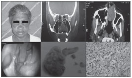
M.F.L.S., 62 years, female, African-descendant, she came to our ENT service complaining of a tumor in her left nasal cavity with six years of evolution. In the three initial years it had a progressive growth, associated with low volume epistaxis episodes. After such period, she developed nasal obstruction on the left side and facial pain. Upon physical exam and fibroscopy we noticed a brown, smooth, pedicled lesion on the left-side septum, well outlined, measuring approximately 4 x 2cm, completely occluding the left nasal cavity and pushing the nasal septum. CT scan of the paranasal sinuses showed a well-outlined soft tissue mass, pushing the septum and the lateral wall. Biopsy reported leiomyoma. Later on, the tumor was endoscopically resected, with a 1cm margin, considered adequate according to anatomical and pathological criteria. Microscopy showed polypoid fragments, coated by a single layer of cylindrical hair cells, typical pseudostratified, showing in the stroma, typical leiomyoma bundles around the thick walls of vessels. (Fig. 1)
Figure 1. Set of photographs from this patient, CT scan, late post-op, macroscopy and microscopy of the lesion.
DISCUSSION AND FINAL REMARKS
This is a slow growth tumor. The most common symptoms are: nasal obstruction, epistaxis, facial pain and headaches. He most frequent treatment for nasal septum angioleiomyoma is endoscopic resection with macroscopic margin, and this was the treatment option for this case - excision with macroscopic and microscopic free margins. Vascular leiomyomas are bundles of smooth muscle cells, relatively organized, and permeated by thick wall vessels4.
The nasal septum vascular leiomyoma is an extremely rare tumor, of uncertain origin5. Resection is the procedure of choice and it bears a high cure rate. The endoscopic procedure is a good option for small to moderate size tumors6.
REFERENCES
1. Ardekian L, Samet N, Talmi YP, Roth Y, Bendet E, Kronenberg J. Vascular leiomyoma of the nasal septum. Otolaryngol Head Neck Surg. 1996;114(6):798-800.
2. Barr GD, More IAR, Path FRC, McCallum HM, Path FRC. Leiomyoma of the nasal septum. J Laryngol Otol. 1990;104:891-3.
3. Bloom DC, Finley JC Jr, Broberg TG, Cueva RA. Leiomyoma of the nasal septum. Rhinology. 2001;39(4):233-5.
4. Campelo VES, Neves MC, Nakanishi M, Voegels RL. Angioleiomioma de cavidade nasal: relato de um caso e revisão de literatura. Braz J Otorhinolaryngol. 2008;74(1):147-150.
5. Singh R, Hazarika P, Balakrishnan R, Gangwar N, Pujary P. Leiomyoma of the nasal septum. Indian J Cancer. 2008;45:173-5
6. Timirlyaleev MKH. Angioleiomyoma of the nasal septum. Vestnik Otorinolaringol. 1973;35:106-10.
1. MD. Resident Physician in ENT and HNS - Santa Casa de Misericórdia da Bahia.
2. MD. ENT and Maxillo-facial surgeon. Preceptor at the ENT Residency Program - Santa Casa de Misericórdia da Bahia.
3. MD. ENT and Maxillo-facial surgeon. Preceptor at the ENT Residency Program - Santa Casa de Misericórdia da Bahia.
4. PhD in Surgery - USP, Head of the ENT Residency Program - Santa Casa de Misericórdia de Bahia.
Santa Casa de Misericórdia da Bahia
Send correspondence to:
Praça Conselheiro Almeida Couto 500 Nazaré
Salvador BA 40050-410
Paper submitted to the BJORL-SGP (Publishing Management System - Brazilian Journal of Otorhinolaryngology) on October 16, 2009
and accepted on December 15, 2010. cod. 6714
Print: ![]()
All rights reserved - 1933 /
2026
© - Associação Brasileira de Otorrinolaringologia e Cirurgia Cérvico Facial