

Year: 2002 Vol. 68 Ed. 2 - (15º)
Artigo Original
Pages: 251 to 253
 PDF PT
 Otosclerosis - stapedotomy results
Author(s):
José Ricardo Gurgel Testa 1,
I. Millas 2,
I.M. De Vuono 2,
M.E.L.R.B.V. Neto 2,
M.F. Lobato 2
Keywords: otosclerosis, stapedotomy, hearing improvement, stapes surgery
Abstract:
Introduction: Otospongiosis or otosclerosis is a common degenerative and hereditary disease of the labyrinthine capsule and occurs mainly in women with age between 20 and 30 years. In recent decades stapedotomy has increasingly tended to become the most used surgical technique to the treatment of otosclerosis. Thus, this study analyzes the results of 59 stapedotomies realized in Paulista Hospital of Otorhinolaryngology on the last 7 years. Study design: Clinical randomized. Materials and Method: Retrospective analysis of 59 patients with otosclerosis who underwent stapedotomy performed by the same surgeon and follow-up clinical and audiological. Results: Hearing improvement proved by audiogram air-bone gap closure in 53 patients (90%). The complications occurred were displaced prosthesis (7%); taste alterations (7%); facial paralysis (3%); vertigo (3%); prosthesis total extrusion (1,5%); tinnitus (1,5%); tympanic membrane perforation (1,5%). Conclusion: Stapedotomy has been seen as good therapeutic choice to the treatment to the conductive hearing loss by otosclerosis seeing that generally has low morbidity and high success rates, providing better life quality to this patients.
![]()
INTRODUCTION
Otosclerosis or otospongiosis is a hereditary degenerative disease of the labyrinthic capsule in which there are bone neoformation foci with increased local vascularization. Clinically, it affects between 0.5 and 1.0% of the population and it is bilateral in 70 to 85% of the cases. It is manifested between adolescence and the 4th decade of life, with the peak incidence between 20 and 30 years. The prevalence is higher in female subjects1.
The region of the otic capsule opposed to the oval window is the most frequently affected structure. The second most common site is around the oval window and the footplate of stapes. Incus and malleus are rarely affected1.
The first description of oval window stapes ankylosis was made by Antonio Valsalva in a necropsy in 1753. The first stapes mobilization was conducted by Kessekl in 1878. Politzer and Sibenmann, in 1900, condemned this kind of surgery, which was forgotten up to 1953, when Rosen used again the technique. It was John Sea in 1956, who introduced the technique of stapedectomy and in 1960 performed the first stapedotomy 1, 2.
In the past decades, stapedotomy has been used as the preferred treatment by many surgeons and it is widely used for treating otosclerosis 3.
Therefore, the present study intends to analyze the results of 59 stapedotomy procedures performed in a 7-year period, considering especially gap closure and postoperative complications and/or events.
MATERIAL AND METHOD
We assessed 59 patients who had otosclerosis and were submitted to stapedotomy in the period between March 1994 and March 2001, at Hospital Paulista de Otorrinolaringologia.
Patients were aged between 20 and 70 years, and the main incidence was between the ages of 31 and 50 years (52%). There were 42 female patients (71%).
Bilateral affection was detected in 48 patients (81%), but only three of them (5%) were submitted to surgery on both ears.
All surgeries were performed by the same otorhinolaryngologist surgeon who used the same surgical technique, as follows:
1. Local or general anesthesia (depending on the level of anxiety of the patient or anatomical abnormalities that complicated the procedure);
2. Infiltration of external acoustic canal skin with about 1.0ml of prilocaine chloridrate with phelipressin;
3. Classical incisions: 6 to 12 hours vertical and horizontal (closing them posteriorly) at about 1cm from the tympanic membrane;
4. Palpation of the ossicle chain to check rigidity;
5. Curettage of posterior-superior bone margin;
6. Mobilization of chorda tympani muscle;
7. Measurement of the height of the prosthesis (incus to plate);
8. Perforation of footplate;
9. Placement of the prosthesis (Teflon piston in 56 surgeries and in the other 3, gold prostheses);
10. De-articulation of the incus/stapes;
11. Removal of the upper portion of the stapes;
12. Placement of gelfoam around the window and the prosthesis;
13. Replacement of tympanic-meatal flap;
14. Application of gelfoam in the external acoustic canal with corticoid ointment and antibiotics, dressing.
Follow-up:
· Hospital discharge 24 hours after the surgery;
· First visit after 7 days;
· Removal of gelfoam after 20 days;
· Conduction of the first postoperative audiometry 30 to 40 days later;
· Bimonthly follow-up in the first 6 post-surgical months and then 6-month and annual follow-ups.
RESULTS
Out of the total of 59 cases, there was auditory improvement confirmed by gap closure in the audiometry of 53 patients (approximately 90%), and preoperative SRT mean was 64dB and postoperative SRT mean was 35dB.
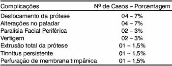
The complications observed are listed below:
In cases in which there was displacement of the prosthesis, we performed re-operation and observed improvement and gap closure in all cases.
There were 3 cases that needed to use hearing aids: two of them because of sensorineural hearing loss and one, to attenuate tinnitus.
Among the patients who did not experience improvement, there were two women who became pregnant right after surgery.
The patients who progressed with peripheral facial paralysis presented anatomical variations, such as facial nerve dehiscence, which were noted only during the surgery, but they progressed to spontaneous recovery after a maximum period of 60 days.
DISCUSSION
Stapedotomy has been continuously performed and described with good results3.
This surgical technique is normally used for the treatment of otosclerosis but it is equally useful for the treatment of maleolar ankylosis, a rare cause of conductive hearing loss that should be submitted to differential diagnosis with otosclerosis 4.
Stapedotomy, compared to stapedectomy, is less traumatic to the cochlea and promotes better long-term hearing (initially in high frequencies) in addition to being easy to perform because of less bleeding, better visualization of the oval window, shorter surgical time and minimum postoperative discomfort; it also offers less risk of prosthesis displacement2, 3, 5.
In patients with advanced otosclerosis, stapedotomy is a very useful procedure, because by improving auditory thresholds, patients may benefit from the use of less powerful hearing aids6.
Although otosclerosis is a disease that normally affects both sides, professionals tend to perform the surgery only on one side to avoid the risk of causing a bilateral sensorineural loss. Since this risk is lower with stapedotomy (instead of stapedectomy), some authors have advocated the benefits of bilateral surgery especially concerning sound localization and significant improvement of binaural hearing7, 8.
There seems to be a consensus that the technique that provides better results is the one the surgeon has mastered the best. Therefore, there are still authors who have better results with stapedectomy 2.
The general complications associated to otosclerosis surgery described in the literature2 are listed below, in decreasing order of frequency:
1. Prosthesis displacement;
2. Granuloma;
3. Total sensorineural hearing loss;
4. Permanent perforation of the tympanic membrane;
5. Vertigo;
6. Partial sensorineural hearing loss;
7. Tinnitus;
8. Facial nerve damage.
Some factors that may influence the postoperative period have also been described5:
· too big a gap, especially in high frequencies, which is more difficult to close;
· thick footplate may cause residual rigidity;
· long-term increased rigidity of ossicle chain because of the progression of the disease or by scaring of a "new" oval membrane.
The low rates of complications and the high rate of success in surgical treatment observed in our sample are in accordance with the data from the literature, showing that stapedotomy is an effective and safe method to surgically treat otosclerosis.
CONCLUSION
Otosclerosis is a hereditary degenerative disease of the labyrinthic capsule and the most effective treatment for the conductive hearing loss is surgery. Stapedotomy has proved to be a good therapeutic option since it normally presents low morbidity and high rates of success, providing better quality of life to otosclerosis patients.
REFERENCES
1. Beales PH. In Kerr AG, Booth JB. Scott-Brown's Disease of Ear, Nose and Throat. 5th edition. London: Butterworth;1987. p. 301-339.
2. Glasscock ME, Storper IS, Haynes DS, Bohrer PS. Twenty-five Years of Experience with Stapedectomy. Laryngoscope 1995;105:899-904.
3. Persson P, Harder H, Magnuson B. Hearing results in Otosclerosis Surgery after Partial Stapedectomy, Total Stapedectomy and Stapedotomy. Acta Otolaryngol 1997; (Stockh) 117:94-99.
4. Vicent R, LopezA, Sperling NM. Malleus Ankylosis: A Clinical, Audiometric, Histologic and Surgical Study of 123 cases. The American Journal of Otology 1999;20:717-725.
5. Ueda H, Miyazawa T, Asahi K, Yanagita N. Factors affecting hearing results after stapes surgery. The Journal of Laryngology and Otology 1999;113:417-421.
6. Ghonim MR, El-Degwy AA, El-Sharabasy AE. Far-Advanced Otosclerosis. ORL 1997;59:332-5.
7. De Bruijn AJG, Tange RA, Dreschler WA. Evaluation of Second-Ear Stapedotomy with the Glasgow Benefit Plot. ORL 1999;61:92-97.
8. De Bruijn AJG, Tange RA, Dreschler WA, Grolman W, Schouwenburg PF. Bilateral stapedotomy in patients with otosclerosis: a disability-oriented evaluation of the benefit of a second ear surgery. Clin Otolaryngol 1998;23:123-7.
1 Ph.D., UNIFESP-EPM.
2 Resident physician, Hospital Paulista de Otorrinolaringologia.
Affiliation: Hospital Paulista de Otorrinolaringologia
Address correspondence to: Rua das Uvaias, 130 ap. 22 - CEP 04055-110 - Tel: (55 11) 5078 9698 - E-mail: iedamillas@ig.com.br
Article submitted on November 14, 2001. Article accepted on February 7, 2002.
Print: ![]()